
Research article · 2022 · Full text
International Journal of Veterinary Sciences and Animal HusbandryHistological structure and evaluation of cellular layers of Large intestine in Guinea pig (Cavia porcellus)
Volume 7(4) · Pages 55–61 · DOI: 10.22271/veterinary.2022.v7.i4a.441 ↗
Research areasAnimal Nutrition and PhysiologyRabbits: Nutrition, Reproduction, HealthGut microbiota and health
Abstract
Author-writtenThe Aim of this study is to determine the histological structure in the wall layers of large intestine of guinea pig. The cecum and colon was characterized by a delicate lining that is moisturized by mucus and also by a gel that is a by-product of bacterial fermentation. This study was performed on the cecum and ascending and descending colon in five health animals. The cecum and colon wall was composed of four tunicae (mucosa, submucosa, muscularis and serosa, the tunica mucosa consist of epithelium, lamina propria and muscularis mucosa. The colon lumen lined by simple columnar epithelium with prominent goblet cells. The epithelial surface of the colon is covered by the openings of tubular intestinal glands which penetrate deep into the thick mucosa. The glands consist of absorptive cells that absorb water and salt, goblet cells were secrete the mucus, and scattered Paneth cells. Crypts of Lie beckon (intestinal glands) are a simple tubular glands is observe in most mammals, which extending from the muscularis mucosa through the thickness of the lamina propria and they open into the intestinal lumen. Muscularis mucosa, consisted of very thin two layers of inner circular and outer longitudinal smooth muscles, the greater thickness of the muscularis mucosa was in the adult type and the submucosa was consists of dense a irregular connective tissue. The muscular layer is consists of internal circular and external longitudinal layers of smooth muscles, which mean their thickness is decreased toward the last parts. Thus, the serous coat is a thin layer of loose connective tissue, the thickness was decreases toward the last parts.
Summary
Source-checkedPlain-language reading of the full paper, prepared with AI and checked against the text. Each point links to the authors' own words.
Why it matters
- Guinea pigs are classified as hindgut fermenters, meaning they process digested nutrients in the posterior part of their digestive tract. View evidence ↗
- The cecum serves as the initial segment of the large intestine, connecting the ileum to the ascending colon. View evidence ↗
- The colon is a muscular tube designed for waste removal and water absorption, consisting of connective tissue, blood vessels, lymphatic tissue, and specialized muscles. View evidence ↗
The question
- The study aims to determine the histological structure of the wall layers within the guinea pig's large intestine. View evidence ↗
What they did
- The research was conducted using five clinically healthy guinea pigs. View evidence ↗
- Tissue samples were collected from the cecum as well as the ascending and descending colon. View evidence ↗
- Tissue sections were cut to a thickness of 5 µm using a rotary microtome. View evidence ↗
- The study employed H&E staining for general histology, along with PAS, Alcian Blue, and Masson Trichrome stains to identify specific components like carbohydrates and connective tissue. View evidence ↗
What they found
- The walls of both the cecum and the colon are composed of four layers: the mucosa, submucosa, muscularis, and serosa. View evidence ↗
- The lumen of the colon is lined with simple columnar epithelium containing prominent goblet cells. View evidence ↗
- The large intestine contains numerous straight tubular glands, approximately 400 µm in size, extending from the muscularis mucosa to the mucosal surface. View evidence ↗
- The flat absorptive surface contains a moderate density of goblet cells, specifically 10 to 25 goblet cells for every 100 epithelial cells. View evidence ↗
- The guinea pig's cecum is a thin-walled, pouch-like organ capable of storing over 65% of the digestive tract's contents. View evidence ↗
- The mucosal glands of the cecum contain fewer goblet cells compared to other segments of the intestine. View evidence ↗
What it means
- The authors conclude that because of its simple structure, the cecum does not perform the majority of digestion but rather completes the process started in the predominant intestinal digestion. View evidence ↗
- Histochemical analysis shows that colonic goblet cells secrete both acidic and neutral mucins, as evidenced by positive PAS and Alcian blue reactions. View evidence ↗
Visual summary
1 visualsIllustrations explain the paper; they do not show study data.
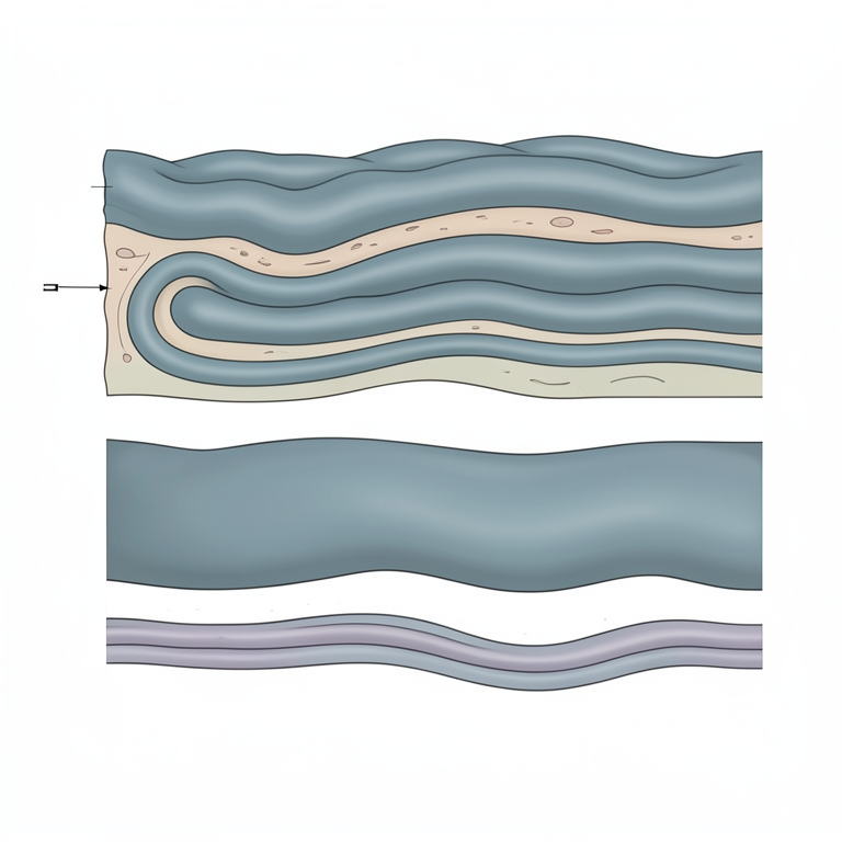
Layers of the Large Intestine Wall
AI-generated conceptual illustration (Guinea pig (Cavia porcellus) large intestine tissue). It explains the idea and does not show study data.
View source evidence ↗Figures & tables
From the original paper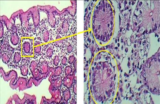 Figure 1
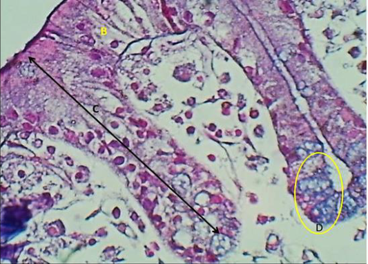 Figure 2
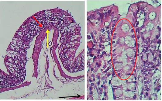 Figure 3
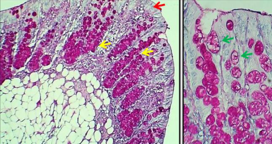 Figure 4
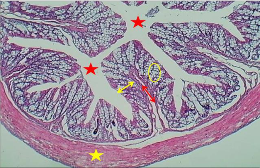 Figure 5
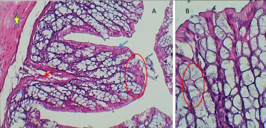 Figure 6
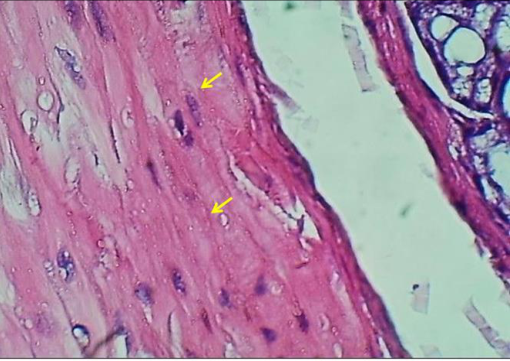 Figure 7
Introduction
Guinea pig, were classified as fermenters of the digested nutrients in the hindgut, guinea pigs are one of the most wonderful pets. The digestive tract in the species Cavia Purcell us, about the structure of pre diaphragmatic digestive segments [1] and the structure of the small intestine [2] . Another topic quite often addressed in the case of guinea pigs is represented by the adnexal glands of the digestive tract [3] As for the caecum, it is described in the literature as the first segment of the large intestine, which makes the connection between the ileum and the ascending colon [4,5,6] the guinea pig was classified as a species at the end of the 17th century and is due to a naturalist named Johann Polycarp Erxleben. The guinea pig is part of the Animalia Kingdom, Cordata, Mammalia Class, Rodentia Order, Hystricomorphic Suborder, Caviidae Family, Caviinae Subfamily, Cavia Genus [7] . The colon is structured as one long continuous hollow tube, and it is a muscular tube composed of lymphatic tissue, blood vessels, connective tissue, and specialized muscles for carrying out the tasks of water absorption and waste removal,The ascending colon is divided into 4 sections. The ampulla coli opens into the first section, approximately 10 cm long and having 3 longitudinal flat bands of muscular tissue (taeniae) that separate rows of haustra or sacculations [8] The mucosa of this section has small protrusions approximately 0.5 mm in diameters that are termed "warzen" or warts. This is unique to lagomorphs and greatly increase the surface is of the colon for absorption. The second section of colon has a single taenia and fewer, smaller haustra [8] there are segmental and haustral contractions that mechanically separates the ingesta into indigestible particles and liquid contents. It is a muscular area about 4 cm long, highly innervated, and vascular. Its mucosal surface has prominent longitudinal folds and goblet cells. It opens into the fourth section of ascending colony that is indistinguishable histologically from the transverse and descending colon [9] . Aim for this research, describe the histological structure of the large intestine cecum and colon in guinea pig. Cecum and Colon histology, the cecum and colon are histologically indistinguishable. Having no villi, the inner surface is smooth and even. The intestinal glands (crypts of Lie berkuhn) are frequent and closely packed together. The glands are simple straight tubular glands and quite long (>600 um). The two major cell types are simple columnar absorptive cells with striated border and numerous goblet cells. Tunicae mucosa: During the research of the gut epithelial lining, this has fascinated many cell biologists due to its dynamism. The first workers in the gut cell biology was [10] , As with the small intestine, the cross-sectional structure of the large intestine consists of four distinct layers, that is, mucosa, submucosa, muscularis, and serosa. The large intestine differs from the small intestine in the following important ways: villi are absent in the large intestine; the microvilli of the large intestine epithelial cells are much less abundant; goblet cells are more prominent in the large intestine; endocrine cells are less prominent in the large intestine; and crypt-to-epithelial migration is a much slower process in the large intestine. The mucosa of the large intestine is a flat absorptive surface area differing from the small intestine in that villi are not present. However, numerous straight tubular glands (400 to 600 µm) are present in parallel cylinders and they extend from the muscularis mucosa to the mucosal surface [11] , the glands are lined by a continuous sheet of columnar epithelial cells, which are separated from the mesenchyme tissue of the lamina propria by a well-defined basement membrane. The epithelium in the lower half of the crypts is composed of proliferating undifferentiated columnar cells, mucus-secreting goblet cells, and at least three types of endocrine epithelial cells [12] , Cellular proliferation is predominantly in the lower part of the crypts in both dogs and cats. The epithelium of the upper half of the crypts consists of differentiating columnar cells, goblet cells, and a few endocrine cells. The flat absorptive surface is lined by many columnar cells as well as a moderate number of goblet cells (10 to 25 goblet cells per 100 epithelial cells) [12] , most of which are largely depleted of their mucous granules. Laminae epithetlialis (Crypts of Lieberkuhn) intestinal glands are a simple tubular glands is observe in most mammals, which extending from the muscularis mucosa through the thickness of the lamina propria and they open into the intestinal lumen at the crypt base. In the lining epithelium of these glands are found the stem cells, absorptive cells, goblet cells, Paneth cells, and some enter endocrine cells [14,15] , In the mouse colon, there are numerous crypts about 700,000 crypts form from the invagination of the epithelial lining containing a total of about 700 million epithelial cells [16] , The types of the cells lining and their size crypts was very in ascending and descending portions of the colon. In the ascending colon, crypts are shorter (about 20 vs. 30 cell positions) and are populated by a smaller number of cells (about 300 vs. 700 cells /crypt) than in the descending colon [17] ,Cell types in the ascending colon, an additional morphologically distinct, the deep crypt secretory cell is located in the crypt base. Here, the vacuolated-columnar and goblet cells are found in the crypt top, and the precursor cells, in the mid crypt [18,19] In the descending colon the cell type include vacuolated-columnar, goblet and enter endocrine cells. These cells are all scattered throughout the crypt wall, contrary to the proliferative precursor cells which are located in the crypt base [20] , Laminae propria, Lamina propria is loose connective tissue in the mucosa layer. Where the mucosal epithelium is extensively evaginated (e.g., intestinal villi) or invaginated (intestinal crypts), the location of lamina propria "beneath" the epithelium amounts to filling-in between nearby epithelial surfaces (i.e., surrounding each crypt, within each villus), which, supports the delicate mucosal epithelium, and allows the epithelium to move freely with respect to deeper structures, and provides for immune defense. When, it compare to other loose connective tissue, the lamina propria is relatively a cellular layer, and it has been called "connective tissue with lymphatic tendencies". Besides, lamina propria contains numerous cells with immune function to provide an effective secondary line of defense, due to the structure of the mucosal epithelium is relatively delicate and vulnerable (i.e., rather easily breached by potential invading microorganisms, in compared with the epidermis). In addition to, lamina propria may be heavily infiltrated with inflammatory cells and may include lymph nodules (i.e., germinal centers where lymphocytes proliferate) at scattered sites along the tract. Such sites are especially characteristic of tonsils, Peyer's patches, [15] . In addition, it has practically no fat cells, and includes a rich bed of capillaries. Finally, lamina propria of intestinal villi may include smooth muscle fibers [15,17] , Laminae muscularis: Generally, the muscular s mucosae is not well studied, which was representing a thin layer of smooth muscle at the boundary between mucosa and submucosa. This layer extended throughout the GI tract from esophagus to rectum. It is thickest in esophagus, where it consists of relatively conspicuous bundles of longitudinal muscle fibers, and thinner in the rest of the tract (stomach, small intestine, colon), although it contains both circular and longitudinal fibers. The collagen and smooth muscle fiber are both share a somewhat similar appearance (eosinophilic fibers with scattered, elongated nuclei), it may take special care to notice the muscularis mucosae. Presumably the functions of this layer is to promote local stirring at the mucosal surface, to improve secretion and the absorption of nutrients [21] Tunicae submucosa: Tunicae submucosa is a dense irregular connective tissue; the larger type is present in this region. Submucosa was located between the muscularis mucosae and the muscularis propria, it is a fibrous connective tissue layer that contains fibroblasts, mast cells, blood and lymphatic vessels, a nerve fiber plexus (Meissner's plexus) is composed of non myelinated, postganglionic sympathetic fibers, and parasympathetic ganglion cells. This layer is similar the mucosa because the lymph node and the gland (submucosal gland) are branched tubuloalveolar and vary in their secretion, which appear serous in procaine and equine, seromucous in cat and mucous in dog and all ruminant species. The intestinal gland in the ruminant can appear throughout the most of the length intestinal submucosa. The blood and lymph vessels, in addition to the lymph nodules, and submucosal gland, all of them are called a submucosal plexus that help submucosal activity. Tunicae muscularis, This layer is consist of two layered smooth muscles arrangement with an inner circular and an outer longitudinal layer, but this region is always only clear in abnormally thickening muscles layer these muscles responsible about peristaltic movement (activity), segmenting contraction of inner circular muscles result in ring like contraction that aid in missing the ingesta for further absorption, actual population of the ingesta farther down in the intestinal tract is contribute by segmenting contraction of the outer longitudinal muscles this contraction is a partially site then followed by inner circular layers, the peristaltic contraction is move the content along the lumen with slow wave frequency, the elastic fibers are also presents, and the muscularis mucosae is penetrate by fine nerve twigs from the submucosal plexus, the fine nerve continuous verticality into the lamina properia this nerve fibre plexus called the myenteric plexus or Auer Bach's plexus, which found between these two muscle layers. The parasympathetic fibres are terminating in parasympathetic ganglion cells, and postganglionic sympathetic fibres are terminating in smooth muscle Tunicae serosa is an outermost layer which consists of connective tissue and covered by a single layer of mesothelial cells. [22] .
Materials and Methods
Materials and Methods
Experimental animals and design
A total of 5 clinically healthy guinea pig were used in this study. Animals were killed by using ketamine dose 1ml/kg before the large intestine (cecum ascending and descending colon) were removed. The ascending and descending colon were washed by the PBS saline to remove all the contents before fixed in 10% neutral buffer formalin
Histological Examination
For light microscopic evaluation, the samples were fixed in 10% neutral buffer formalin for 24 hours in room temperature. After fixation, the tissue processing (dehydration with an ascending grade of alcohol, clearing in xylene and impreg nation (embedding) in paraffin wax) was done. Immediately after embedding, the samples were blocked with paraffin wax and then sectioned using standard histological techniques, exactly 5 µm thick using rotary microtome. [23] . Finally the sections were mounted on glass slides and stained with hematoxylin and eosin (H&E) staining techniques as described by for general histology [23] . As well as Periodic Acid Schiff (PAS), Alcian Blue, Masson Trichrome as described by [24] .and Jones, [24] , for demonstration of the carbohydrate, mucoprotein, glycoprotein and basement membrane for (PAS) and Alcine Blue, connective tissue and muscle fibers and collagen fiber and smooth muscles for Masson Trichrome. The slides were then dipped in xylene and mounted with cover slip using DPX mounting medium. The slides were examined under light microscope (Olympus).
Result
Cecum Histology In the guinea pig, the caecum has a thin wall. The mucosa is the most developed component of the cecal wall and contains the surface epithelium, glands, lamina propria, and muscularis mucosae. The mucosa is the most developed component of the cecal wall and contains the surface epithelium, mucosal glands, lamina propria, and muscularis mucosae. At this level, there are crypts similar to the ones of the stomach. In the thickness of the mucosa exists typical intestinal glands, arranged from the bottom of the crypts to the muscular mucosae? The surface epithelium consists of enterocytes (Fig. 1). Glandular epithelium forms glove finger-like structures that generate mucosal glands that are arranged from the surface epithelium to the muscularis mucosae, on which they are aligned with the fundic portion. The first part of the glands appears with a wide lumen, that narrows visibly so that the opening of the glands (Fig. 1). We noticed that the cells that line these small crypts seem identical to those on the surface of the mucosa. The wall of the glands is made up of morphologically similar cells that make a simple columnar epithelium. These glands are of medium length and appear arranged at a certain distance from each other so that the lamina propria between them is well represented and has a prominent cellular infiltrate. In the studied individuals, the walls of intestinal glands presented isolated goblet cells along with enterocytes. The muscularis mucosae is thin but continuous, the submucosa contains a loose connective tissue, and muscularis external appears arranged in two layers, circular internally and longitudinal externally (Fig. 2).
Colon Light Microscopy Analysis
In general, the histological examination show that the lining epithelium of the entire colon is simple columnar with large number of goblet cells (fig. 3.5). The glands are simple straight tubular with large number of goblet cells scattered mainly in the upper portion of the glands
Ascending and Descending Colon
The mucosa of the large intestine is a flat absorptive surface area differing from the small intestine in that villi are not present. However, numerous straight tubular glands (400µm) are present in parallel cylinders and they extend from the muscularis mucosa to the mucosal surface. The glands are lined by a continuous sheet of columnar epithelial cells, which are separated from the mesenchymal tissue of the lamina propria by a well-defined basement membrane. The epithelium in the lower half of the crypts is composed of proliferating undifferentiated columnar cells, mucus-secreting goblet cells, and at least three types of endocrine epithelial cells. Cellular proliferation is predominantly in the lower part of the crypts in both dogs and cats. The epithelium of the upper half of the crypts consists of differentiating columnar cells, goblet cells, and a few endocrine cells. The flat absorptive surface is lined by many columnar cells as well as a moderate number of goblet cells (10 to 25 goblet cells per 100 epithelial cells) (Fig. 3) most of which are largely depleted of their mucous granules. The innermost layer of the mucosa is separated from the submucosa by the muscularis mucosae, a layer of smooth muscle cells roughly eight to 10 cells (or 70 to 80 µm) thick. The submucosa of the colon resembles the submucosa of the other tubular digestive organs. It contains many blood and lymph vessels, dense connective tissue sparsely infiltrated by cells (fibroblasts, lymphocytes, plasma cells, mast cells, macrophages, and eosinophils), and the unmyelinated nerve fibers and ganglion cells that form the submucosal plexus. (Fig. 3) The muscularis is composed of an inner circular muscular layer forming a tight spiral circumferentially along the course of the colon and an incomplete outer longitudinal muscle layer. The ganglion cells of the myenteric plexus of Auerbach are found between the circular and longitudinal muscle layers. Unmyelinated postganglionic fibers are also found in the circular muscle layer and communicate with the submucosal (Meissner) plexus. (Fig. 3)
Descending colon
In the present investigation, goblet cells were found interspersed between columnar absorptive epithelial cells (Fig. 5). The lamina propria beneath the epithelium formed collagen and reticular fibers. The lamina muscularis consisted of smooth muscle fibers separating the tunica mucosa from the underlying submucosa. The mucosal glands (crypts of Lieberkuhn) in the lamina propria were found to open between the bases.
Tunica submucosa consisted of collagen and reticular fibers, fibrocytes, lymphocytes and blood vessels (Fig. 2 &7). The mucosa is covered by simple columnar epithelium with two cellular types absorptive cells, which are simple columnar cells and goblet cells, who are arranged among the enterocites, increasing in number towards the final portion of the intestine. The lamina propria-submucosa, formed by dense vascularizated connective tissue, the lamina propia spreads up to the part distally, forming the axis of this structure. The tunica muscularis of the descending colon, consisted of the wide inner circular layer, and the outer longitudinal; between both layers (Fig. 7)
The serosa is composed of mesothelial cells and covers only the portions of the large bowel that lay within the peritoneal cavity. Several classification systems for colonic mucosal architecture and cellularity have been propose. It should be emphasized that there are important procedure-related differences in the cellularity and architecture of the colonic mucosa, and these differences must be taken into account when interpreting colonic histology. For example, the protein and fiber content of the diet have significant effects on colonic mucosal morphology (e.g., crypt depth and cellularity). (Fig. 5,6)
Discussion
The caecum is located on the left side of the abdomen, is 15-20 cm long in the guinea pigs, and has three muscular bands.
To graphical aspects reported by us are similar to those reported by Stan, 2018 and Snipes's, 1982. Another species with a relatively similar arrangement of the caecum is the pig [5] and [26] . Unlike in rats [4] the caecum is arranged on the right side, but more cranial. In the species Cavia porcellus, the caecum is a pouch-like organ, with thin walls and can store more than 65% of the contents of the digestive tract [27] Some authors claim that in guinea pigs, most of the digestion takes place in the caecum, and the gastrointestinal transit time in guinea pigs occurs in about 13 to 30 hours [7] The caecum in the guinea pigs has a thin wall, with a very simple structure and no villi. In addition to enterocytes, the walls of the cecal mucosal glands present a smaller number of goblet cells than in the other intestinal segments, but from a histo chemical point of view, they behave similarly concerning the PAS and Alcian blue reactions. This proves that the goblet cells in the caecum of guinea pigs are identical to the ones from the other intestinal segments.
Overall, the cecal mucosa in guinea pigs has a relatively simple structure, which cannot fully support the statement of some researchers, namely that in rodents, the digestion takes place mostly at the level of the caecum [11] It is known that certain components of feed cannot be digested in the small intestine due to their more particular structure. We refer here to lignin, cellulose, etc., which can be disintegrated only at the level of the caecum mostly by fermentative rather than by enzymatic processes. Accordingly, such food components are decomposed in the caecum, and the resulting principles are absorbed here. The simple structure of the caecum in guinea pig may suggest that local digestion can't decompose the vast majority of the food [7] following these aspects, we can say that cecal digestion in guinea pigs only completes the intestinal digestion, which is predominant In general colon showed that the relationship between thickness of mucosa and submucosa was almost in positive pattern as proved. Increasing thickness of tunica mucosa is need to associate higher thickness in submucosa to increase the absorptive capacity. Whereas, the tunica submucosa contained concessive network of blood vessels which are important to absorption, these observations were in agreement with the results of investigations carried out by [28] indeed, the increasing thickness of tunica mucosa in the particular lamina epithelial is very important for absorption of microbial products in the caecum and colon. [29] Who reported that the dietary fibre might stimulate multiplication of the mucosal epithelial cells, thus increasing the mucosal secretion? Also, the different sources of fibre influenced different mucosal cells. In fact, the mucosa is a major site for digestion and absorption of nutrients but also for interaction with the normal and pathogenic flora [30] , in rabbits, the ceacal and colon mucosa wall change from 16 days of age with the appearance of ridges [31] , Therefore, mucosal morphology may be related to digestive physiology [32] . In accordance with the present trend of increase in thickness of tunica mucosav [33] . Found that the intestinal mucosa wall developed gradually from birth until the rabbits were weaning. These may be attributed to the increased intake of high dietary fibre causing distension in all segments of the intestine. Such observations were also reported by [34] . Thus, the observation was increased in the colon wall with age progress, and this may be caused by some kind of toxin which produced in the distal end of the intestine and ceacum by microbes. These toxins thicken the wall and the mucosal membrane of the colon. These observations were in agreement with the results of investigations carried out by [35,36] . Moreover, the thickness of tunica mucosa and thickness of lamina epithelialis mucosa are dramatic to noting the increase between 3 and 4 weeks of age. This may mainly attribute to that bunnies start to feed on solid feeds at 3 weeks of age. However, In contrast to the pronounced increase in colon wall thickness during the age progress. The finding was disagreed with [34] .he reported that there is no significant differences in mucosal depth of colon between 8 and 16 weeks of age. It is assumed that the mucosa increases in depth as grow Also, there are increasing in the thickness of lamina propria, T. submucosa, T. muscularis and T. serosa. The development of tunica musculosa may be in relation to dietary factors rather than age progress, in this respect, [34,34] .found that adding pectin or cellulose to the diet of rabbits thickened the colon muscle layer. Whereas, a supplement of lignin thinned the colon muscle layer. However, [34] .found that after 4 weeks, the muscle layer becomes thinner which may be attributed to the increased intake of high dietary fibre causing distension in the colon. However, the trend of development of the thickness of tunica musculosa may be related to the changes of the diet as found by Khalil [35] .in lambs and it was affected by the nature of diet as reported by [36,37,38] .
During the period of the age progress the mitotic activity is very prominent the population of the mitoses cells, Therefore, the fermentation products, (VFA) will have a stabilizing effect on the colonic mucosal cells. Ceacal fermentation could also produce hydrogen ions that would increase the rate of cell division. The concentration of VFA has been suggested as being a simulative factor of mucosal growth. These results were in accord with those of Also, the goblet cells show significance too in the increases rapidly [35] 5. Conclusion Enterocytes, a small number of goblet cells, which are better represented in the deep part of the glands. Colon Histo chemically it has been observed that goblet cells are PAS and Alcian blue positive, which shows that they secrete both neutral and acidic mucins. The intensity of these two histo chemical reactions is similar to that of goblet cells from other intestinal segments, proving that they are typical goblet cells. The large volume of the caecum suggests that this is an important section for the digestion process, although the relatively simple structure of the caecal mucosa suggests that the digestion here is not preponderant,
Acknowledgements
The researcher wishes to send their gratitude to department of anatomy and histology, college of veterinary medicine of University of Baghdad
Figures and tables from the paper
References
26 in the original paper- Matosz B, Luca V, Dezdrobitu CC, Martonos C, Damian A. Major salivary glands topography in Guinea Pigs (Cavia porcellus), Agriculture, Science and Practice. 2016;1-2:97-98.
- Stan FG. A Comparison Between the Macroscopic Anatomy of the Cecum in Laboratory Rat and Guinea Pig, Bulletin UASVM Veterinary Medicine. 2018;75(1):131-132.
- Barone R. Anatomie comparée des mammifères domestiques. Splanchnologie I. Appareil digestif, appareil respiratoire. 3th ed. Paris: Vigot Fréres, 1997;3.
- Snipes RL. Anatomy of the guinea-pig cecum. Anatomy and Embryology. 1982;165:97-111. Available from: http://dx.doi. org/10.1007/BF00304586.
- Carleton MD, Musser GG. Order Rodentia, In Mammal Species of the World, 3rd edition. 2005;3:745.
- Stan FG, Damian A, Gudea A, Dezdrobitu C, Bob D, Martonoş C, Bochis I, Pogana B. Comparative Anatomical Study of the Large Intestine in Rabbit and Chinchilla, Bulletin UASVM Veterinary Medicine. 2014; 71(1):208-212.
- Bensley RR. The structure of mammalian gastric glands. Quart J Micro scop Sci 1898;41:361-89.
- Spinato MT, Barker IK, Houston DM. A morphometric study of the canine colon: comparison of control dogs and cases of colonic disease. Can J Vet Res. 1990;54(4):477.
- Peranzi G, Lehy T. Endocrine cell populations in the colon and rectum of cat, dog, and monkey: fine structure, immunocytochemistry, and distribution. Anat Rec. 1984;210(1):87-100.
- German AE, Hall EJ, Day MJ. Analysis of leucocyte subsets in the canine intestine. J Comp Pathol. 1999;120(2):129-45.
- Seeley RR, Stephens TD, Tate p. Textbook of anatomy and physiology, 7th ed. McGrow. Hill Companies, 2006:900-902.
- Cheng H, Bjerknes M. Whole population cell kinetics and post-natal development of the mouse intestinal epithelium. Anat Rec. 1985;211:420-6.
- Karam SM. CP Leblond. Dynamics of epithelial cells in the corpus of the mouse stomach V. Behavior of enteroendocrine and caveolated cells. General conclusions on cell dynamics in the oxyntic epithelium. Anat Rec. 1993;236(2);333-340.
- Altmann GG. Morphological observations on mucussecreting non goblet cells in deep crypts of the rat ascending colon. Am J Anat. 1983;167:95-117.
- Altmann GG. Renewal of intestinal epithelium: New aspect as indicated by recent ultra-structural observations. J Ele Mic. Tech. 1990;16:2-14.
- Chang WWL, Leblond CP. Renewal of the epithelium in the descending colon of the mouse. I. Presence of three cell populations: vacuolated-columnar, mucous and argentaffin. Am. J Anat. 1971;131:73-100.
- Kohles M. Gastrointestinal Anatomy and Physiology of Select Exotic Companion Mammals, Vet Clin Exot Anim. 2014;17:165-178.
- Kohles M. Gastrointestinal Anatomy and Physiology of Select Exotic Companion Mammals, Vet Clin Exot Anim. 2014;17:165-178.
- Jacobs LR, Lupton JR. Effect of dietary fibre on rat large bowel mucosal growth and cell proliferation. Am. J Physiol. 1984;246(4):378-385.
- Gallois MT, Gidenne L, Fortun-Lamothe IL. HuerouLuron, Lalles JP. Weaning age and development of the small intestine mucosa in the young rabbit. Proceeding of the 8 th Rabbit Congress, Sept., World Rabbit Science Association, Puebla, Mexico. 2004;7-10:1079-1085.
- Sabatakou O, Xylouri-Frangiadaki E, Paraskevakou E, Papantonakis K. Scanning electron microscopy of large intestine (caecum and colon) of rabbit during foetal and pos natal life. J Submicrosc. cytol Pathol. 1999;31:231236.
- Yu B, Chiou WS. The morphological changes of intestinal mucosa in growing rabbits. Lab. Anim. 1997;31:254-263.
- Johanson IT, Gee JM, Mahoney RR. Effect of dietary supplements of guar gum and cellulose on intestinal cell Proliferation enzyme levels and sugar transport in the rat. Br. J Nutr. 1984;52:477-487.
- Klalek AE, kalaba ZM, El-Gogary MR. Functional Anatomical and Histological Development of Caecum in Rabbits.Current research in Poultry science. 2011;1(2):54-65.
- AL-Taai, SAH Hasan MS. Histomorphological Study of Proventricular and Gizzard in Adult Starling Bird (Sturnus Vulgaris). Plant Archives. 2020;20(1):16711678.
- AL-Taai SAH, Nsaif RH, Almayahi MS. Histomorphological study of esophagus in squirrel (Sciurus anomalus). Biochemical and Cellular Archives. 2021;21:1391-1394.