
Research article · 2022 · Full text
International Journal of Veterinary Sciences and Animal HusbandryHistomorphological study of trachea in starling birds (Sturnus vulgaris)
Volume 7(5) · Pages 06–09 · DOI: 10.22271/veterinary.2022.v7.i5a.435 ↗
Research areasAnimal Nutrition and PhysiologyComparative Animal Anatomy StudiesBird parasitology and diseases
Abstract
Author-writtenThe current study was included 10 birds of Sturnus valguris, divided into two groups, each group has 5 birds for using of the study. Ages of that birds were average between (6-9 months), and they were weight (123-131 g). The present study was had two sides, the first side was represented morphological results and second side was histological target. The trachea was musculo-membranous cartilaginous tube, located between the larynx and runs away toward left and right lungs, inside the thoracic cavity and continuously dorsally with esophagus. The study expressed the trachea in S. valguris decreased in diameter towards posterior progressively, where the study showed tracheal ring, characterized complete ring, the trachea was characterized flexible, non-folded rings and series of cartilage rings, this ring numbers were range average between (70 to 100) in different numbers between types. Second target included the histological results, where it's showed tracheal rings were complete and observed mucosa of trachea was composed pseudostratified columnar ciliated epithelium which lined internal surfaces of trachea with distribution of goblet cells, either the second layer represented lamina propria was a loose connective tissue which rich a blood vessels. Either others layers represented by submucosa, muscularis and adventitia.
Summary
Source-checkedPlain-language reading of the full paper, prepared with AI and checked against the text. Each point links to the authors' own words.
Why it matters
- In birds, the trachea is a flexible, cylindrical cartilaginous tube that can be arranged in coils or loops. View evidence ↗
- Unlike mammals, which have C-shaped tracheal rings, avian tracheal rings are typically complete. View evidence ↗
- The number of cartilage rings in a bird's trachea varies depending on the length of its neck. View evidence ↗
The question
- The study aimed to describe the morphological and histological characteristics of the trachea in starling birds (Sturnus vulgaris). View evidence ↗
What they did
- The researchers used 10 starling birds, divided into two groups of 5 birds each. View evidence ↗
- The birds used in the study were between 6 and 9 months old on average and weighed between 123 and 131 g. View evidence ↗
- Histological samples were analyzed using Hematoxillin and Eosin (H&E) and periodic acid schafit (PAS) stains. View evidence ↗
What they found
- The diameter of the starling's trachea progressively decreases toward the posterior end. View evidence ↗
- Starlings possess flexible, non-folded, complete cartilage rings, with the number of rings averaging between 70 and 100. View evidence ↗
- The trachea divides into primary bronchi before entering the left and right lungs. View evidence ↗
- The tracheal mucosa is composed of pseudostratified columnar ciliated epithelium containing goblet cells. View evidence ↗
- The lamina propria, the second layer of the tracheal wall, consists of loose connective tissue rich in blood vessels. View evidence ↗
- The tracheal wall consists of four distinct layers: the mucosa, submucosa, muscularis, and the adventitia. View evidence ↗
- The submucosa is composed of dense connective tissue located beneath the mucosa, and the muscularis consists of smooth muscle fibers. View evidence ↗
What it means
- The authors conclude that starling birds have a well-developed trachea characterized by complete rings. View evidence ↗
Visual summary
1 visualsIllustrations explain the paper; they do not show study data.
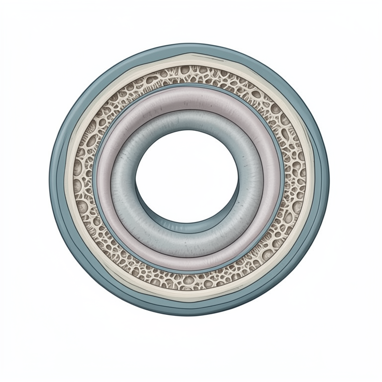
Layers of the Starling Tracheal Wall
AI-generated conceptual illustration (starling bird (Sturnus vulgaris) trachea). It explains the idea and does not show study data.
View source evidence ↗Figures & tables
From the original paper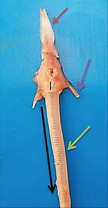 Figure 1
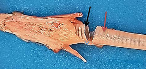 Figure 2
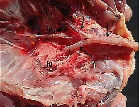 Figure 3
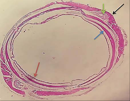 Figure 4
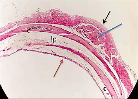 Figure 5
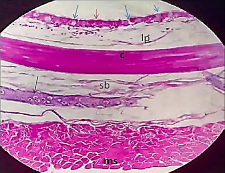 Figure 6
Introduction
Respiratory system begins at the nares, passages in the head that lead to pump gas to the larynx. Trachea extends from the larynx to left and right lungs. Trachea is different in some birds may tortuous or coiled, as in the trumpet bird [1,2] . The trachea branches into two extra pulmonary primary bronchi, each of that goes to a lung and its associated air sacs [3] . Birds considered as one of the most important nutritional sources. They constitute a high rate of human consumption specially poultry meat which is characterized by depression of cholesterol percent. The trachea in birds appears as cylindrical tube-like flexible cartilaginous structure, with an empty interior arranged in loops or coils. So that the length of the trachea is much longer than the length of the neck and variable dimension in different bird species [4,5,6] . It passes from the right side of the neck ventral to the esophagus and ventro-lateral to the cervical vertebrae as it extends caudally, therefore, most of the trachea lies on the right side of the neck and can easily be palpate in the live bird. As it approaches the thoracic inlet returns to the midline-ventral position. The trachea bifurcate into two parts, left and right. Primary bronchi, which enters the hilus of the lung on the septal surface [7,8] . The trachea based on a series of tracheal rings, which tends to ossify in large species. The form of these rings is rather constant in birds generally. Each is complete and not C-shaped as in mammals. This shape is like a signets ring, the broad part forming the left and right halves alternately of successive rings. Each broad part overlaps externally the narrow parts of the two adjacent rings [9] . The trachea follows the larynx and is made up of the tracheal cartilage ring [10,11] . In birds; the number of cartilage rings in the trachea varies due to the length of the neck. The trachea usually made up of 108-126 cartilages in dove penguins [12,13] . Histologically, the tracheal wall is composed of pseudostratified ciliated columnar epithelium and goblet cells, the propria-submucosa under the basal cells of ciliated respiratory epithelium is consist from the loose connective tissue with mucous and serous tracheal glands.
The hyaline cartilage may be complete (O-shaped) or noncomplete (U or C shaped) rings which are supported the tracheal wall. Periphery longitudinal oriented striated muscle which is located and surrounded externally tunica adventitia [14, 15, 16] . In Bursa roller pigeon (Columba livia) and Turkey the epithelium lining of the trachea is mucous type that is secreted by simple alveolar mucus glands intraepithelial or goblet cells [17,18] . In birds, the anterior portion of epithelium lined trachea contains numerous, simple alveolar mucous glands while in the posterior portion of epithelium the goblet cells are lieu mucous glands which form 'intraepithelial glands [18] . Whereas [19] , mention that goose have plentiful goblet cells constituted the 'intraepithelial glands' which is contain mucin with a predominance of sulphate ester salt.
Aim of the study
The study was designed to describe the morphological and histological results of trachea during starling birds Sturnus vulgaris
Materials and Methods
We brought 10 birds of starlings from Al-Amiriyah market in baghdad & kept them at home for short period, before starting to dissect them for the purpose of studying histology & morphology of trachea, the weights of birds were approximately the same, we use a surgical set, formalin 37%, blades, normal saline 0.9%w/v, containers, blades, blue paper, cylinder tube. *we diluted the formalin to 10% so the tissue doesn't damage. Starting to dissect the bird by killing the animal through sloughing it gently and we placed the bird on his back, pull the legs to the side and the back. Cut the skin between the legs and abdomen on each side, Hold the legs from near the hip and then lift it to the top so inseparable, Raise the skin over the thigh and chest. Complete raise the skin from the director and even the beak, Cut the abdomen gently in a cross-sectional area at the end of the sternum, in order to gain access to internal organs (trachea), Cut the ribs on the right side and the left until it reaches the shoulder, Raise the chest at this stage, lift the trachea carefully, and then put each of trachea in containers that contain diluted formalin 10%, then send the samples to get dissected histologically in the histology lab. Stains have been used on samples is Hematoxillin and Eosin (H&E) stain and periodic acid schafit (PAS) stain. The staining procedure for H&E follows a basic protocol [20].
Results and Discussion
Results and Discussion
Morphological results
The current study was included two sides, the first side was represented morphological results and second side was histological target. The trachea is an organ of the respiratory system. Define, its musculo-membranous cartilaginous tube, located between the larynx and runs away toward left and right lungs inside the thoracic cavity (figure 1). So the trachea is continuously dorsally with esophagus. The study expressed the trachea in starling birds decreased in diameter towards posterior progressively (figure 1). These results corresponding with [1,23] . Whom said the craniocaudal width of the rings progressively increase throughout the cranial third of the trachea and then progressively decrease throughout the caudal third of the trachea. These results was accepted with said [21] , in Iraqi pigeons, the trachea was long tube extended from larynx until bifurcate into left and right primary bronchio which enter inside the lungs. where the study showed tracheal ring of starlings, characterized complete ring, the trachea was characterized flexible, non-folded rings and series of cartilage rings, this ring numbers were range average between (70 to 100) in different numbers between types of the birds (figure 2).these results accepted with [9]. The present study showed the division of trachea before enter into the left and right lungs is called primary bronchi (figure 3). This result accepted with [22] , whom stated the trachea bifurcates into two primary bronchi dorsal to the base of the heart. These enter the ventral surface of the lungs after short course. In penguins, a median septum divides the trachea into left and right tubes, making it very easy to intubate a primary bronchus by mistake.
Histological results
Histologically, the current study was included histological structures, where the study showed tracheal rings of starlings were complete (figure 4). Also the study observed mucosa of trachea was composed from epithelia called pseudostratified columnar ciliated epithelium which lined internal surfaces of trachea with goblet cells, either the second layer represented lamina propria was a loose connective tissue which rich a blood vessels (figure 5). These study accepted with [14,23] , said the tracheal wall is composed of pseudostratified ciliated columnar epithelium and goblet cells, the propria-submucosa under the basal cells of ciliated respiratory epithelium is consist from the loose connective tissue with mucous and serous tracheal glands. The hyaline cartilage may be complete (O-shaped) or non-complete (U or C shaped) rings which are supported the tracheal wall. Periphery longitudinal oriented striated muscle which is located and surrounded externally tunica adventitia. These results accepted with [21] , whom stated the trachea is included from four tunicus, mucosa, submucosa, muscularis and adventitia, where epithelia lined by psudostratified columnar epithelium ciliated. So the study showed all layers of trachea. Mucosa, submucosa, muscularis as well as adventitia is last layer. The mucosa consist of epithelia with goblet cells, and lumina properia is loose connective tissue and mucosa muscularis. Either submucosa was beneath mucosa dense connective tissue, third tunica is called muscularis is smooth muscle fibers (Figure 6). These results parallel with [14,17,18] in Bursa roller pigeon (Columba livia) and Turkey the epithelium lining of the trachea is mucous type that is secreted by simple alveolar mucus glands intraepithelial or goblet cells. In birds, the anterior portion of epithelium lined trachea contains numerous, simple alveolar mucous glands while in the posterior portion of epithelium the goblet cells are lieu mucous glands which form 'intraepithelial glands [18] . Whereas [19], mention that goose have plentiful goblet cells constituted the 'intraepithelial glands' which is contain mucin with a predominance of sulphate ester salt.
Acknowledgement
AcknowledgementsThe author wishes to send their gratitude to department of anatomy and histology, college of veterinary medicine of University of Baghdad.
References
17 in the original paper- Podos J, Huber SK, Taft B. Bird song: The interface of evolution and mechanism. Annu. Rev. Ecol. Evol. Syst. 2004;35:55-87.
- Fedde MR. Relationship of Structure and Function of the Respiratory System to Disease Susceptibility. Poult. Sci. 1998;77:1130-1138.
- Miller EH, Seneviratne SS, Jones IL, Robertson GJ, Wilhelm SI. Syringeal Anatomy and Allometry in Murres (Alcidae: Uria). J Ornithol. 2008;49:545-554
- Onuk B, Haziroglu RM, Kabak M. Gross Anatomy of the Respiratory System in Goose (Anser anser domesticus): Bronchi and Sacs Pneumatics. Ankara Univ. Vet. Fak. Derg. 2009;56:165-170.
- Brown RE, Brain JD, Wang N. The Avian RespiratorySystem: A unique Model for Studies of Respiratory Toxicities and for Monitoring Air Quality. Environ Health Prospect. 1997;105:188-200.
- Pesek L. The Avian Respiratory System. Winged Wisdom Pet Bird Magazine. 2000;1:1-3.
- Getty R. The Anatomy of the Domestic Animals Vol. 2.5th edition, Saunders Company; c1975. p. 1896-1902.
- Yersin AG, Eders FW, Simmons DG. Tracheal Cilia Response to Exogenous Niacin in Drinking Water of Turkey Poults Infected. North Carolina State Uni. 1991;33:674-680. (Abstract).
- Aughey E, Frye FL. Comparative Veterinary Histology with. Clinical Correlates. Manson Ltd. Lon; 2001. p. 9394.
- Bacha WJ, Bacha LM. Color Atlas of Veterinary Histology 2 nd (ed.): Lippincott Williams & Wilkins; 2000. p. 175-190.
- Jeffery PK. Structure and Function of Mucus-secreting Cell of Cat-and Goose Airway Epithelium. Exper. Eye. Res. 2004;78(2):187-96. (Abstract).
- Luna LG. Manual of histologic staining method of armed forces institute of pathology. 3ed. New York, U.S.A.; 1968. p. 123.
- AL-Taai Suhaib AH. Microscopic and morphometric study in trachea and lungs of adult Iraqi pigeon (Columba livia), sys.rev.pharm. 2021;12(2):342-346.
- Dyce KM, Sack WO, Wensing CJG. Textbook of Veterinary Anatomy. 2nd ed.; 2002. p. 812-815. W.B. Saunders Co., Philadelphia.
- AL-Taai SA, Khalaf AS. Histomorphological study of the tongue in adult starling birds (Sturnus valguris). Iranian Journal of Ichthyology. 2022;9:116-122. Retrieved from http://www.ijichthyol.org/index.php/iji/article/view/785
- AL-Taai SAH, Hasan MS. Histomorphological Study of Proventricular and Gizzard in Adult Starling Bird (Sturnus vulgaris). Plant Archives. 2020;20(1):16711678.
- Samuelson DA. Text book of Veterinary Histology. Saunders-Elsevier; 2007. p. 246-248.
Cited by 1
From OpenAlex, OpenCitations and reference lists read by Scinovex
- Anatomical, pathological, and histological features of experimental respiratory infection of birds by biofilm-forming bacteria Staphylococcus aureus ↗
Е. М. Ленченко, Сачивкина Надежда Павловна, Olesya Anatolyevna Petrukhina, Nikolay Vladimirovich Petukhov, А. А. Жаров, Natallia Zhabo et al. · Veterinary World · 2024
Background and Aim: The pathogenesis of staphylococcal infections is mediated by virulence factors, such as enzymes, toxins, and biofilms, which increase the resistance of microorganisms to host immune system evasion. Testing and searching for standardized multi-level algorithms for the indication and differentiation of biofilms at the early stages of diagnosis will contribute to the development of preventive measures to control…
❝ 5PDF