
Research article · 2021 · Full text
International Journal of Advanced Biochemistry ResearchBiomarkers in leukemia
Volume 5(2) · Pages 46–47 · DOI: 10.33545/26174693.2021.v5.i2a.85 ↗
Research areasComputational Drug Discovery MethodsGDF15 and Related BiomarkersCancer and biochemical research
Abstract
Author-writtenCancer, particularly leukemia is one of the major causes of death amongst the cancer patients. So solving the mystery behind underlying causes and abnormalities that leads to the leukemia has become a challenge for he researchers. Again in each type of the leukemia, which kind of the biological marker is associated is a puzzle. Biological marker and tumour markers are measurable biochemicals that are associated with a malignancy. A wide range of these biological markers known as cluster differentiation (CD markers) markers have been found to be associated with leukemia. These include the Cd2, CD5, Cd7, CD10, CD20, CD33, CD34, etc. The expression of all these CDs has been associated in different types of leukemia. So, studying the level of expression of these different CDs can be a major breakthrough in the treatment strategies of leukemia.
Summary
Source-checkedPlain-language reading of the full paper, prepared with AI and checked against the text. Each point links to the authors' own words.
Why it matters
- Leukemia is a significant cause of mortality among cancer patients, making the understanding of its underlying causes a priority for researchers. View evidence ↗
- Leukemia is characterized as a heterogeneous disease involving the uncontrolled growth of myeloid or lymphoid blood precursor cells. View evidence ↗
- Acute leukemia is the most frequent type of malignancy found in children. View evidence ↗
The question
- The authors aim to address the challenge of identifying which specific biological markers are associated with different types of leukemia. View evidence ↗
What they did
- The study focuses on biological and tumor markers, which are biochemicals that can be measured to identify malignancy. View evidence ↗
- The research specifically examines cluster differentiation (CD) markers as the primary biological markers associated with leukemia. View evidence ↗
- Flow cytometry is identified as a routine, automatable method for the rapid quantitative analysis of individual cell characteristics. View evidence ↗
- Confocal reflectance microscopy is described as a technology capable of providing real-time, noninvasive images of living tissue architecture and cellular morphology. View evidence ↗
What they found
- Acute myelogenous leukemia (AML) is the most common adult form, typically affecting men more than women with an average diagnosis age of 65 years, and is associated with CD13, CD14, and CD15. View evidence ↗
- Chronic Myelogenous Leukemia (CML) is a myeloproliferative disorder characterized by bone marrow cell proliferation outside the marrow and the expression of CD33. View evidence ↗
- Acute Lymphocytic Leukemia (ALL) involves abnormal growth of early lymphocytes and is associated with markers including CD2, CD3, CD4, CD7, CD8, CD10, CD22, CD23, and CD79a. View evidence ↗
- CD34 is identified as a critical marker because it is present in hematopoietic pluripotential stem cells. View evidence ↗
- An increased presence of CD38 is observed in multiple myeloma as well as certain types of acute lymphoblastic and myeloblastic leukemia. View evidence ↗
What it means
- The authors conclude that various CD markers are utilized across different types of leukemia to aid in diagnosis. View evidence ↗
- The authors note that the field is rapidly evolving, with new CD markers being discovered monthly, meaning current marker panels will likely change as more specific information emerges. View evidence ↗
Visual summary
1 visualsIllustrations explain the paper; they do not show study data.
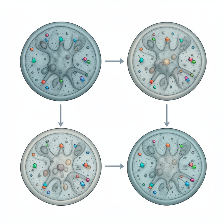
Leukemia Marker Mapping
AI-generated conceptual illustration (human hematopoietic cells). It explains the idea and does not show study data.
View source evidence ↗Introduction
Leukemia is a heterogeneous disease, characterized by the deregulated proliferation of blood precursor cells of myeloid or lymphoid origin. Cancer involving the blood forming cells acute leukemia is the most common malignancy in children. It is due to a clonal proliferation of hematopoietic cells (usually white blood cells). In leukemia, abnormal immature white cells increase greatly and invade other tissues and organs. These white cells are not able to function at their normal task of fighting disease which makes the leukemic child vulnerable to infection or haemorrhage (Mohr A et al. 2004) [1] . A safety biomarker is a treatment-emergent finding that substitutes for or translates into a clinically relevant adverse outcome. Environmental studies and toxicology defined a biomarker as any cellular or molecular indicator of toxic exposure, adverse health outcome or susceptibility. That definition paved the way for a further delineation of biomarkers into three categories-biomarkers of exposure, effect and susceptibility; these categories are often cited for their potential utility in environmental, occupational or other related toxicological applications. Currently, the most important application of biomarkers is in drug discovery and development. The role of biomarkers in various therapeutic areas particularly cancer, cardiovascular disease and disorders of the central nervous system, is described. Biomarkers are useful not only for diagnosis of some of these diseases but also for understanding the pathomechanism as well as a basis for the development of therapeutics. Mature functional B-and T-lymphocytes express a number of characteristics cell surface proteins including receptors for antigens. Thus, they can be distinguished on the basis of these surface proteins, which are often referred to as CD for cluster of differentiation, followed by unique identifying numbers, A specific antibody can be raised against each of these CD cell-surface markers; therefore they are called CD antigens (Anne L et al. 2003) [2] . This system of nomenclature was originally developed for membrane proteins or protein complexes that could be identified by their physical properties, such as molecular weight, or their interaction with specific antibodies. Glycosylated CD molecules act as binding sites for lectins, histamine, and receptors for insulin and growth hormones. An individual CD usually consists of a single or dimeric polypeptide chain and may be glycosylated; accordingly, the molecular weight of different CDs varies for example, 24 kDa for CD9 and kDa for CD22 [3] . Myeloid markers: CD 13, CD 14, CD 15, CD 33,
Flow cytometry
Flow cytometry can rapidly and quantitatively analyze the characteristics and responses of individual cells and is easily automatable. Therefore, as a well-established and routine method, it is suited for the analysis of individual patient material in order to arrive at optimized personalized antitumor drug therapies in the clinical setting or to aid the identification of novel compounds and combination treatment by high-throughput screening methods Howard (Fingert et al. 2006) [5] .
Confocal reflectance microscopy
Confocal reflectance microscopy is a new technology that can provide detailed images of tissue architecture and cellular morphology of living tissue in near real In concept, in vivo confocal imaging resembles histological tissue evaluation, except that three-dimensional subcellular resolution is achieved noninvasively and without stains. In epithelial structure, resolution of 1 µm has been achieved with a 200-400-µm filed of view and a penetration depth of up to 500 µm recently, flexible reflectance confocal micro endoscopes have been described that can obtain highresolution confocal images of tissue in vivo in near real time. Use of this instrument provides the potential to image oral epithelial tissues with subcellular resolution in a clinical setting (Martyn TS et al. 2005) [5] . CD 34 is one of the most important markers for it is seen in the hematopoietic pluripotential stem cell (PSC) which is the cell that is "wanted" in bone marrow or peripheral blood stem cell transplants. CD 38 is a receptor that is found in plasma cell, some thymocytes (early lymphocytes still in the thymus), NK cells, and in very early B cells. Increased presence of CD 38 has been found in cells in multiple myeloma, and certain acute lymphoblastic and Myeloblastic leukemia. CD38 is also thought to be of predictive value in determining the clinical course that will CLL will take (David M. Thomas, 2004) [4] .
Types of
Types of
Conclusion
As leukemia is one of the most death causing disease amongst all types of cancer. A number of diagnostic methods are available to diagnose different types and severity of cancer. Biomarkers are the most acceptable diagnostic aid used in cancer. There are different CD markers are identified for different condition in cancer. It is very difficult to identify particular CDs that are used for particular type of Leukemia. The above study shows some CDs are used in a different type of Leukemia.
References
4 in the original paper- Mohr A, Zwacka RM, Debatin KM, tahnke KS. A Novel method for the combined flow cytometric analysis of cell cycle and cytochrome c release. Cell Death and differentiation. 2004;11(10):1153-1154.
- Anne L, Ann M, Thomas G, Reza A, Adel K, Rebecca R. Confocal Microscopy for Real-Time Detection of oral Cavity Neoplasia, Clinical Cancer Research. 2003;9(13):4714-4721.
- Howard Fingert, Mary Varterasian. Safety Biomrkers and the Clinical Development of Oncology Therapeutics: Considerations for cardiovascular Safety and Risk Management, the AAPS Journal. 2006;8(1):10.
- Martyn TS, Cliona MM, Joseph L, et al. Molecular biomarkers for the study of childhood leukemia, Toxicology and Applied Pharmacology. 2005;206(2):237-245.