
Research article · 2022 · Full text
International Journal of Advanced Biochemistry ResearchStudy of muscle biomarkers in hypothyroid patients
Volume 6(1) · Pages 61–66 · DOI: 10.33545/26174693.2022.v6.i1a.90 ↗
Research areasAdipose Tissue and MetabolismDiet and metabolism studiesHigh Altitude and Hypoxia
Abstract
Author-writtenBackground: Hypothyroidism is characterized by a decline in basal metabolic rate which can lead to glycogen build up and decreased enzyme activity in energy synthesis and other regulatory systems. Thyroid hormones have a number of key targets including skeletal muscle. Thus, in hypothyroid patients altered glycolysis, glycogenolysis and mitochondrial metabolism have an impact on skeletal muscle function. Timely assessment of muscle biomarkers Creatine Kinase (CK) (NAC) [N-acetyl-cystein], CK-MM, Lactate dehydrogenase (LDH), Aspartate Transaminase (AST) and Aldolase in patients of hypothyroidism can estimate the usefulness of these enzymes as screening tests to detect muscle damage associated with hypothyroidism. Objective: To estimate the serum CK(NAC), CK-MM, LDH, AST and Aldolase activity and compare with normal healthy controls. Material and Methods:The study was conducted in the Department of Biochemistry, Teerthanker Mahaveer Medical College & Research Centre Moradabad. CK (NAC), CK-MM, LDH, AST and Aldolase were measured in 60 patients with hypothyroidism under the age group of (20-60) years using automated method and their levels were compared with 60 healthy individuals as controls. Results: Serum CK (NAC), CK-MM and Aldolase were significantly higher in cases than the controls (p
Summary
Source-checkedPlain-language reading of the full paper, prepared with AI and checked against the text. Each point links to the authors' own words.
Why it matters
- Hypothyroidism causes a decrease in the basal metabolic rate, which can result in the accumulation of glycogen and reduced activity of enzymes involved in energy synthesis. View evidence ↗
- Because thyroid hormones target skeletal muscle, hypothyroidism can impair muscle function by altering mitochondrial metabolism, glycolysis, and glycogenolysis. View evidence ↗
- Measuring muscle biomarkers like CK (NAC), CK-MM, LDH, AST, and Aldolase may serve as a screening method to detect muscle damage in patients with hypothyroidism. View evidence ↗
The question
- The study aimed to measure the activity of serum CK(NAC), CK-MM, LDH, AST, and Aldolase in hypothyroid patients and compare these levels to healthy controls. View evidence ↗
What they did
- CK, LDH, and AST were measured using the UV-Kinetic method, while Aldolase was measured using ELISA. View evidence ↗
- Data analysis involved calculating the mean and standard deviation, with comparisons performed using student's t-tests and Pearson's coefficient correlation. View evidence ↗
What they found
- Serum CK(NAC) activity was significantly higher in hypothyroid cases (282.70±60.21 IU/L) compared to controls (150.4±30.65 IU/L), with p < 0.001. View evidence ↗
- CK-MM levels were significantly elevated in hypothyroid patients (45.49±16.78 IU/L) compared to healthy controls (19.95±5.52 IU/L), with p < 0.001. View evidence ↗
- Aldolase activity was significantly higher in the hypothyroid group (16.86±9.10 IU/L) than in the control group (4.59±2.69 IU/L), with p < 0.001. View evidence ↗
- LDH activity was slightly increased in hypothyroid patients (249.68±42.01 IU/L) compared to controls (244.95±16.31 IU/L), but this difference was not statistically significant (p = 0.418). View evidence ↗
- AST levels showed no significant difference between hypothyroid cases (43.00±26.37 IU/L) and controls (40.06±7.06 IU/L), with p = 0.406. View evidence ↗
- The highest percentage of hypothyroid patients in the study (25%) were in the 50-60 age group, suggesting the condition is more common in this range. View evidence ↗
What it means
- The authors conclude that biochemical muscle markers can assess disease severity and that early detection of these enzymes can improve the management of comorbidities and the quality of life for hypothyroid patients. View evidence ↗
- The authors suggest that Creatine kinase may be a more effective biochemical diagnostic tool for detecting muscle injury in hypothyroidism than other indicators. View evidence ↗
Visual summary
2 visualsIllustrations explain the paper; charts are replotted from the paper's own table values.
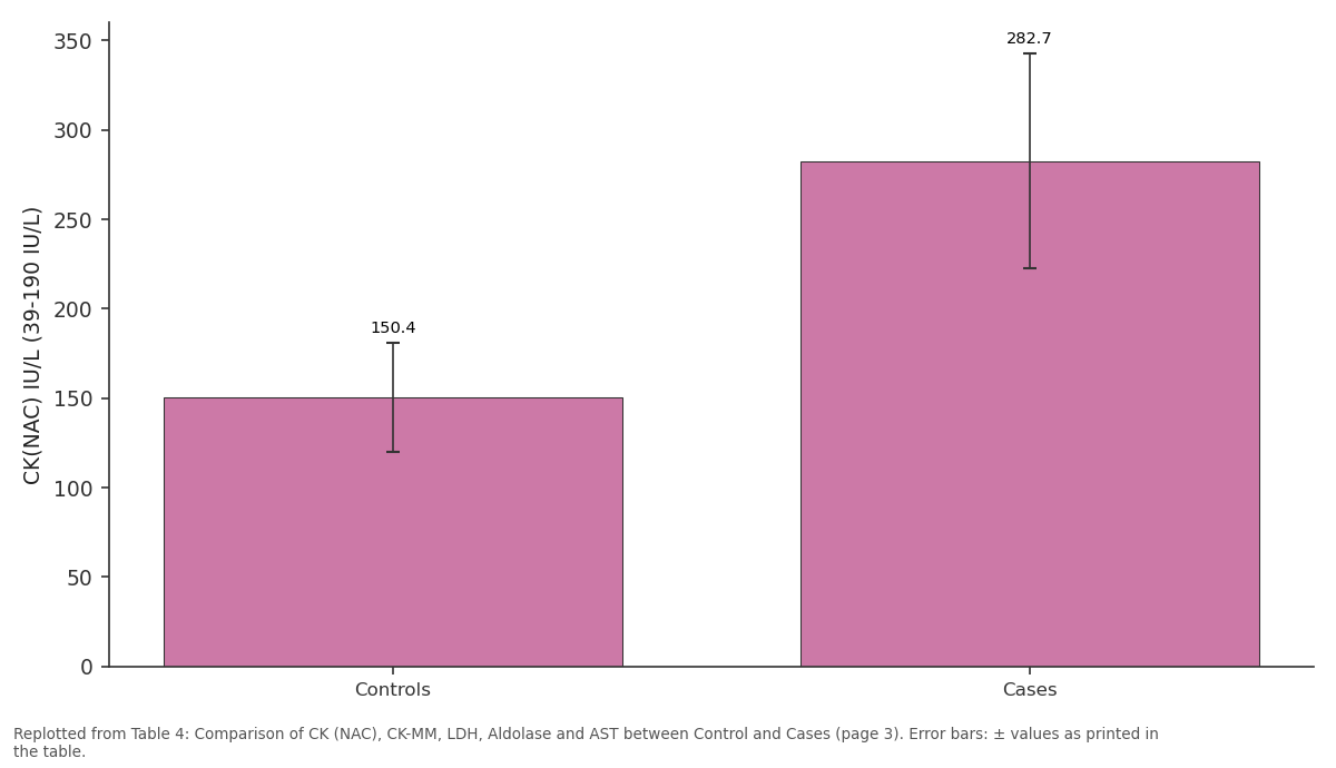
CK(NAC) IU/L (39-190 IU/L): Controls vs Cases
Replotted from Table 4 (page 3) of the original paper. Every bar is the paper's own table value.
View source evidence ↗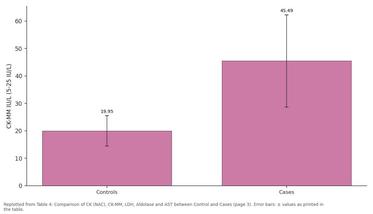
CK-MM IU/L (5-25 IU/L): Controls vs Cases
Replotted from Table 4 (page 3) of the original paper. Every bar is the paper's own table value.
View source evidence ↗Figures & tables
From the original paper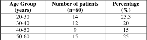 Table 1
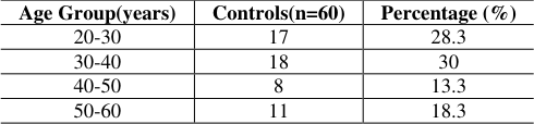 Table 2
 Table 3
 Table 4
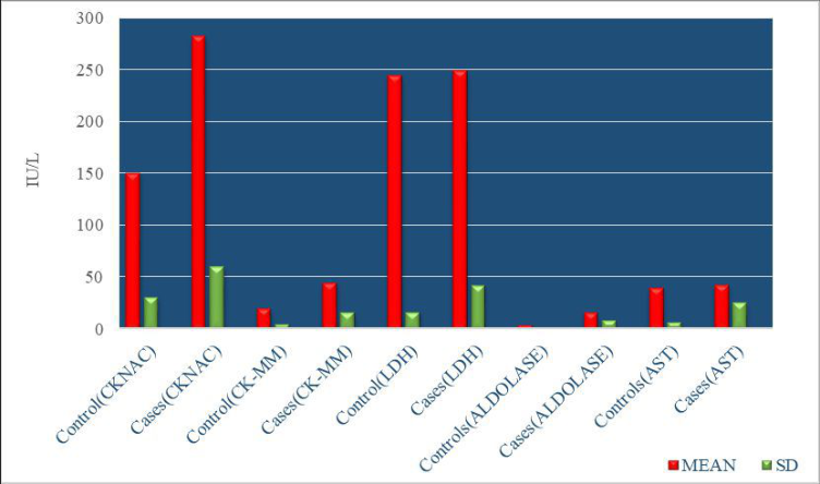 Figure 1
Introduction
Thyroid hormones T 3 (triiodothyronine), T 4 (tetraiodothyronine) and Thyroid stimulating hormone (TSH) are produced by the thyroid gland and play a major role in the control of BMR, tissue differentiation and growth. T 3 & T 4 hormones are produced with the help of TSH and Thyrotropin releasing hormones (TRH) [1,2] . Hypothyroidism (related to a malfunctioning thyroid gland) and hyperthyroidism (due to an overreactive thyroid gland) are the two types of thyroid disorders [3] . Primary hypothyroidism is a clinical illness caused by decreased T 3 , T 4 and an increase in TSH hormones [4] . It is a widespread disorder that affects 4 to 15% of the world's population in mild to severe forms. In India, females are more often affected than males. Hypothyroidism affects approximately 11% of women and the risk of acquiring it increases as one reaches maturity [5] . Muscle's soreness, pale dry skin, a bloated face, mild fatigue, cold intolerance, reduced joint mobility, carpel tunnel syndrome, weight gain and some severe manifestation like cardiomyopathy and myxedematous coma are all signs of hypothyroidism [6] . Hypothyroidism is characterised by a decrease in baseline metabolism rate which can lead to glycogen build up and decreased enzyme activity in energy synthesis and other regulatory systems. Hypothyroidism lowers adenosine triphosphate (ATP) levels below a hazardous threshold by reducing glycolysis and oxidative phosphorylation [7] . Thyroid hormones have a number of targets, including skeletal muscle. Hypothyroidism has systemic effects due to metabolic disturbances or myxoedematous infiltration of connective tissue which includes the build-up of glycosaminoglycans in the tissues of many organs such as skeletal muscles, kidneys and other organs resulting in multiple organ dysfunction. As a result, skeletal muscle performance in hypothyroid adults is affected by changes in glycogenolysis and mitochondrial metabolism [8,9] . In hypothyroidism, clinical indicators of muscle damage include CK, Aldolase, AST and LDH [10] . The enzymes CK is found in skeletal muscles, cardiac muscles and the brain. In healthy people serum CK activity is affected by age, race, lean, body mass and physical activity [11,12] . In people with primary hypothyroidism, the amount of CK in their blood can vary. In skeletal muscle, only a modest quantity of CK-MB isoenzyme is detected, ranging from 0.2 to 0.15 percent of total enzyme activity. In skeletal muscles and heart tissue, CK activity is highest. As a result, total CK activity may be one of the sensitive diagnostic markers for various muscle diseases [13] .
Aldolase is an enzyme that catalyses a variety of reactions in distinct pathways. For instance, fructose 1-6 bisphosphate is converted to glyceraldehyde and dihydroxyacetone phosphate during glycolysis. Aldolase concentration has been demonstrated to be affected by myotonic muscle diseases including progressive muscular dystrophy. As a result, Aldolase could be used as a useful and specific biomarker for muscle injury [14,15] . Lactate dehydrogenase is an enzyme which interconverts pyruvate and lactate as well as NADH and NAD+. Pyruvate, lactate, NADH and NAD+ are all converted to NADH and NAD+ by lactate dehydrogenase. LDH activity has been observed to be changed in hypothyroid conditions according to several previous investigations. In several disorders the LDH isoenzymes are increased in blood [16] . LDH elevation could indicate either increased or decreased hepatic clearance. It has been seen that patients with hypothyroidism produce more lactate during exercise than healthy people, a finding that may be associated with poor mitochondrial oxidative dysfunction [17] . Aspartate aminotransferase is a transaminase that catalyses the reaction between aspartate and amino acids. This process happens in the mitochondria and cytoplasm which gives energy to tissue [18] . AST has a substantially longer half-life than CK and is found to have a much wider distribution of tissues. It's a cytoplasmic and mitochondrial enzyme that catalyses aspartate deamination to produce oxaloacetate which can then enter the Kerb's cycle and so contribute to ATP synthesis [19,20] . Previous research works on the activities of CK, Aldolase, LDH and AST in people with hypothyroidism have yielded mixed results. Some scientists have noticed that there is no change in the activity of these enzymes in hypothyroid patients while, others have found that the activity of these enzymes increases in hypothyroid patients. Hence, this study was intended with the aim to estimate the changes in activity of serum enzymes CK (NAC), CK-MM, LDH, Aldolase and AST in patients with hypothyroidism and to estimate the usefulness of measurement of these enzymes as a screening method to detect muscle damage associated with hypothyroidism. This study was also intended to find whether timely estimation of muscle biomarkers CK (NAC), CK-MM, LDH, AST and Aldolase in hypothyroidism could help in better management of comorbidities associated with hypothyroidism and bring about an improvement in the quality of life of these patients.
Materials and Methods
The present study has been done in the Department of Biochemistry, Teerthanker Mahaveer College and Research Centre, General Medicine OPD in Moradabad, Uttar Pradesh from January 2021 to December 2021. Total 120 subjects were selected and divided into two groups. Group I included 60 diagnosed cases with hypothyroidism. They were recruited for the study after taking their due written consent. The subjects were in the age group of 20 to 60 years [3] including both males and females. Group II included 60 healthy control subjects who were age and sex matched with the cases.
Exclusion Criteria
Muscle ailments with causes other than hypothyroidism [3] , Cardiac disorders [3] , Patients on long term medications affecting muscle biomarkers [21] , Hepatic disease [21] Crush injury (Rhabdomyolysis) [21] . After overnight fasting for 12 hrs, blood sample (8 ml) was obtained from all the subjects & dispensed into plain vials under proper aseptic conditions. After centrifugation 3000 RPM for 5 min, serum sample was used for analysis. Various methods were used - Creatine Kinase (CK(NAC), CK-MM) by UV -Kinetic Method [22,23] LDH by (UV Kinetic Method) [24] AST by (UV Kinetic Method) [25] Aldolase by ELISA (Enzyme linked immunosorbent assay) [26] T 3 , T 4 & TSH by ELFA (Enzyme Linked Fluorescent Assay) [27] Statistical analysis: SPSS version 28.0 for data analysis and Microsoft Word and Excel for graph generation. Mean and standard deviation was assessed & comparison was done using student's t-test & Pearson's coefficient correlation (r-value).
p-value of less than 0.05 was considered as significant [S]. p-value of less than 0.001 was considered as highly significant [HS].
Results
The results of the measured by different biochemical parameters in the research were listed below for hypothyroid patients and healthy subjects. The activity of CK (NAC) in patients with hypothyroidism was more in cases than in healthy controls. The mean value of CK-MM was raised in hypothyroid cases in comparison to normal subjects. The analysis showed that the mean value of LDH was slightly increased in cases as compared to healthy individuals but the difference is not statistically significant. The concentration of Aldolase was elevated in hypothyroid patients as compared to the healthy controls.
There is no significant difference in the value AST between cases and controls.
Discussion
The thyroid gland is a primary metabolic endocrine gland as T 3 and T 4 hormone secreted from thyroid gland are essential for the growth, development, and various other metabolic functions of the entire body organs. Hypothyroidism is a common endocrine condition caused by inadequate thyroid hormone production or decreased thyroid hormone activity [3] .
Hypothyroidism can be caused by a variety of reasons including autoimmune disease, thyroiditis, iodine deficiency and hereditary conditions. Thyroid hormone production is impaired which impacts the functions of all other organs in the body [7] . These hormones have an impact on normal growth, metabolism and other processes as well as O 2 intake at cellular level. Weakness, fatigue, swollen face, cramps, weight gain, cold intolerance, exertional myalgias, muscle weakness and hair thinning are all symptoms of hypothyroidism. The severity of the hormone deficiency determines the symptoms of hypothyroidism [5] .
In overt hypothyroidism, the skeletal muscle damage is more common compared to subclinical hypothyroidism. Thyroid dysfunction results in a wide range of muscular aliments. It improves muscular sensitivity to circulating catecholamines and raises demand of micronutrients for muscles. Proximal muscular weakness, muscle pain and delayed tendon jerk relaxation are some of the heralding symptoms of muscle damage [13] . Therefore, this study was conducted to assess the effect of muscle markers such a CK (NAC), CK-MM, LDH, Aldolase and AST in patients with hypothyroidism.
In this cross-sectional study total of 120 patients were recruited of which 60 were healthy controls and 60 were patients suffering from hypothyroidism.
In table 1 In Table 3 the levels of T 3 , T 4 and TSH in hypothyroid patients with that of controls has been compared. It was seen that the mean of T 3 levels in hypothyroid patients was 0.36±0.30 ng/dl whereas in healthy controls the mean value was 1.12±0.77 ng/dl. We have found significant changes in the levels of T 3 in patients than healthy controls. The mean value of T 4 in cases of hypothyroid was 1.88±0.88 μg/dl and controls were 7.53±4.13 μg/dl. There was a significant decrease in the levels of T 4 in patients with hypothyroidism as compared to the healthy subjects. Our findings of mean value of TSH in cases of hypothyroid patients were 13.91±7.00μ IU/ml and 2.53±1.23 μIU/ml was in the healthy subjects. It shows that the levels of TSH in hypothyroid cases are high than the healthy controls (p<0.001).
| Thyroid Profile | Hypothyroid Controls (Mean±SD) | Hypothyroid Cases (Mean±SD) | p Value | Significance Level |
|---|---|---|---|---|
| T3 ng/dL (0.60-1.85 ng/dL) | 1.12±0.77 | 0.36±0.30 | <0.001 | HS |
| T4 µg/dL (4.80-11.80 µg/dL | 7.53±4.13 | 1.88±0.88 | <0.001 | HS |
| TSH µIU / mL (0.390-6.160 µIU/mL) | 2.53±1.23 | 13.91±7.00 | <0.001 | HS |
Table text extracted from the original PDF; superscript letters are shown inline. The original table image is below.
View original table image
| Age Group (years) | Number of patients (n=60) | Percentage (%) |
|---|---|---|
| 20-30 | 14 | 23.3 |
| 30-40 | 12 | 20 |
| 40-50 | 9 | 15 |
| 50-60 | 15 | 25 |
Table text extracted from the original PDF; superscript letters are shown inline. The original table image is below.
View original table image
In Table 4 the activity of CK (NAC), CK-MM, Aldolase, AST and LDH in hypothyroid patients was compared with that of controls. It has been assessed that the activity of CK (NAC) in patients with hypothyroidism was 282.70±60.21 IU/L and in the healthy subjects was 150.4±30.65 IU/L. There was a significant increase in the activity of CK (NAC) in hypothyroid cases as compared to the subjects. These findings are similar with Mushtaq S, et al. 2014 [7] who found an increase in activity of CK in patients with hypothyroidism with mean value 108.79±34.92 IU/L as compared to controls 232.9±91.0 IU/L. There were 30 cases (25 females and 6 males) and 30 controls (25 females and 5 males). They concluded that T 3 , T 4 and TSH along with muscles enzymes can be used to determine the severity as well as for screening and early diagnosis of hypothyroidism associated with myopathy.
| Muscle Markers | Controls | Cases | p-value | Significance Level |
|---|---|---|---|---|
| CK(NAC) IU/L (39-190 IU/L) | 150.4±30.65 | 282.70±60.21 | <0.001 | HS |
| CK-MM IU/L (5-25 IU/L) | 19.95±5.52 | 45.49±16.78 | <0.001 | HS |
| LDH IU/L (140-250 IU/L) | 244.95±16.31 | 249.68±42.01 | 0.418 | NS |
| ALDOLASE IU/L (1.0- 7.5 IU/L) | 4.59±2.69 | 16.86±9.10 | <0.001 | HS |
| AST IU/L (5-40 IU/L) | 40.06±7.06 | 43.00±26.37 | 0.406 | NS |
Table text extracted from the original PDF; superscript letters are shown inline. The original table image is below.
View original table image
In another study R. Shanti M, et al. 2017 [6] found elevation in CK activity in cases 224.94±76. 2018 [29] who had found increased activity of LDH in cases with hypothyroidism 428.70±146.39 IU/L than controls 343.64±93.75 IU/L. They studied 50 hypothyroid female patients and 50 healthy euthyroid females between the ages of 30 and 60. Hypothyroidism can produce a considerable rise in CK and LDH activity, according to their findings. Similarly, Growder Mc DA, et al. 2011 [9] also found the high activity of LDH in subclinical 340.38±153.38 IU/L and in overt hypothyroidism patients 421.00±203.91 IU/L as compared healthy control 202.85±36.06 IU/L. In their study, they included 18 people with overt hypothyroidism 50 people with subclinical hypothyroidism and 99 healthy people as controls. They saw a considerable rise in LDH activity.
The LDH enzyme is found in all cells, although it is particularly abundant in the liver, muscle, and kidney. In the heart, LDH1 is the most important isoenzyme. LDH2 is found in both the heart and the muscles. The increased level of LDH could be attributed to a decrease in liver clearance. It can also rise as a result of myositis which is a skeletal muscle inflammatory disease seen in hypothyroidism.
Similarly, LDH5 (M4) is elevated in muscular dystrophies. LDH1 (H4) levels are higher in myocardial infarction than LDH2.
In the present study the activity of Aldolase in hypothyroid cases was 16.86±9.10 IU/L and in control was 4.59±2.69 IU/L. There was a significant increase in the activity of Aldolase in hypothyroid cases compared with the healthy subjects.
Aldolase is a biomarker for muscle injury that is both important and specific. Myotonic muscle disorder, such as progressive muscular dystrophy causes a high concentration of Aldolase [10] .
AST levels were 43.00±26.37 in patients of hypothyroidism and in controls 40.06±7.06 IU/L. There is no significant difference in the activity of AST between cases and controls. The findings of our study are different from Taywade O, et al. 2019 [5] who found elevation in the patients with hypothyroidism 57.97±19.33 IU/L as compared to controls 21.28±8.31 IU/L. They took 50 hypothyroid cases with 50 controls as study group. They also noticed an increase in blood enzymes, which can be used to screen for myopathy in hypothyroid patients and estimation of these enzymes can help to predict and monitor the outcome of hormone replacement therapy. AST is found in all tissues except bone, with the liver, heart, kidneys, brain and skeletal muscles having the highest levels. Thyroid dysfunction is associated with an increase in AST levels. The half-life of AST is substantially longer than that of CK and is detected in a wider range of tissues than CK. Thus, the study included all the parameters CK-NAC, CK-MM, LDH, Aldolase and AST were increased in hypothyroid patients as compared with healthy subjects. The findings in the study are regarded as an important step to observe the relationship between muscle markers and hypothyroidism. They can be used as a diagnostic tool to prevent complications associated with increased activity in patients with hypothyroidism.
Conclusion
The activity of CK-NAC, CK-MM, LDH, Aldolase and AST was shown to be raised in individuals with hypothyroidism as compared to healthy controls in this study. Hypothyroidism is more common in the 50-60 age group according to this study. Males were not as affected as females. In hypothyroidism, skeletal muscles are affected which is reflected by an increase in these muscle enzymes. Myopathy in hypothyroidism is a significant cause of morbidity. Creatine kinase may be a better biochemical diagnostic for detecting muscle injury in hypothyroidism than other biochemical indicators. As a result, we conclude that biochemical muscle markers can be used to assess disease severity. Early detection and diagnosis of these enzymes can help with the management of comorbidities associated with hypothyroidism as well as to improve the quality of life in these patients.
Acknowledgement
AcknowledgementI am grateful to all the faculty members and technical staff of the Biochemistry department and laboratory for their kind support.
Figures and tables from the paper
| Age Group(years) | Controls(n=60) | Percentage (%) |
|---|---|---|
| 20-30 | 17 | 28.3 |
| 30-40 | 18 | 30 |
| 40-50 | 8 | 13.3 |
| 50-60 | 11 | 18.3 |
Table text extracted from the original PDF; superscript letters are shown inline. The original table image is below.
View original table image
References
25 in the original paper- Panag K Singh G, Goyal S. Evaluation of Creatine Kinase as a Diagnostic Tool for Thyroid Function. Indian Journal of Contemporary Politics. 2012;23(4):221-223.
- Vaitla P, Managasvalasa S, Vani N. Levels of Serum Creatine Phosphokinase in thyroid disorders. International Journal of Biochemistry & Cell Biology. 2017;4(1):16-18.
- Parwaj S, Tiwari D, Pandey S, Pandey SK. A Study on Creatine Kinase and LDH activity in Hypothyroidism. International Journal of Recent Scientific Research. 2021;12(4):1-6.
- Taywade O, Patil N. A Biochemical Elevation of Serum Enzymes CK, LDH, & AST in Patients of Hypothyroidism. International Journal of Biochemistry. 2019;11(2):60-63.
- Shanti R, Vijaylaskshmi M, Mahalakshmi R. Creatine Kinase and Lactate dehydrogenase activity in Patients with Hypothyroidism. International Journal of Clinical Biochemistry and Research. 2017;4(2):182-186.
- Mushtaq S, Tilak AM, Malik RR, Shinde SA, Phalak PJ. Biochemical Evaluation of Myopathy in Patients of Hypothyroidism. Indian Journal of Basic and Applied Medical Research. 2014;3(2):364-372.
- Cakir M, Samanci N, Balci N, Balci MK. Musculoskeletal Manifestation in Patients with Thyroid disease. Journal of Clinical Endocrinology and Metabolism. 2003;59(2):162-167.
- Growder Mc DA, Fraser YP, Gordon L, Crawford TV, Rawlins JM. Serum Creatine Kinase and LDH activities in Patients with Thyroid Disorders. Nigerian Journal of Clinical Practice. 2011;14(4):454-459.
- Islam D, Akter K, Hoque A, Hossain S, Hossain M. Serum Levels of Muscle Enzymes in Hypothyroidism. International Journal of Medical Research Professionals. 2020;6(3):39-43.
- Duyff RF, Vanden BJ, Laman DM, Vonloon BJ, Linssen WH. Neuromuscular findings in Thyroid Dysfunction. Journal of Neurology, Neurosurgery, and Psychiatry. 2000;68(6):750-755.
- Archana P, Lal AK, Negi KS. Serum Creatine activity in thyroid disorders. Journal of Medical Education & Research. 2007;9(1):25-26.
- Tejomani M, Meera KS, Vasudha KC. Relevance of Creatine Kinase Activity and Serum Creatinine Levels in Hypothyroidism. International Journal of Recent Trends in Science and Technology. 2013;8(3):263-269.
- Brancaccio P, Lippi G, Maffuli N. Biochemical markers of muscular damage. Clinical Chemistry and Laboratory Medicine. 2010;48(6):1-11.
- Hart E, Finsterer J, Grossegger C, Kroiss A, Stollberger C. Relationship Between Thyroid Function and Skeletal Muscle Involvement in Subclinical and Overt Hypothyroidism. Journal of Endocrinology. 2001;11(1):217-221.
- Koukourakis MI, Giatromanolaki A, Sivridis E. Lactate dehydrogenase isoenzymes 1 & 5 differential expression by neoplastic and stromal cells in non-small cell lung Cancer and other epithelial malignant tumors. Journal of Tumor Biology. 2003;24(7):199-202.
- Ozer J, Ratner M, Shaw M, Bailey W, Schomarker S. The current State of serum biomarkers of hepatotoxicity. Journal of Toxicology. 2008; 245(3):194-205.
- Beth A, Valentine DVM, Julia T, Shelley SM. Increased Serum Alanine Aminotransferase activity associated with muscle Necrosis in the Dog. Journal of Veterinary Internal Medicine. 1990;4(3):140-143.
- Kanwar G, Shekhawat M, Chandra MM, Neeraj S. A Comparative Study of Serum AST/SGOT Levels among the Patients of Thyroid Disorders. IOSR Journal of Biochemistry and Biotechnology. 2017;3(1):66-72.
- Tandra S, Vuppalanchi R. Use of statins in patients with liver disease. Current Treatment Options in Cardiovascular Medicine. 2008;11(4):272-278.
- Horder M, Elser RC, Gerhardt M. Approved Recommendation on IFCC Methods for the Measurement of Catalytic Concentration of Enzymes. IFCC Method for Creatine Kinase. European Journal of Clinical Chemistry and Clinical Biochemistry. 1991;29(10):435-456.
- Apple FS. Diagnostic markers for detection of acute myocardial infarction and reperfusion. Laboratory Medicine. 1992;97(2):217-226.
- Bergmeyer HU, Horder M. Approved recommendation on IFCC method for the measurement of catalytic concentration of Enzymes. IFCC method for LAspartate aminotransferase. Journal of clinical chemistry and clinical biochemistry. 1986;24(7):497510.
- Klee GG, Hay ID. Assessment of Sensitive Thyrotropin Assays for an expanded role in Thyroid function testing proposed Criteria for Analytical Performances and Clinical Utility. Journal of Clinical Endocrinology and Metabolism. 1987;64(3):461-71.
- Prakash. Serum Creatine Kinase An adjunct biochemical index of subclinical Thyrotoxicosis. Annals of Clinical Biochemistry. 2001;38(1):57-58.
- Jaiswal N, Nirwan SD. Study of Serum Creatinine Levels in Hypothyroidism. Journal of Mahatma Gandhi University of Medical Sciences and Technology. 2018;3(3):79-81.