
Research article · 2022 · Full text
International Journal of Veterinary Sciences and Animal HusbandryHistomorphological study of small intestine during one day age in local pigeon Columba livia
Volume 7(2) · Pages 17–20 · DOI: 10.22271/veterinary.2022.v7.i2a.408 ↗
Research areasComparative Animal Anatomy StudiesAnimal Nutrition and Physiology
Abstract
Author-writtenThe study was included 8 local pigeons Columba livia for macroscopic and microscopic findings. Ages were 24 hour after hatching. They were weight (9-10 g). Our study showed the entire small intestine, they were included three loops, duodenum, jejunum and ileum. The present study expressed the duodenum U-shape, have two arms; ventral (descending) and dorsal (ascending) loops, that loops included between them the pancreas. Shape of small intestine characterized a white in color and very thin wall, therefor we gently must deal during dissection. The study showed jejunum and ileum of one day age were recognized by appearance of Meckel's diverticulum connected with yolk sac. The diverticulum separated between jejunum and ileum. It's located at the end jejunum. The jejunum was characterized spiral coil like, and anastomosing with upper (dorsal) part of duodenal arm and occupied almost the posterior part of body cavity. Part of jejunum and duodenum were covered by right lobe of liver. The light microscope showed the duodenal mucosa had finger structure of villi. First part of mucosa was called the epithelium, simple columnar with oval nuclei, lined surface of villi. Lamina properia was loose connective tissue. Third layer of mucosa was muscularis. In jejunum, the upper surfaces of villi had a few distribution of the goblet cells, these cells were appeared positive reaction with Alcian blue stain. Lengths and shapes of villi were different between three entire segments. Firstly the study was recorded absence of intestinal glands in entire the small intestine during one day age pigeon. Either crypts of Lieberkuhn was less developed or absence during the current study.
Summary
Source-checkedPlain-language reading of the full paper, prepared with AI and checked against the text. Each point links to the authors' own words.
Why it matters
- The small intestine serves as the primary location for the digestion and absorption of nutrients. View evidence ↗
- Avian intestinal morphology differs from that of mammals, which facilitates rapid food intake and absorption. View evidence ↗
The question
- The study aims to describe the morphological and histological characteristics of the small intestine in one-day-old local pigeons (Columba livia). View evidence ↗
What they did
- The researchers examined 8 local pigeons (Columba livia) weighing 9-10 g, all of which were 24 hours old after hatching. View evidence ↗
- Pigeons were anesthetized with ketamine and xylazine before dissection, and the small intestine samples were preserved in 10% formalin. View evidence ↗
- Specimens were analyzed using routine histological staining, specifically Hematoxylin and Eosin (H&E) and Alcian blue. View evidence ↗
What they found
- The small intestine consists of three loops, the duodenum, jejunum, and ileum, with the U-shaped duodenum encompassing the pancreas between its descending and ascending arms. View evidence ↗
- In one-day-old pigeons, the entire small intestine is white in color and possesses a very thin wall. View evidence ↗
- Meckel's diverticulum, connected to the yolk sac, was observed at the end of the jejunum, separating it from the ileum. View evidence ↗
- The jejunum is spiral-coil shaped, occupies most of the posterior body cavity, and anastomoses with the dorsal part of the duodenal arm. View evidence ↗
- Histological analysis of the duodenum revealed finger-like villi lined by a simple columnar epithelium with oval nuclei, a loose connective tissue lamina propria, and a muscularis layer. View evidence ↗
- The upper surfaces of the jejunal villi contained a few goblet cells, which reacted positively to Alcian blue staining. View evidence ↗
- Intestinal glands were absent throughout the small intestine of one-day-old pigeons, whereas they were present in adult pigeons. View evidence ↗
- The crypts of Lieberkuhn were either absent or underdeveloped across all three segments of the small intestine in the one-day-old pigeons. View evidence ↗
What it means
- The authors conclude that the small intestine of one-day-old local pigeons differs from some previous studies, specifically regarding the presence of Meckel's diverticulum and the absence of intestinal glands in the mucosa. View evidence ↗
Visual summary
1 visualsIllustrations explain the paper; they do not show study data.
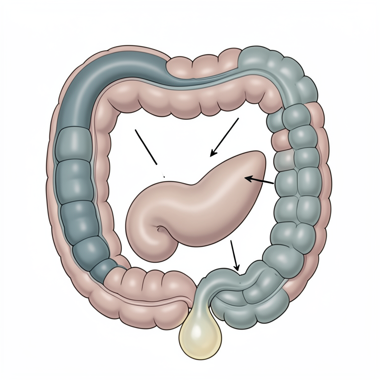
Anatomy of the One-Day-Old Pigeon Small Intestine
AI-generated conceptual illustration (one-day-old pigeon (Columba livia) small intestine). It explains the idea and does not show study data.
View source evidence ↗Figures & tables
From the original paper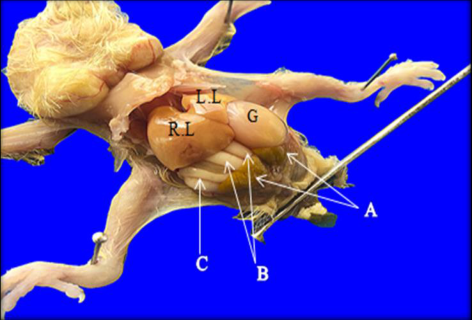 Figure 1
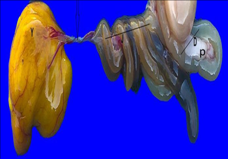 Figure 2
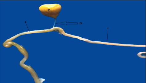 Figure 3
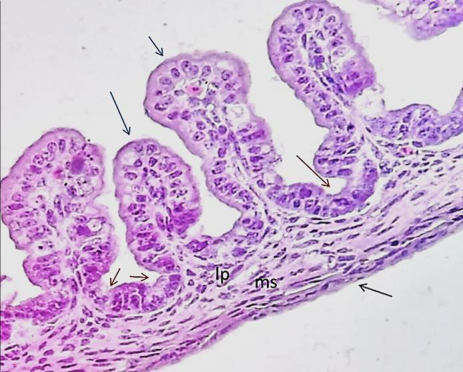 Figure 4
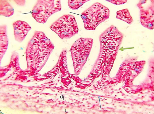 Figure 5
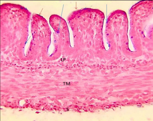 Figure 6
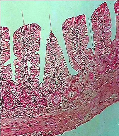 Figure 7
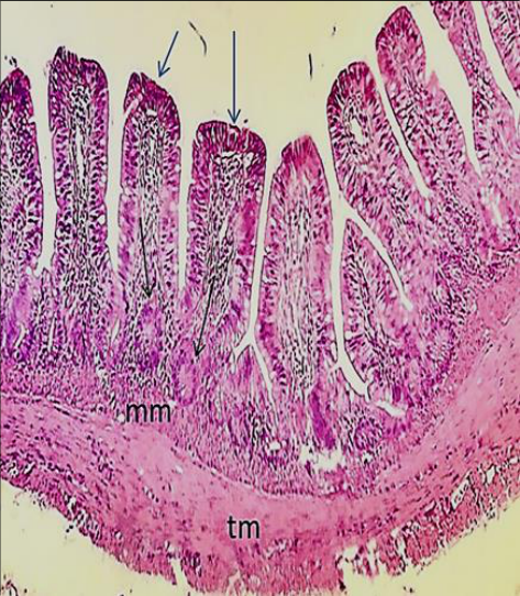 Figure 7
Introduction
The birds are considered the second order after mammals. The numbers of birds are widespread around the world and vary in species and shapes [1] . The digestive system is playing an important role of converting food of animals consume, it's provided nourishment essential for the growth and maintenance of life [2] . The small intestine is the main site of digestion and absorption of food [3] . The birds have a high metabolism, therefore all birds consume a lot of food [4] . There are many individual variations that characterizes the elementary tract during environmental conditions as adaptation [5,6] . Morphology, shape of the intestine in birds are differ from mammalians, therefore the birds have rapid intake and content food absorption [7] . The small intestine is divided into 3 parts; duodenum, jejunum and ileum in order. The duodenum is located between the gizzard at above and jejunum, a loop encompass large part of the pancreas. The second loop is called jejunum, between the duodenum and the ileum. When is jointed by the Mackle's diverticulum. Either ileum is the last part of the small intestine which it's extends from the diverticulum to the ileocaecal junction [8] . Histologically, the small intestine is consisted of four tunics, mucosa, submucosa, muscularis and adventitia [9] . The study is conducted to describe the morphological, histological sections of the small intestine for one day of local pigeon Columba livia.
Materials and Methods
Materials and Methods
Experimental design
The experiment included 8 pigeons C. livia, these birds were bought at the local market & then they are put inside cages.
The weights of birds were range average (9-10 g). Ages were 24 hours after hatching. Surgical instruments were used for anatomy. All pigeons were given ketamine and xylazine anesthesia. Then, the pigeons were putting on dissected board for dissection. All parts of the small intestine were put inside 10% formalin container. The intestines were hollow and spotless of food particles. The specimens were sent to histological technique lab. Then the stains were used for routine staining of specimens. Hematoxylin and Eosin (H&E) and Alcian blue stain according to the procedure [10] .
Results and Discussions
Our study showed the small intestine during one day C. livia. Generally, the small intestine included three loops, duodenum, jejunum, and ileum. The present study expressed the duodenum U-shape, which has two arms; descending and ascending loops. They included between them the pancreas (figure 2). There are some differences between small intestine during one day and other ages. The entire small intestine was characterized as white in color and very thin wall, therefor we gently must deal during dissection (figure 1). The study showed jejunum and ileum of one day age local pigeons were recognized by the appearance of Meckel's diverticulum connected with yolk sac (figure 2,3). The diverticulum separated between jejunum and ileum. It's located at the end jejunum (figure 3). These results of morphology were no parallel with [11,12] , who said of avian species such as quail, herbivorous birds. But, the study accepted with [13] in local pigeons C. livia during one and twenty-day whom said appearance of Meckel's diverticulum between jejunum and ileum, clearly in one day age. The structure began resolving during 20 day age, until adult age. The jejunum was characterized spiral coil like, and anastomosing with the upper (dorsal) part of duodenal arm. The jejunum is occupied almost the posterior part of body cavity. Part of jejunum and duodenum were covered by right lobe of liver (Figure 1). These results corresponding with [14,15,16] . The final part of small intestine was ileum. The histological sections were showed the duodenal mucosa had finger structure of villi. First part of mucosa was called the epithelium, simple columnar with oval nuclei, it's lined surface of villi. Lamina properia was loose connective tissue. Third layer of mucosa was muscularis (figure 4). In jejunum, upper surfaces of villi was a few distribution of the goblet cells, these cells were appeared positive reaction with Alcian blue stain (figure 5). Lengths and shapes of villi were different between three entire segments (Figures 4,5,6). These results accepted with [16] whom showed the tunics of small intestine in striated scope owls. The differences between present studies with previous studies, intestine glands were no found during one day age within lamina properia. The study recorder absence of intestinal glands firstly in entire the small intestine (figure 4,5,6). While in old pigeon was presented the intestine glands (figure 7). These results varied with previous studies about appearance of intestinal glands during old ages. [9,17] , whom said the intestinal glands appeared in lamina properia of small intestine. The present study showed crypts of Lieberkuhn was less developed or absence during three segments (figure 4,5,6). These results were not accepted by [16], who said about crypts of glands were well developed.
Conclusions
The small intestine of one day age of local pigeons doesn't appear similar to previous studies. There are some variations during morphology, where they have presented Meckel's diverticulum. During the histological sections, the light microscope showed, that the intestinal glands were not found in mucosa. Note, the study was indexed the figure 7 during the study of adult-age local pigeons for explanation only.
Acknowledgments
The study was supported by the Department of Anatomy and Histology and the College of Veterinary Medicine, University of Baghdad.
Figures and tables from the paper
References
15 in the original paper- Lavin SR, Karasov WH, Ives AR, Middleton KM, Jr TG. Morphometric of the avian small intestine compared with that of non-flying mammals: Aphylogenetic approach. Physiological and Biochemical Zoology. 2008;81(5):526550.
- Moţ M. Morphological aspects of digestive apparatus in owl (Asio flammeus) and dove (Columba livia). Lucrari Stiintifice Medicina Veterinara. 2010;8(2):364-367.
- Sivakumar MN, Vijayaragavan HD. Gross and Microscopic Changes in the ileum of Male and Female Domestic quail. New York Sci. J. 1989;3(10)108-111.
- Igwebuike UM, Eze UU. Morphological characteristics of the small intestine of the African pied crow (Corvus albus). Animal Research International. 2010;7(1):11161120.
- Yamauchi KE, Incharoen T, Yamauchi K. The relationship between intestinal histology and function as shown by compensatory enlargement of remnant villi after mid-gut resection in chickens. Anat. J Rec. 2010;293:2071-2079.
- AL-Muhammdi HH, Al-Taai SAH. Histological study of small intestine during twenty day age in Iraqi pigeon (Columba livia). Plan. Arch. 2020;20(2):5323-5327.
- Luna LG. Manual of histologic staining method of armed forces. Institute of pathology. 3ed. New York, U.S.A, 1968, 123. https://doi.US. 1968;123:201300459165.
- Taylor M. Anatomy and physiology of the gastrointestinal tract for the avian practitioner. In Birds. Post Grad Found in Vet. Sci. Uni. of Sydney, Aust. Proc. 2000;334:107-113.
- Rana J, Dhote BS, Ambwani TK, Kumar S. Histochemical studies on small intestine of Uttara Fowl. International Journal of Science, Environment and Technology. 2016;5(3):1181-1188.
- Al-Mohammadi, Harith Hummadi. Histomorphological and Histochemical Study of Small Intestine of Local Pigeon (Columba livia) in Three Ages after Hatching. A thesis. Submitted to the council of Veterinary Medicine College, University of Baghdad in partial Fulfillment of Requirements for the Degree of master of Science in Veterinary Medicine (Anatomy and Histology), 2020.
- Usendi IL, Okafor CL, Adaka N, Onyiche ET, Nwaogu CI, Ezeasor ND. Gross and Histomorphometric Changes in the Small Intestine of Anak and Marshal Broiler Hybrids. Indian Journal of Veterinary Anatomy. 2013;25(2):76-78.
- Hamdi H, El-Ghareeb A, Zaher M, Abu Amod F. Anatomical, Histological and Histochemical Adaptations of the Avian Alimentary Canal to Their Food Habits: IIElanus caeruleus. Internat. J Sci. and Engineering Research. 2013;4(10):1355-1364.
- Al-Saffar FJ, Al-Samawy ERM. Histomorphological and histochemical study of the small intestine of the striated scope owls (Otus scors brucei). Singapore J Chem. Biol. 2016;5(1):1-0.
- Nasrin M, Siddiqi MNH, Masum MA, Wares MA. Gross and histological studies of digestive tract of broilers during postnatal growth and development. J Bangladesh Agril. Univ. 2012;10(1):69-77. ISSN 1810-3030.
- AL-Taai SAH, Hasan MS. Histomorphological Study of Proventricular and Gizzard in Adult Starling Bird (Sturnus vulgaris). Plant Archives. 2020;20(1):16711678.