
Research article · 2022 · Full text
International Journal of Veterinary Sciences and Animal HusbandryDystocia due to Schistosoma reflexus in a Nellore brown EWE: A case report
Volume 7(3) · Pages 42–43 · DOI: 10.22271/veterinary.2022.v7.i3a.421 ↗
Research areasCongenital Anomalies and Fetal Surgery
Abstract
Author-writtenSchistosoma reflexus is a fetal congenital disorder seen most commonly in cattle but rarely observed in small ruminants. A Schistosoma reflexus presented with its extremities delivered successfully pervaginally in a pleuriparous Nellore Brown ewe was reported. All clinical parameters were within normal limits. Upon per-vaginal examination, the fetal head was present at the pelvic brim. Based on this, a tentative diagnosis of dystocia due to malpresentation was made.
Summary
Source-checkedPlain-language reading of the full paper, prepared with AI and checked against the text. Each point links to the authors' own words.
Why it matters
- Schistosoma reflexus is a congenital fetal disorder that occurs most frequently in cattle and is rare in small ruminants. View evidence ↗
- This condition is characterized by exposed thoracic and abdominal viscera, rigid and ankylosed limbs, and a fetal head located near the sacrum. View evidence ↗
- The disorder is primarily caused by the transfer of autosomal recessive genes with incomplete penetrance during embryonic development. View evidence ↗
The question
- The authors report the successful per-vaginal management of a Nellore Brown ewe presenting with a Schistosoma reflexus fetus by its extremities. View evidence ↗
What they did
- The subject was a pleuriparous Nellore Brown ewe presented at the Veterinary Clinical complex in Proddatur due to straining. View evidence ↗
- Initial per-vaginal examination revealed the fetal head at the pelvic brim, leading to a tentative diagnosis of dystocia caused by malpresentation. View evidence ↗
- The procedure involved administering epidural anesthesia, repelling rigid limbs to access the fetal head, lubricating the birth canal, and using careful traction to remove the dead fetus. View evidence ↗
- Post-delivery care for the ewe consisted of parenteral doses of antibiotics, anti-inflammatories, and antihistamines for five days. View evidence ↗
What they found
- At presentation, the ewe was standing with four fetal limbs protruding from the vulva. View evidence ↗
- The ewe's clinical parameters remained within normal limits during the case. View evidence ↗
- The removed fetus had a normal head, an underdeveloped abdominal wall with all viscera exposed, a curved spine, and ankylosed limbs. View evidence ↗
- The case was considered exceptional because the fetus, presented by four ankylosed limbs, was successfully delivered per-vaginally due to a relaxed and lubricated birth canal. View evidence ↗
What it means
- The authors conclude by suggesting that further research is needed regarding this condition. View evidence ↗
- The authors note that when Schistosoma reflexus presents by extremities with joint ankylosis, manipulative delivery is typically prevented. View evidence ↗
Visual summary
1 visualsIllustrations explain the paper; they do not show study data.
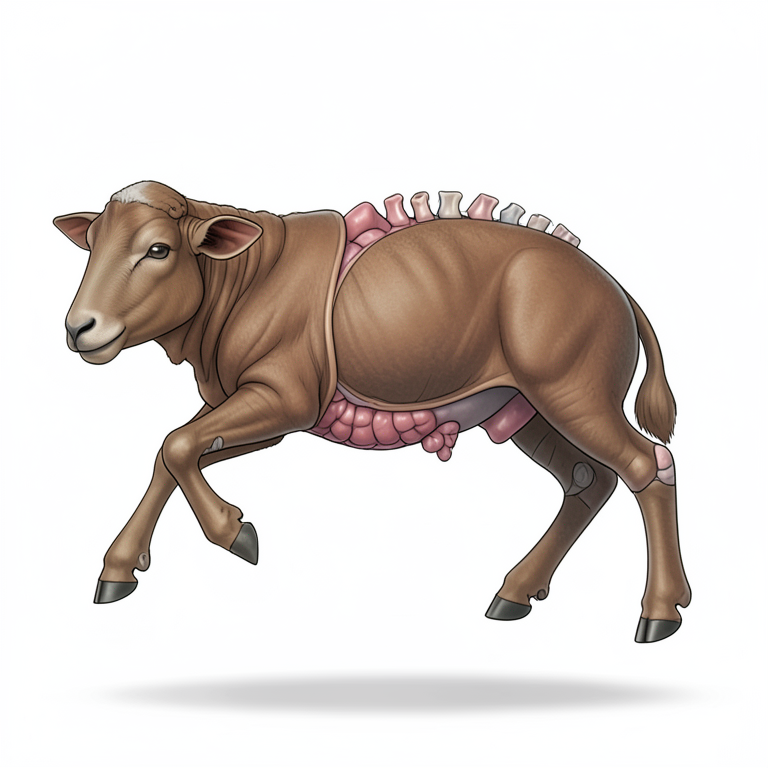
Anatomical Characteristics of Schistosoma Reflexus
AI-generated conceptual illustration (Nellore brown ewe fetus with Schistosoma reflexus). It explains the idea and does not show study data.
View source evidence ↗Figures & tables
From the original paper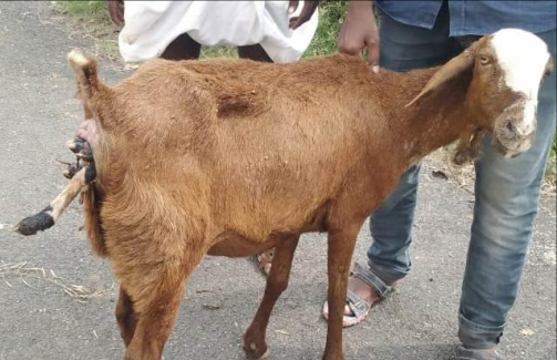 Figure 1
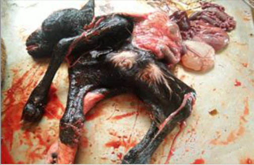 Figure 2
Introduction
Schistosoma reflexus is a fetal congenital disorder seen most commonly in cattle but rarely observed in small ruminants (Roberts, 1971) [8]. The thoracic and abdominal viscera are exposed and fetal head lies near its sacrum are the characteristics of Schistosoma reflexus. The limbs are usually ankylosed and rigid. A true Schistosoma is classified as a case with both viscera exposed and inversion of the spine.
This congenital anomaly generally occurs during embryonic development of the fetus. Schistosoma occurs mainly due to the transfer of autosomal recessive genes having incomplete penetrance to the developing embryo (Laughton et al., 2005; Balamurugan et al., 2020) [3, 1]. Successful per vaginal management of Schistosoma reflexus presented with its extremities in Nellore Brown ewe is reported. Case history and clinical observations A pleuriparous Nellore Brown ewe was presented to the Veterinary Clinical complex, College of Veterinary Science, Proddatur with a complaint of straining since early hours of the day.
The ewe was standing and four fetal limbs were protruded from the vulva (Figure 1). All clinical parameters were within normal limits. Upon per vaginal examination fetal head was present at pelvic brim. Based on this, a tentative diagnosis of dystocia due to malpresentation was made.
Treatment and Discussion After giving epidural anaesthesia, limbs with rigid joints were repelled to take out fetal head. The birth canal was lubricated sufficiently to remove the forelimbs and by careful traction removed Schistosoma dead fetus (Figure 2). The head of the monster was normal and abdominal wall was not fully developed and all viscera remained outside with curved spine and ankylosed limbs. The ewe was treated with antibiotic, anti-inflammatory and antihistamines in prescribed doses parenterally for five days.
Schistosoma is a rare kind of fetal monstrosity seen primarily in cattle. Schistosoma reflexus may be presented viscerally or by its extremities. Natural birth in visceral presentation is fairly common (Noakes et al., 2009) [5]. Suthar et al., (2011) [11], Singh et al., (2017) [9] and Promod et al., (2020) [7] reported per vaginal delivery and Tsuma and Abuom (2008) [10] recorded correction by caesarean section in visceral presentation of SR in ewes and does.
However Motunrayo et al., (2015) [4] published pervaginal delivery of Schistosoma reflexus conjoined twins in ewe. VET 2022; 7(3): 42-43 K Jyothi Assistant Professor and Head, Department of Veterinary Gynaecology and Obstetrics, Proddatur, Kadapa, Andhra Pradesh, India YV Pridhvidhar Reddy Assistant Professor, Department of VCC, College of Veterinary Science, Proddatur, Kadapa District, Andhra Pradesh K Sai Gunaranjan Assistant Professor, Department of Veterinary Gynaecology and Obstetrics, Proddatur, Kadapa, Andhra Pradesh, India K Jyothi Assistant Professor and Head, Department of Veterinary Gynaecology and Obstetrics, Proddatur, Kadapa, Andhra Pradesh, India http://www.veterinarypaper.com in a nondescript buffalo: A case report. The Pharma Innovation Journal. 2020;9(4):57-58. 2. Kumar B, Yadav D, Saxena A.
Schistosomus Reflexus in a Goat-A Case Report. Theriogenology Insight-An International Journal of Reproduction in all Animals. 2016;6(3):119-21. 3. Laughton KW, Fisher KR, Halina Partlow WG. Schistosomus reflexus syndrome: A heritable defect in ruminants.
Anatomia, histologia, embryologia. 2005;34(5):312-8. 4. Motunrayo AH, Ogechi NR, Bire GD, Tagwai KJ, When a Schistosoma reflexus presents by its extremities together with ankylosis of joints, is likely to prevent manipulative delivery. However, the present case was exceptional as Schistosoma presented by its four ankylosed limbs, could be delivered per vaginally using mutational operations as the birth canal was sufficiently relaxed and well lubricated. The cesarean operation was suggested for this type of presentation unless the fetus is small by Noakes et al., (2009) [5].
Like the present case, Kumar et al., (2016) [2]; Prabhakaran et al., (2020) [6] recorded ventro transverse presentation and per vaginal delivery in does and Kumar et al., (2016) [2] opined that extremities presentation common in does compare to visceral presentation. Omolathebu OJ. Dystocia due to schistosomus reflexus (Cojoined twins) in a Yankassa ewe. Journal of Coast Life Medicine. 2015;3(4):333-5. 5.
Noakes DE, Parkinson TJ, England GCW. Veterinary Reproduction and Obstetrics, 9th edn. W.B Saunders Company, Philadelphia; c2009. 6. Prabaharan V, Palanisamy M, Rajkumar R, Raja S.
Successful per vaginal delivery of schistosomus reflexus in a non-descriptive doe. International Journal of Science, Environment and Technology. 2020;9(3):456-459. 7. Pramod K, Sasi G, Banu P, Rajendra M, Tipu, S. Dystocia due to Schistosomus reflexus in a Marwari ewe.
Haryana Veterinarian. 2020;59(2):285-286. 8. Roberts SJ. Veterinary Obstetrics and Genital Diseases, 2nd edn. SJ Roberts, Ithaca, NY. 1971;605:70-73. 9.
Singh D, Kumar P, Kumar A, Kumar A, Ruhel S. Fig 1: Nellore Brown ewe suffering from dystocia with exposed fetal Dystocia due to schistosomus reflexus (co-twin) in a Marwari goat-a case report. Life Sciences Leaflets. 2017;94:10-12. 10. Tsuma VT, Abuom TO.
A case report of Schistosomus extremities Reflexus in a Lamb. The Kenya Veterinarian. 2008;32(1):41-43. 11. Suthar DN, Sharma VK, Dabas VS, Bhoi DB. Per- vaginal handling of Schistosomus reflexus as a cause of dystocia in a Goat.
Veterinary World. 2011;4(7):330-331. Fig 2: Schistosoma reflexus with exposed viscera and ankylosed limb
Conclusion
All clinical parameters were within normal limits. Upon per vaginal examination fetal head was present at pelvic brim. Based on this, a tentative diagnosis of dystocia due to malpresentation was made. It is suggested that further research should be done in this area.
Acknowledgement
The Authors are thankful to Sri Venkateswara Veterinary University for providing facilities to carry out this work.
References
1 in the original paper- Balamurugan B, Reddy YP, Jyothi K. Successful pervaginal delivery of Schistosoma reflex foetal monster