
Research article · 2021 · Full text
International Journal of Advanced Biochemistry ResearchEvaluation of qualitative phytochemical constituents, antimicrobial activity and in vitro anti-inflammatory activities of Mappianthus iodoides methanol leaf extracts
Volume 5(2) · Pages 33–39 · DOI: 10.33545/26174693.2021.v5.i2a.74 ↗
Research areasPhytochemistry and Biological ActivitiesEthnobotanical and Medicinal Plants StudiesTraditional and Medicinal Uses of Annonaceae
Abstract
Author-writtenMappianthus iodoides is a plant remedy exploited in the management of various microbial infections and it is also useful in reducing pain. The present study screened the leaf extracts of Mappianthus iodoides for its phytochemical constituents, anti-microbial and in vitro anti-inflammatory activities. Heat- and hypotonic induced haemolysis of red blood cells were used to access the anti-inflammatory activities and the anti-microbial activity was determined by agar well diffusion method. The plant extracts were screened for qualitative phytochemical constituents and the presence of alkaloids, flavonoids, tannins, cardiac glycosides, phenols, steroids, amino acids, anthraquine and soluble carbohydrates were observed. The results showed that the extracts had significant inhibition of microbial growth against all the organisms tested. The minimum inhibitory concentration (MIC) of the extracts showed that almost all the organisms tested were responsive to the extracts at a given concentration. Also, the extracts significantly (P
Summary
Source-checkedPlain-language reading of the full paper, prepared with AI and checked against the text. Each point links to the authors' own words.
Why it matters
- Mappianthus iodoides is traditionally used as a remedy for microbial infections and pain reduction. View evidence ↗
- Unresolved acute inflammation can lead to organ dysfunction and contribute to chronic conditions such as arthritis, diabetes, and cardiovascular diseases. View evidence ↗
- Phytochemicals are bioactive principles in plants responsible for various medicinal properties, including antimicrobial and anticancer effects. View evidence ↗
The question
- The study aims to evaluate the phytochemical constituents of Mappianthus iodoides methanol leaf extract to validate its traditional use for pain and infection management. View evidence ↗
What they did
- Leaf extracts were prepared by soaking crushed, dried leaves in methanol, cold water, and hot water for 24 hours. View evidence ↗
- The antimicrobial activity was tested against five bacterial strains: E. coli, P. aeruginosa, S. typhi (Gram-negative), and B. cerus and S. aureus (Gram-positive). View evidence ↗
- Antimicrobial activity was measured using the agar well diffusion method and the micro dilution method to determine the minimum inhibitory concentration (MIC). View evidence ↗
- Anti-inflammatory activity was assessed by measuring the stabilization of human erythrocyte membranes against heat and hypotonic-induced haemolysis. View evidence ↗
What they found
- The methanol extract contained high concentrations of phenols, saponins, flavonoids, tannins, and steroids, whereas these were low or absent in the aqueous extract. View evidence ↗
- The methanol extract showed antimicrobial potency with the lowest MIC of 125 mg/ml against S. typhi and the highest MIC of 500 mg/ml against P. aeruginosa. View evidence ↗
- At a concentration of 500 mg/ml, the methanol extract produced inhibition zones ranging from 8.76±1.0 mm (P. aeruginosa) to 20.33±0.47 mm (S. typhi). View evidence ↗
- The methanol extract inhibited heat-induced haemolysis in a dose-dependent manner, reaching 89% inhibition at 800 µg/ml. View evidence ↗
- The methanol extract prevented hypotonic-induced haemolysis, with inhibition percentages of 66%, 31%, 36%, 100%, and 65% for doses from 100 to 800 µg/ml, respectively. View evidence ↗
What it means
- The observed antimicrobial and anti-inflammatory activities support the traditional medicinal claims for Mappianthus iodoides. View evidence ↗
- The authors recommend further research to isolate the specific active components responsible for the plant's pharmacological properties. View evidence ↗
Visual summary
3 visualsIllustrations explain the paper; charts are replotted from the paper's own table values.
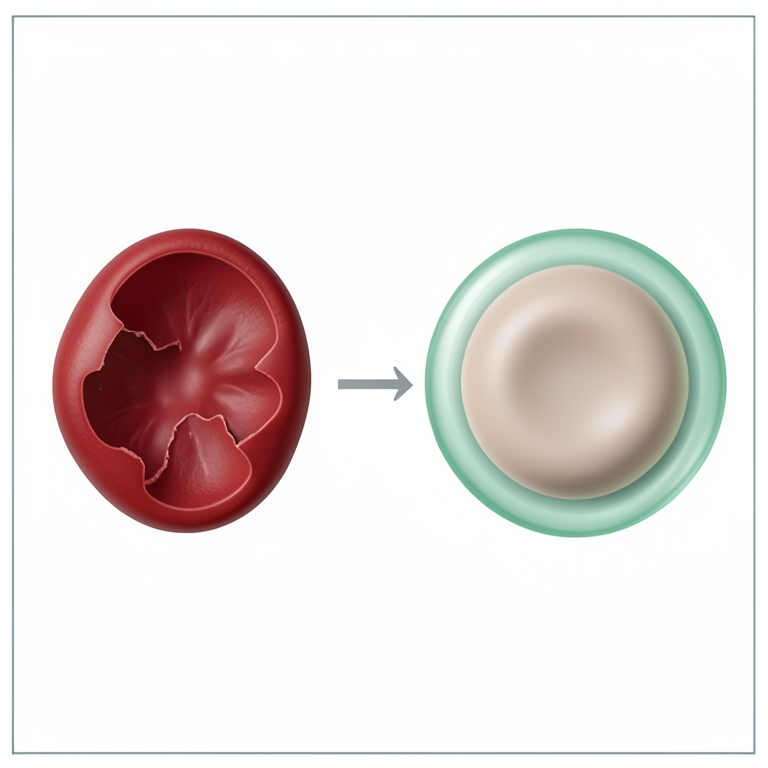
Membrane Stabilization Concept
AI-generated conceptual illustration (human red blood cells). It explains the idea and does not show study data.
View source evidence ↗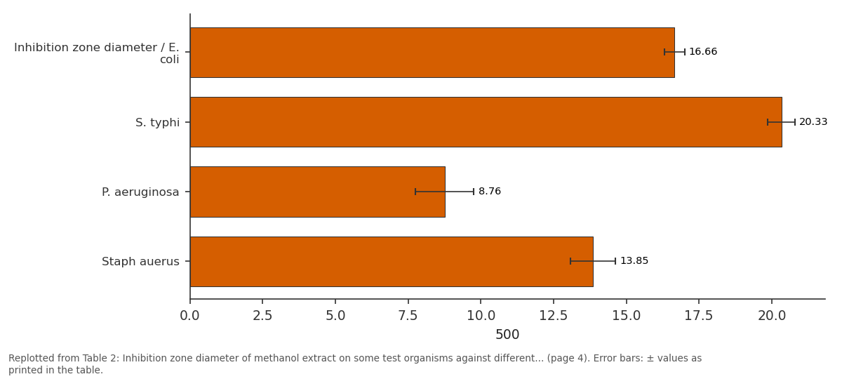
500: Inhibition zone... vs S. typhi vs P. aeruginosa vs Staph auerus
Replotted from Table 2 (page 4) of the original paper. Every bar is the paper's own table value.
View source evidence ↗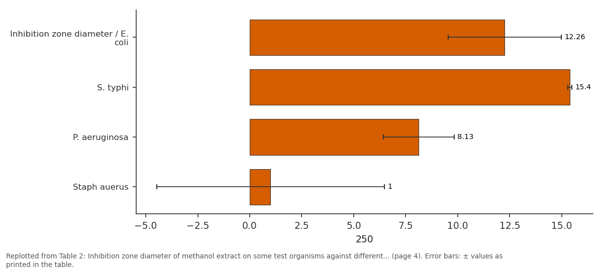
250: Inhibition zone... vs S. typhi vs P. aeruginosa vs Staph auerus
Replotted from Table 2 (page 4) of the original paper. Every bar is the paper's own table value.
View source evidence ↗Figures & tables
From the original paper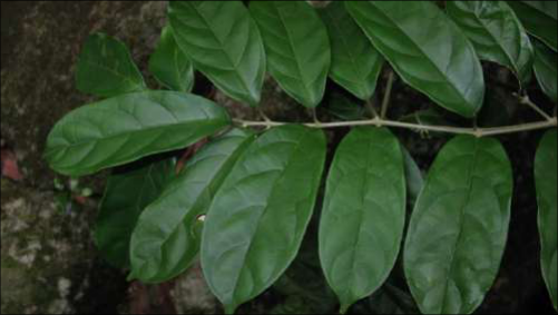 Figure 1
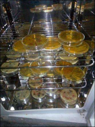 Figure 2
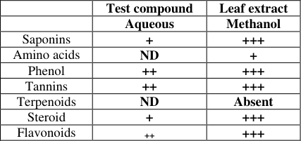 Table 1
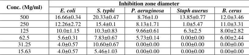 Table 2
 Table 3
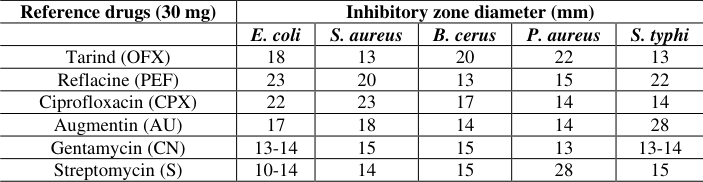 Table 4
 Table 5
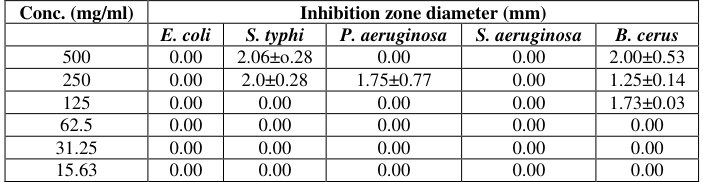 Table 5
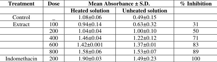 Table 6
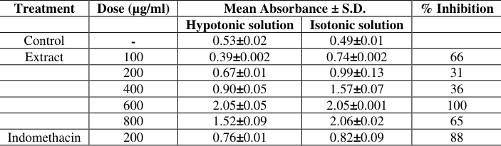 Table 7
Introduction
The first phase of the body's defense against invading pathogens, foreign bodies and/or injury is known as the acute inflammatory phase [1] . The acute inflammatory phase involves the immune cells, blood vessels and a host of other agents that mediate infection. The purpose of these reactions is to get rid of the initial cause of cell injury, evacuate dead cells and to initiate tissue repair [1] . However, if the acute phase of the inflammatory process is not resolved, it can be a contributing factor to organ dysfunction and aggravate many widely occurring chronic inflammatory clinical manifestations which include arthritis, asthma, allergy, diabetes, inflammatory process of aging and cancers, organ fibrosis, metabolic syndrome, neurodegenerative diseases, cardiovascular and periodontal diseases [3][4][5][6][7][8][9][10][11][12][13] . Phytochemicals are known to contain important bioactive principles. They are present in different parts of plants in varying amounts. They are important bioresource of drugs used mainly in traditional medicine, modern medicine, pharmaceutical intermediates and this leads to the synthesis of drugs [14] . They have been reported to be responsible for the antimicrobial, antibiotic, anticancer, antihelminthic and antisickling properties of many products used in medicine [15] . Mappianthus species are a group of flowering plant belonging to the family Icacinaceae. The genus Mappianthus comprises of three important species which are widely spread in subtropical and tropical zone. It is used by traditional people for the treatment of traumatic injury, rheumatalgia, arthralgia etc. [16] . However, considering the myriad uses of this plant, no significant work has been done on Mappianthus iodoides leaf extract; especially so far, there is no scientific report on its antiinflammatory properties, phytochemical constituents and anti-microbial properties of the International Journal of Advanced Biochemistry Research extract. Therefore, the present study seeks to evaluate the phytochemical constituents of the methanol extract of Mappianthus iodoides to ascertain the folkloric use of the extract in the management of pain and microbial infections.
Materials and Methods
Materials and Methods
Chemicals and materials
All chemicals used in this study were of analytical grade and were purchased from Sigma Aldrich Chemical Co. Ltd. (USA).
Plant material
Mappianthus iodoides was collected from the environment of Osusu Umuikpeghi Village in Obingwa L.G.A of Abia State. The plant material was confirmed by Mr. Alfred Ozioko of Bioresources Development and Conservation Programme (BDCP), Aku Road, Nsukka, Nigeria in November, 2020, where, the voucher specimen was kept for future reference.
Test Microorganisms
The gram negative bacteria (Escherichia coli, Pseudomonas aeruginosa and S. typhi and gram positive bacteria (Bacillus cerus and Staphylococcus aureus) were obtained from Microbiology Laboratory, Biological Sciences Department, Clifford University, Owerrinta, Abia State.
Methods
Methods
Extraction of plant
The leaves were plucked fresh from the stalk and were dried at room temperature for about two weeks and crushed. The crushed leaf was weighed and divided into three equal parts and was soaked in 200 ml of methanol, 200 ml of cold water and 200 ml hot water respectively for 24 hours with intermittent shaking and covered to avoid the growth of mould. Filtration was carried out using Whatman No 1 filter paper and a funnel into a 100 ml volumetric flask. The filtrate was evaporated to dryness using a water bath at 45 °C. The crude extracts were kept in a refrigerator at 4 °C until further use. The extraction yield was calculated using the relation; % yield = (weight of extract (g)/weight of plant material (g) × 100.
Preparation of media
A known amount of 38 g of the Mueller-Hinton agar was measured using weighing balance with a spatula and aluminum foil, and dissolved in 1000 ml of distilled water in a conical flask. The dissolved solution was homogenized by heating gently on a Bunsen burner, the conical flask was then covered with a non absorbent cotton wool and sealed with an aluminum foil and then autoclaved at 121 °C for 15 minutes.
Sterilization of plates
Fifty (50) petri dishes were thoroughly washed, cleaned and allowed to dry. Thereafter, the 50 petri dishes were carefully packed in five nylon bags and sterilized using the autoclave at 121 °C for 15 mins.
Sterilization of media and plate pouring
Homogenized Mueller-hinton agar was autoclaved at 121 °C for 15 minutes. The sterilized media was poured into the 50 petri dishes, each plate containing 20 ml of agar and the plate pouring process was done close to a Bunsen burner, then allowed to cool and the gel and the plate were inverted to avoid drop of moisture on the agar which may contaminate it. The plates were incubated at 37 °C for 12 hours to ascertain its sterility and then it was ready to be used for anti-microbial screening.
Preparation of normal saline
A known amount of 0.9 g of NaCl, was dissolved in 100 ml of distilled water, 4 ml each was pipetted into 6 clean test tubes the test tubes were then sealed with non-absorbent cotton wool and then autoclaved at 121 °C for 15 minutes.
Preparation of McFarland's standard
The McFarland's standard provides the turbidity reference for checking the approximate number of cells in dilution. They are sealed tubes of Barium sulphate suspension prepared as described by McFarland for use as standard for adjusting densities of bacterial suspensions and other turbid suspension. McFarland's standard helps researcher to have an idea of the approximate microbial activity against a given approximate number of test organisms. To prepare McFarland's standard 0.05 ml of 1% Barium chloride was added to 9.95 ml of 1% Sulphuric acid to obtain a turbidity of 0.119 at (600 nm) which was compared to the turbidity of the bacterial suspensions.
Inoculant dilution
The organisms inoculated and incubated at 37 °C were removed after 24 hours. A Finn micropipette was used to prepare a dilution of the organism in sterilized normal saline to achieve turbidity equivalent to MCF 0.119. The five different organisms with the turbidity as McFarland's standard were then used to seed the well labeled Petri dishes (labeled with names of the test organisms) using the spread plate method and with the help of a Finn micropipette. Using a sterilized cork borer of 6 nm; five wells were cut round in each of the agar plates already seeded with the test organism.
Preparation of 20% dimethyl sulfoxide (DMSO)
Known amount of DMSO, 20 ml was pipette into a 100 ml volumetric flask and 80 ml of water was added to the volumetric flask to make it up to 100 ml.
Serial dilution of the extract
Known amount of the extract (0.5 g) was weighed and added into the test tube, 1 ml of 20% DMSO was added into the first test tube which is the stock (500 mg) and it was shaken to dissolve the plant extract. One (1) ml was taken
International Journal of Advanced Biochemistry Research out from the first test tube into the second test tube with dilution rate (250 mg), the procedure continued until the fifth test tube to achieve concentrations of the extract equivalent to 500, 250, 125, 62.5, and 31. 25 mg/ml. This procedure was repeated for hot water and cold water extracts.
Antimicrobial studies
All tests were performed in triplicate and standard antibiotics discs were used as a positive control while 20% DMSO was used as a negative control. The six wells made round the plates with 6 mm cork borer were well labeled (using concentrations of the extract; 500, 250, 125, 62.5, and 31. 25 mg/ml), the different concentrations were introduced into appropriate well using a Finn micropipette. The set up was left to stand for 30 minutes and then the plates were incubated at 37 °C for 24 hours. At the completion of the incubation, the plates were brought out and the formation of clear zones were measured using a meter rule and recorded in millimeter (mm) and the results were recorded as mean diameter of zones of growth (with name of the test organism).
Agar well diffusion test
A loopful of bacterial culture was placed on a solidified MHA media. With a sterile cork borer, agar well of 6 mm diameter was dug on agar surface. Each well was aseptically filled with respective extracts of 20 μL. The plates were allowed to soak the extracts and incubated at 37 °C for 24 h. Zone of inhibition was measured.
Determination of the minimum inhibitory concentration (MIC)
The minimum inhibitory concentration can be regarded as the least concentration of the anti-microbial agent found to stop the growth of a particular test organism. The MIC value explains the potency of the antimicrobial agent, it may be interpreted that the organism is ''susceptible'' intermediate'' or ''resistant'' to the antimicrobial agent [17] . The lower the MIC value, the more susceptible the organism and the more potent the antimicrobial substance. The MIC was accessed using the micro dilution method. The MIC samples were found to be active by the dilution method, the test extracts were dissolved in 20% DMSO to give the stock concentration of 500 mg/ml and this was subjected to serial dilution in a series of test tubes to a working concentration ranging from (500 to 15.63 mg/ml) and the following concentrations were obtained, (500, 250, 125, 62.5, 31.25, and 15.63 mg/ml). The broth was prepared by weighing 1.3 g of agar broth in a volumetric flask and making up to 100 ml, the broth was pipetted into 10 test tubes each for aqueous and methanol extracts respectively. The diluted extract for aqueous and methanol were transferred into the test tubes according to their concentrations respectively (500, 250, 125, 62.5, 31.25, 15.63 mg/ml). The five organisms were introduced into the test tubes and were incubated at 37 °C for 24 hrs.
Phytochemical analysis
The crude extracts were subjected to phytochemical analysis using the method described by Harbourne, 1998 [18] .
Membrane Stabilization Studies
Heat and hypotonic-induced haemolysis of human erythrocyte were used to determine the stabilization of membrane and this was done using the method described by Shinde et al., 1989 [19] with some modifications, Anosike et al., 2018 [20] .
Heat-Induced haemolysis of Red Blood cell
A known quantity of the extracts was dissolved in isotonic phosphate buffer solution. Centrifuge tubes were arranged in quadruplicate containing about 5 ml doses of the extracts (100, 200, 400, 600, 800 µg/ml). Control experiment was set up by arranging two sets of centrifuge tubes containing 5 ml of isotonic phosphate buffer solution and 5 ml of 200 µg/ml of indomethacin respectively. Erythrocyte suspension (0.1 ml) was added to each tube and mixed gently. Two of the tubes from each set were incubated at 54 °C for 20 minutes in a water bath. Thereafter, the tubes were centrifuged at 1300 g for 3 minutes and the haemoglobin content of the supernatant was determined using spectronic 21D (Milton Roy) spectrophotometer at 540 nm. The percentage inhibition of haemolysis of the extract was calculated thus Percentage Inhibition of haemolysis =
Hypotonic-induced Haemolysis of Red Blood Cell
A known quantity of the extracts was dissolved in hypotonic solution (distilled water). Duplicate pairs (per dose) of the centrifuge tubes contained 5 ml graded doses (100, 200, 400, 600, 800 µg/ml). However, an isotonic phosphate buffer solution containing about 5 ml graded doses of the extracts (100, 200, 400, 600, 800 µg/ml) were put into the other duplicate pairs (per dose) of the centrifuge tube. Control experiment was set up by arranging two sets of centrifuge tubes containing 5 ml of distilled water and 5 ml of 200 µg/ml of indomethacin respectively. Erythrocyte suspension (0. Where, OD 1 = Absorbance of test sample in isotonic solution OD 2 = Absorbance of test sample in hypotonic solution OD 3 = Absorbance of control sample in hypotonic solution.
Preparation of the Erythrocyte Suspension
Fresh blood sample (3 ml) was collected from healthy volunteers and were put in heparinized bottles and centrifuged at 3000 rpm for 10 minutes. A volume of saline similar to the volume of the supernatant was used to dissolve the red blood pellets. The volume of the dissolved red blood pellet was measured and reconstituted as 40% v/v suspension with isotonic buffer solution (10 nM sodium Phosphate buffer, pH 7.4). The buffer solution contained 0.2 g of NaH 2 PO 4 , 1.15 g of Na 2 HPO 4 and 9 g of NaCl in 1 litre of distilled water. The reconstituted red blood cell (resuspended supernatant) was used as such.
Statistical analysis:
Results were presented in tables are expressed as mean ± S.D. The level of significance was tested using one-way ANOVA. Results were regarded as significant when p<0.05 that is there is greater than 95% probability that the result is not by chance. All statistical analyses were performed using SPSS software.
Results
Results
Phytochemical screening
The result of the phytochemical screening of the methanol extract of the leaf of Mappianthus iodoides is shown in Table 1. The result shows the presence of alkaloids, flavonoids, tannins, saponins and cardiac glycosides, anthraquinones, reducing sugar, steroids, phenol, soluble carbohydrate, proteins and amino acids by qualitative methods. Terpenoids were absent. The qualitative phytochemical composition of Mappianthus iodoides presented in Table 1 shows high concentrations of some bioactive phyto-active principles such as phenols, saponins, flavonoids, tannins and steroids in the methanol extract while the concentrations were low or absent in the aqueous extract. Amino acids were in low concentration in the methanol extract and absent in the aqueous extract. Terpenoids were absent in both aqueous and methanol extract.
| — | Test compound | Leaf extract |
|---|---|---|
| Aqueous | Methanol | |
| Saponins | + | +++ |
| Amino acids | ND | + |
| Phenol | ++ | +++ |
| Tannins | ++ | +++ |
| Terpenoids | ND | Absent |
| Steroid | + | +++ |
| Flavonoids | ++ | +++ |
Table text extracted from the original PDF; superscript letters are shown inline. The original table image is below.
View original table image
Antimicrobial activity
The zone diameters of inhibition of the crude extracts at different concentrations are shown in Table 2. From the result of the zones of inhibition of methanol extract of M. iodoides, it can be seen that it displayed credible activities on the organisms tested when compared to the standard drugs used. Table 3 shows the minimum concentration of the crude extract required to inhibit the growth of the organism. The lowest (125 mg/ml) for the methanol extract of M. iodoides was recorded against S. typhi, while the highest MIC (500 mg/ml) was recorded against P. aeruginosa.
| Extract | MIC values in (mg/ml) | — | — | — | — |
|---|---|---|---|---|---|
| E. coli | S. aureus | B. cerus | P. aureus | S. typhi | |
| Methanol | 125 | 250 | 250 | 500 | 125 |
| Aqueous | 0.0 | 0.0 | 500 | 250 | 0.0 |
Table text extracted from the original PDF; superscript letters are shown inline. The original table image is below.
View original table image
| Conc. (Mg/ml) | Inhibition zone diameter | — | — | — | — |
|---|---|---|---|---|---|
| E. coli | S. typhi | P. aeruginosa | Staph auerus | B. cerus | |
| 500 | 16.66±0.34 | 20.33±0.47 | 8.76±1.0 | 13.85±0.77 | 12.0±3.46 |
| 250 | 12.26±2.72 | 15.4±0.1 | 8.13±1.71 | 1.0±5.47 | 11.0±3.31 |
| 125 | 10.0±1.15 | 10.3±0.83 | 9.66±0.61 | 6.3±2.5 | 8.00±2.82 |
| 62.5 | 5.6±0.31 | 7.83±0.67 | 5.73±0.14 | 0.00±0.00 | 6.00±2.44 |
| 31.25 | 4.0±0.57 | 10.60±0.67 | 0.00±0.00 | 0.00±0.00 | 0.00±0.00 |
| 15.63 | 4.0±0.57 | 5.46±1.03 | 0.00±0.00 | 0.00±0.00 | 0.00±0.00 |
Table text extracted from the original PDF; superscript letters are shown inline. The original table image is below.
View original table image
Membrane stabilization of human red blood cell Effect of methanol extract of M. iodoides on heatinduced haemolysis of human red blood cell
Table 6 shows that the extracts significantly (p<0.05) shielded the human red blood cell membrane against breakdown induced by heat in a dose dependent manner. The result also shows that 800 µg/ml of the extract produced the highest percentage inhibition of haemolysis of 89% and this is comparable to the standard drug used. Effect of methanol extract of Mappianthus iodoides on hypotonicity-induced haemolysis of human red blood cell.
| Treatment | Dose | Mean Absorbance ± S.D. | — | % Inhibition |
|---|---|---|---|---|
| Heated solution | Unheated solution | |||
| Control | 1.08±0.06 | 0.49±0.15 | ||
| Extract | _ 100 | 0.94±0.14 | 0.63±0.32 | 31 |
| 200 | 1.04±0.04 | 1.00±0.10 | 50 | |
| 400 | 1.46±0.04 | 1.22±0.12 | 71 | |
| 600 | 1.42±0.001 | 1.37±0.01 | 83 | |
| 800 | 1.58±0.06 | 1.53±0.07 | 89 | |
| Indomethacin | 200 | 1.90±0.03 | 1.49±0.23 | 100 |
Table text extracted from the original PDF; superscript letters are shown inline. The original table image is below.
View original table image
Table 7 shows that Mappianthus iodoides extract significantly (p<0.05) prevented lysis induced by water (hypotonic solution). This is evident by the high percentage respective inhibition of 66, 31, 36, 100, and 65 obtained at doses of 100-800 µg/ml. This result is very comparable to that of indomethacin (200 µg/ml) which produced percentage inhibition of 100%.
| Treatment | Dose (µg/ml) | Mean Absorbance ± S.D. | — | % Inhibition |
|---|---|---|---|---|
| Hypotonic solution | Isotonic solution | |||
| Control | - | 0.53±0.02 | 0.49±0.01 | |
| Extract | 100 | 0.39±0.002 | 0.74±0.002 | 66 |
| 200 | 0.67±0.01 | 0.99±0.13 | 31 | |
| 400 | 0.90±0.05 | 1.57±0.07 | 36 | |
| 600 | 2.05±0.05 | 2.05±0.001 | 100 | |
| 800 | 1.52±0.09 | 2.06±0.02 | 65 | |
| Indomethacin | 200 | 0.76±0.01 | 0.82±0.09 | 88 |
Table text extracted from the original PDF; superscript letters are shown inline. The original table image is below.
View original table image
Discussion
In this present study, we evaluated the phytochemical constituents and its antimicrobial and anti-inflammatory activities. Though there are so many extracts with antimicrobial and anti-inflammatory activities, Mappianthus iodoides extracts have not been well studied. Anti-inflammatory activities can be evaluated by many methods but this study focused on heat-induced membrane stabilization and hypotonic-induced human erythrocyte membrane stabilization. The phytochemical screening of the leaf extract of Mappianthus iodoides shows the presence of alkaloids, flavonoids, tannins, terpenes, saponins, and cardiac glycosides by quantitative methods, terpenoids were found to be absent in the crude extract. These secondary metabolites possess several medicinal properties which have been documented elsewhere [21] . Five (5) of the bacterial strains tested (Gram-positive and Gram-negative) were sensitive to the methanol extracts of Mappianthus iodoides and this was determined by agar well diffusion method. Though agar well diffusion method is not efficient because the antimicrobial activity may be affected by various factors the type of agar used, salt concentration, incubation temperature and the molecular size of the antimicrobial agent. However, it does not distinguish between the agents that can kill the bacteria and those than can stop the growth of the bacteria [22] . The MIC values in Table 3 showed that the methanol extract had antimicrobial activity against all the bacteria tested while the aqueous extract exhibited activity against B. cerus and P. aureus only.
Table 6 shows that the extract significantly (p<0.05) prevented heat-induced haemolysis of HRBC. This is evidence that stabilization of the membrane is a possible method of the anti-inflammatory activity of the extract. Hypotonic solutions are known to have haemolytic effect. Haemolysis occurs when there is excess accumulation of fluid in the cell and this result in the swelling of the membrane and eventual bursting of the erythrocyte membrane. When the erythrocyte membrane gets ruptured, it will make the cell liable to secondary damage. This damage occurs by free radical-induced lipid peroxidation [23] . Membrane stabilization prevents the leakage of serum proteins into the tissue fluid. This process goes on by inflammatory mediators especially when there is an increase in membrane permeability [24] . The methanol extract of Mappianthus iodoides leaf could stabilize the erythrocyte membrane induced by hypotonic solution by inhibiting the discharge of enzymes that could disintegrate the cell and other active inflammatory mediators. At 600 µg/ml of the extract, it showed 100% inhibition of haemolysis and this result is similar to the standard drug, indomethacin and this result is in agreement with work of Anosike et al., 2018 [20] . Another possible mechanism for the membrane stabilizing activity of Mappianthus iodoides extract could be due to an increase in the surface area/volume ratio of the cell, which could be brought about by increase in membrane size or decrease in cell size and interaction with membrane proteins [25] . However, it has been well documented that the cell volume of red blood cells are closely related to the calcium content inside the cell [20] . This is good evidence that the ability of the extracts to protect the human red blood cell could be due to the ability of the Mappianthus iodoides to change the influx of calcium into the red blood cell membrane.
Conclusion
The present study reveals that the extracts possess some phyto-active principles of therapeutic importance and these possess biological activity against a number of diseases. Therefore, further studies are required to discover and isolate the active components of the pharmacological properties of the plant.
Acknowledgement
AcknowledgementsThe authors are grateful to the administration of Clifford University, Owerrinta, Abia State, Nigeria led by the President/Vice-Chancellor, Prof. A.C. Omeonu for providing the materials needed for this research. We are also grateful to Mr. Ben Ikoro, the laboratory technologist in charge of the Biochemistry Laboratory for assisting us with the available materials and equipment.
Figures and tables from the paper
| Reference drugs (30 mg) | Inhibitory zone diameter (mm) | — | — | — | — |
|---|---|---|---|---|---|
| E. coli | S. aureus | B. cerus | P. aureus | S. typhi | |
| Tarind (OFX) | 18 | 13 | 20 | 22 | 13 |
| Reflacine (PEF) | 23 | 20 | 13 | 15 | 22 |
| Ciprofloxacin (CPX) | 22 | 23 | 17 | 14 | 14 |
| Augmentin (AU) | 17 | 18 | 14 | 14 | 28 |
| Gentamycin (CN) | 13-14 | 15 | 15 | 13 | 13-14 |
| Streptomycin (S) | 10-14 | 14 | 15 | 28 | 15 |
Table text extracted from the original PDF; superscript letters are shown inline. The original table image is below.
View original table image
| Nalicidic Acid (NA) | 29 | 17 | 10 | 15 | 16 |
|---|---|---|---|---|---|
| Septrin (SXT) | 16 | 16 | 19 | 11 | 10 |
| Ampicillin (PN) | 12-13 | 14 | 12 | 28 | 13 |
Table text extracted from the original PDF; superscript letters are shown inline. The original table image is below.
View original table image
| Conc. (mg/ml) | Inhibition zone diameter (mm) | — | — | — | — |
|---|---|---|---|---|---|
| E. coli | S. typhi | P. aeruginosa | S. aeruginosa | B. cerus | |
| 500 | 0.00 | 2.06±o.28 | 0.00 | 0.00 | 2.00±0.53 |
| 250 | 0.00 | 2.0±0.28 | 1.75±0.77 | 0.00 | 1.25±0.14 |
| 125 | 0.00 | 0.00 | 0.00 | 0.00 | 1.73±0.03 |
| 62.5 | 0.00 | 0.00 | 0.00 | 0.00 | 0.00 |
| 31.25 | 0.00 | 0.00 | 0.00 | 0.00 | 0.00 |
| 15.63 | 0.00 | 0.00 | 0.00 | 0.00 | 0.00 |
Table text extracted from the original PDF; superscript letters are shown inline. The original table image is below.
View original table image
References
22 in the original paper- Jeremy RP, David KD. The total mesorectal excision specimen for rectal cancer: A review of its pathological assessment. J. Clin Pathol. 2007 Aug;60(8):849-855. https//doi.org/ 10.1136/jcp.2006.043802.
- Chukwunelo CC, Anosike CA, Nnamonu EI, Ekpo DE, James PO, Okonkwo TI. Evaluation of Leukocyte Mobilization and platelet aggregatory Effects of Ciprofloxacin, Lincomycin and Erythromycin in Wistar Albino Rats. Notulae Scientia Biologicae. 2019;11(4):345-351.
- Chiurchiù V, Leuti A, Maccarrone M. Bioactive lipids and chronic inflammation: Managing the fire within. Front. Immunol, 2018;9:38. https://doi.org/ 10.3389/fimmu.2018.00038.
- Doyle R, Sadlier DM, Godson C. Pro-resolving lipid mediators: Agents of anti-ageing. Semin. Immunol. 2018;40:36-48. https://doi.org/10.1016/j.smim.2018.09.002.
- Fullerton JN, Gilroy DW. Resolution of inflammation: A new therapeutic frontier. Nat. Rev. Drug Discov. 2016;15(8):551-567. https://doi.org/10.1038/ nrd.2016.39.
- Grivennikov SI, Greten FR, Karin M. Immunity, Inflammation, and Cancer. Cell. 2010;140(6):883-899. https://doi.org/10.1016/j.cell.2010.01.025.
- Libby P, Tabas I, Fredman G, Fisher EA. Inflammation and its resolution as determinants of acute coronary syndromes. Circ. Res 2014;114(12):1867-1879. https:// doi.org/10.1161/CIRCRESAHA.114.302699.
- Medzhitov R. Origin and physiological roles of inflammation. Nature 2008;454(7203):428-435. https://doi.org/10.1038/nature07201.
- Ortega-G´omez A, Perretti M, Soehnlein O. Resolution of inflammation: An integrated view. EMBO Mol. Med 2013;5(5):661-674. https://doi.org/10.1002/ emmm.201202382.
- Perretti M, Cooper D, Dalli J, Norling LV. Immune resolution mechanisms in inflammatory arthritis. Nat. Rev. Rheumatol. 2017;13(2):87-99. https://doi.org/ 10.1038/nrrheum.2016.193.
- Shimizu T. Lipid mediators in Health and disease: Enzymes and receptors as therapeutic targets for the regulation of immunity and inflammation. Annu. Rev. Pharmacol. Toxicol. 2009;49(1):123-150. https://doi.org/10.1146/annurev. pharmtox.011008.145616.
- Van Dyke TE, Serhan CN. Resolution of inflammation: A new paradigm for the pathogenesis of periodontal diseases. J. Dent. Res. 2003;82(2):82-90. https://doi.org/ 10.1177/154405910308200202.
- Viola J, Soehnlein O. Atherosclerosis -a matter of unresolved inflammation. Semin. Immunol. 2015;27(3):184-193. https://doi.org/10.1016/j.smim.2015.03.013.
- Ncube NS, Afolayan AJ, Okoh AI. Assessment techniques of antimicrobial properties of natural compounds of plant origin: Current methods and future trends. African Journal of Biotechnology. 2008;7(12):1797-1806.
- Mamta S, Jyoti S, Rajeev N, Dharmendra S, Abhishek G. Phytochemistry of medicinal plants. Journal of Pharmacognosy and Phytochemistry. 2013;1(6):168182.
- Shinde UA, Phadke AS, Nari AM, Mungantiwar AA, Dikshirt VJ, Saraf MN. Membrane stabilization activity-A possible mechanism of action for antiinflammatory activity of Cedrus deodora wood oil. Fitoterapia. 1989;70(3):251-257.
- Anosike CA, Ako AC, Nwodo OFC. Anti-inflammatory and membrane stabilization activities of methanol extract of Cissus aralioides leaves. Research Journal of Pharmaceutical, Biological and Chemical Sciences. 2018;9(4):18-28.
- Morsy N, Sherif EAA, Abdel-rassol TMA. Phytochemical analysis of Calotropis procera with anti-microbial activity investigation. Main Group Chemistry. 2016;15(3):267-273.
- Eloff JN. A sensitive and quick method to determine the minimal inhibitory concentration of plant extracts for bacteria. Planta Medica. 1998;64(8):711-713.
- Bukhari IA, Alhumayyd MS, Mahesar AL, Gilani AH. The analgesic and anticonvulsant effects of piperine in mice. Journal of Physiology and Pharmacology. 2013;64(6):789-794.
- Chaitanya R, Sandhya S, David B, Vinod KR, Murali S. HRBC membrane stabilizing property of root, stem and leaf of Glochidion velutinum. International Journal of Research in Pharmacology and Biomedical Science. 2011;2(1):256-259.
- Awe EO, Makinde JM, Olajide OA, Wakeel OK. Membrane stabilizing activity: A possible mechanism of action for the anti-inflammatory and analgesic properties of Russelia equisetiformis. International Journal of Pharmacology. 2006;2(4):447-450.