
Research article · 2020 · Full text
International Journal of Advanced Biochemistry ResearchRole of serum levels of vitamin D in patients of breast Cancer
Volume 4(1) · Pages 37–41 · DOI: 10.33545/26174693.2020.v4.i1a.69 ↗
Research areasVitamin D Research Studies
Abstract
Author-writtenBackground: Breast cancer is the most frequently diagnosed cancer and the leading cause of cancer death among females. Breast cancer is a hormone dependent disease which occurs due to hyperestrogenic states.Aim and Objectives: To study the role of serum levels of vitamin D in patients of breast cancer and age matched control subjects along with comparison of lymph node positive and negative patients of breast cancer.Material and Methods: A total of 30 newly diagnosed patients of breast cancer with thirty age matched subjects as controls were included in the study. Serum was analysed for vitamin D levels by RIA Analyser SR300 method in both the groups.Results: The mean serum vitamin D level was significantly lower in cases as compared to controls (p
Summary
Source-checkedPlain-language reading of the full paper, prepared with AI and checked against the text. Each point links to the authors' own words.
Why it matters
- Breast cancer is the most common cancer diagnosed in women and the primary cause of cancer-related deaths in this population. View evidence ↗
- The development of breast cancer is linked to hormone dependency, specifically occurring in hyperestrogenic states. View evidence ↗
- Vitamin D is thought to influence breast tissue because of its role in regulating the growth and differentiation of both normal and malignant cells. View evidence ↗
The question
- The study aimed to evaluate serum vitamin D levels in breast cancer patients compared to age-matched controls and to compare these levels between patients with and without lymph node involvement. View evidence ↗
What they did
- Serum 25(OH) vitamin D levels were measured using a fully automated RIA Analyser SR300 with a Beckman Coulter total kit. View evidence ↗
- Participants underwent a systemic evaluation including clinical exams, lymph node status, height, weight, serum vitamin D measurement, and routine biochemical and hematological tests. View evidence ↗
- Data analysis was performed using SPSS version 20, with continuous variables reported as mean ± standard deviation and categorical variables as percentages. View evidence ↗
What they found
- A weak negative correlation was observed between BMI and vitamin D levels in the cancer cases (r = -0.312, p = 0.093). View evidence ↗
- Cancer cases had significantly higher mean weight (70.4 ± 4.4 kg vs 60.8 ± 4.1 kg) and BMI (26.4 ± 2.0 kg/m² vs 22.7 ± 1.9 kg/m²) than controls (p = 0.0001). View evidence ↗
What it means
- The authors conclude that vitamin D may have a protective, anticarcinogenic effect by regulating cell proliferation, differentiation, and apoptosis in breast cells. View evidence ↗
- The authors recommend that women, particularly those with a family history of breast cancer, maintain a normal BMI, check vitamin D levels regularly after age 40, and take supplements. View evidence ↗
- The study is limited by its small sample size and the use of a single vitamin D measurement, which may not reflect long-term status. View evidence ↗
Visual summary
2 visualsIllustrations explain the paper; charts are replotted from the paper's own table values.
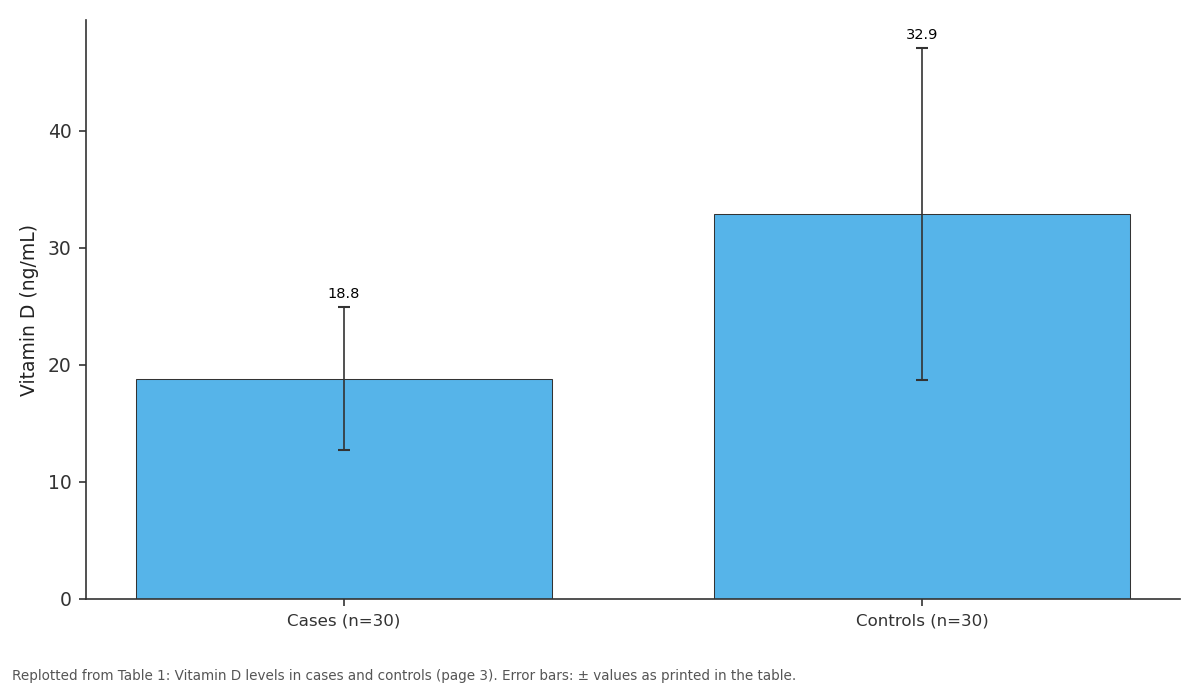
Vitamin D (ng/mL): Cases (n=30) vs Controls (n=30)
Replotted from Table 1 (page 3) of the original paper. Every bar is the paper's own table value.
View source evidence ↗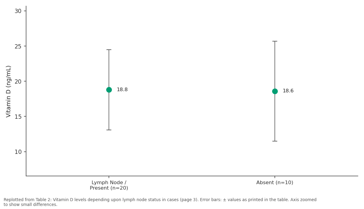
Vitamin D (ng/mL): Lymph Node / Present... vs Absent (n=10)
Replotted from Table 2 (page 3) of the original paper. Every bar is the paper's own table value.
View source evidence ↗Figures & tables
From the original paper Table 1
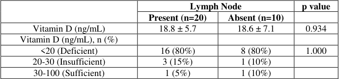 Table 2
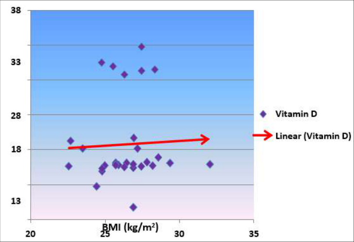 Figure 1
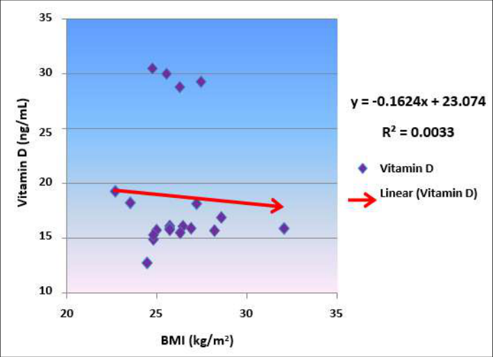 Figure 2
Introduction
Breast cancer is the most common diagnosed cancer and worldwide, it is the leading cause of cancer death among females. Approximate 1.7 million cases and 521,900 deaths occurred in 2012. Breast cancer alone accounts for 25% of all cancer cases and 15% of all cancer deaths among females. More developed countries account for about one-half of all breast cancer cases and 38% of deaths. Breast cancer incidence rates have been rising in many countries in South America, Africa, and Asia. The reason is not completely understood, but likely reflects changing reproductive patterns, increasing obesity, decreasing physical activity and some breast cancer screening activity. Mortality rates in these countries are also increasing, most likely due to changes associated with westernization compounded by delayed introduction of effective breast cancer screening programs and, in some cases; limited access to treatment [1,2] . Breast cancer accounts for 14% of cancers in Indian women. It is reported that with every four minutes, an Indian woman is diagnosed with breast cancer. Breast cancer is on the rise, both in rural and urban India. In a 2018 report of Breast Cancer statistics recorded 1,62,468 new registered cases and 87,090 reported deaths. Cancer survival becomes more difficult in higher stages of its growth, and more than 50% of Indian women suffer from stage 3 and 4 of breast cancer. Post cancer survival for women with breast cancer was reported 60% for Indian women, as compared to 80% in the U.S [3] . Less than 10% of breast cancers have been directly linked to germ line mutations. Several mutations have been implicated in familial cases. Various genes such as Breast Cancer 1,2 (BRCA-1, BRCA-2) } p53, Phosphatase and Tensin Homolog (PTEN) are involved in this. The convergence of these genes in a shared role reveals underlying biology of these illnesses. Thus all women with strong family histories of breast cancer should be referred to genetic screening programs [4] . The occurrence of breast cancer in the females are dependent upon the various other factors also like early menarche, late menopause, nulliparity, history of previous radiation exposure and high estrogen exposure state like polycystic ovarian disease. Dietary and food habits like Intake of alcohol, fatty food, oral contraceptives etc. also play an important role in development of breast cancer while breast feeding, folic acid and aspirin have got positive protective action upon it [5] . Breast lesions account for one of the largest group of conditions necessitating pathological, radiological and surgical intervention. A palpable mass is the most common symptom of underlying malignancy and must be distinguished from the normal nodularity of the breast. 6 Vitamin D: Vitamin D (1, 25-dihroxycalciferol or calcitriol) is a steroid hormone. Due to its endogenous synthesis and functioning as the ligand for the vitamin D receptor (VDR) this vitamin and its metabolites are hormone and hormone precursors rather than being simple vitamins. 7dehydroxycholesterol is absorbed from the diet and converted to vitamin D in the skin in the presence of sunlight. It is transported to the liver and undergoes 25hydroxylation by cytochrome p450 like enzymes in the mitochondria and microsomes to form 25-hydroxycholecalciferol which is the major storage form of vitamin D. 1-α -hydroxylation is the final step in hormone activation in presence of 1-α-hydroxylase and parathyroid hormone, taking place in kidney. Vitamin D also affects the immune system and VDRs are expressed in several white blood cells including monocytes and activated T and B cells, thereby having an effect on the immune system. Vitamin D increases expression of the tyrosine hydroxylase gene in adrenal medullary cells. It is also involved in the biosynthesis of neurotrophic factors, synthesis of nitric oxide synthase and increased glutathione levels. All these varied roles emphasize the importance of vitamin D in our body. More recently, it has become clear that receptors for vitamin D are present in a wide variety of cells such as brain, heart, gonads, prostate and breast and the hormone has got biological effects which extend far beyond its classical role in mineral metabolism. Due to its important role in regulation of cellular growth and differentiation in normal and malignant cells, the study was planned which focuses on the role of effects of vitamin D on breast tissue under physiological conditions and in breast cancer patients [7] . Thus, the present study was conducted to study the role of serum levels of vitamin D in patients of breast cancer and age matched control subjects along with comparison of lymph node positive and negative patients of breast cancer.
Material and Methods
The present study is a hospital based cross-sectional study conducted in the Department of Biochemistry in collaboration with the Department of Surgery PGIMS, Rohtak. Thirty newly diagnosed patients with breast cancer were taken for the study. Thirty age matched subjects were taken as controls.
Exclusion Criteria 1) Females on medications like oral contraceptives and hormone replacement therapy 2) Females taking drugs that alter vitamin D, like antiepileptic, diuretics and vitamin D supplement. 3) Females suffering from chronic liver, renal or endocrinal disease.
4) Those with other risk factors like malabsorption and hypovitaminosis (post gastric or bowel resection patient). 5) Pregnant and lactating females 6) Patients of osteoporosis 7) Any kind of metabolic disorder or tumor lysis syndrome.
A systemic evaluation comprising explained detailed history, clinical examinations along with the lymphnode status, complete medical history, general physical, local examination, height and weight measurement, measurement of serum vitamin D, hematological investigations and routine biochemistry along with complete haemogram was done in cases and controls. Clinical assessment of lymphnode status was done in cases and later eventually classified as clinically lymphnode positive and negative. BMI was calculated by formula BMI (kg/m2)=weight/(ht) 2 . BP was measured in both the groups.
Specimen Collection and Storage
Under all aseptic conditions 6 ml of Venous blood sample was taken from ante cubital vein, 4 ml in a plain red capped evacuated blood collection tube and 2 ml in purple capped (EDTA) evacuated blood collecting tube. Samples were processed within 1 hour of collection. Serum of cases and controls was separated by centrifugation at 2000 rotation per minute for 10 minutes after clotting. Separated serum was analysed for routine biochemistry and serum for vitamin D, was stored at -20 ºC till further analysis. Whole blood was subjected to hematological analysis. Various routine biochemical investigations were performed on autoanalyser using standard kits.
Estimation of serum vitamin D
Serum 25 (OH) vitamin D level was estimated on fully automated RIA Analyser SR300 by Stratec Biomedical AG, Germany using Beckman Coulter 25OH vitamin D total kit. 8
Assay Procedure
All reagents were allowed to reach room temperature, and mixed thoroughly by gentle inversion before they were used. The content of vial was reconstituted with the volume of the tracer buffer vial indicated on the label. After the reconstitution tracer was stable maximum for one week if kept at 2-8 degree C or at -20 degree C until expiry date. The contents of standard and control vials were reconstituted with 500 mL of distilled water, indicated on the vial label. Contents were mixed gently after 10 minutes to avoid foaming. Wash solution was prepared by adding 69 volumes of distilled water to 1 volume of wash solution (70x).
Biomedical waste
All the biomedical waste generated during this study in laboratory was discarded as per biomedical waste management and handling rules latest 2016 guidelines.
Statistical Analysis
The data was analyzed by using SPSS version 20. Categorical variables were presented as number and percentage while continuous variables were presented as mean and standard deviation. Linear correlation between vitamin D level, anthropometric and biochemical parameter was done using by Pearson correlation. For comparison between two groups, t-test was used for normally distributed continuous variables, and Mann Whitney U test was used for non-normally distributed continuous variables. Chisquare test or Fischer exact test were used for categorical variables. p-value < 0.05 was considered to be statistically significant.
Results
The mean age was 48.5±10.4 years in the cases and 48.4 ± 9.9 years in the controls (p=0.970). The mean weight and BMI was significantly higher in cases compared to controls with p value 0.0001. Haemoglobin level in controls was significantly higher than cases (p=0.0001). The mean blood urea and serum creatinine were also significantly higher in controls than in cases (p=0.040, p=0.0001 respectively), while the mean serum uric acid level (5.4 ± 0.83 mg/dL) was significantly higher in cases than controls (5.0 ± 0.51 mg/dL) (p=0.028). However all the parameters were within their respective reference range in both the groups? Mean serum vitamin D level was comparable between lymph node positive and negative (p=0.934). The mean vitamin D level was comparable between lymph node positive and lymph node negative cases (p=0.934). Vitamin D was found to be comparable between lymph node positive and lymph node negative cases (p=1.000). A weak negative correlation was observed between vitamin D levels and BMI in cases (r= -0.312, p=0.093) (Figure 1).
Discussion
Various studies are available regarding the role of vitamin D in breast cancer worldwide. In the present study, the mean age was 48.5±10.4 years in the cases and 48.4±9.9 years in the controls (p=0.970). This is in comparison with study done by Kamath et al. which states that the highest incidence of breast cancer is above the age of 40 years and the age standardized incidence rate varies between 9 to 32 per lakh women. 5 Four patients out of 30 gave positive family history of breast cancer which is an established risk factor for the disease. Study done by Fauci et al. also showed the involvement of BRCA I gene in inheritance of breast cancer [9] . Of the 30 established cases of breast cancer, 60% (n=18) were from rural background and 40% ( 12) from urban while 53.3% ( 16) from control group belonged to rural areas and 46.7% (14) were from urban area. The distribution from rural and urban areas was comparable (p=0.79). 22 (73.3%) out of 30 cases were literate while 8 (26.7%) females were illiterate. In control group 25 (83.3%) subjects were literate while 5 (6.7%) were illiterate. The difference in literacy status between cases and controls was not statistically significant (p=0.53). Higher incidence in literate population can be attributed to their awareness and timely consultation with their physician leading on to early diagnosis. Mean + SD of BMI (kg/m 2 ) value was higher in the case group as compared to control group and the difference was statistically significant (P= 0.004). Increased BMI is probably because of increased weight in cases as compared to controls which could be due to their irregular dietary habits. Systolic BP and diastolic BP we.re comparable between cases and controls (p=0.31 & p=0.18 respectively) [10] . Mean Hb was significantly lower in cases which could be due to their decreased appetite in the disease. Mean level of Blood urea and serum creatinine was lower in cases being 35.4 mg/dL and 0.78 mg/dL respectively as compared to controls in which it was 39.7 mg/dL and 0.94 mg/dL. However the values in both the samples were in the respective reference range. Similarly mean + SD of Alkaline phosphatase was high in cases as compared to the controls. This could be explained on the basis of some bony involvement because of the course of the disease. Alkaline phosphatase (ALP) is an enzyme that is associated with the osteoblastic activity during the remodeling of bone. It is processed in the liver and excreted into the digestive tract in the bile. Therefore increased serum ALP points towards some associated bone or liver pathology. In cancer both liver and bone can be involved. In the present study there is an increase in serum ALP in cases as compared to controls with the p value of 0.363 which is statistically nonsignificant [11] .
Levels of serum vitamin D in both groups
In the present study level of 25(OH) D was significantly low in cases than controls mean (± SD) value being 18.8 (±6.1) ng /mL and 32.9(±14.2)ng /mL respectively with a p value of 0.0001. 80% of the cases had deficient vitamin D levels, 13.3% had insufficient levels while 6.7% had sufficient levels of vitamin D. In contrast 60% of controls had sufficient levels. This points towards the potential protective role of vitamin D in breast cancer risk. A cross sectional study conducted by Mawer et al. reported a significant decline in serum levels of 1,25(OH)2 D with the progression of breast cancer. 12 This also points towards the possible protective association of Vitamin D and breast cancer. Details about the signaling pathways mediating vitamin D induced cell death still needs to be explored. The inhibitory effects of 1, 25 (OH)2 D on breast cancer cell growth could be mediated by inducing the expression of cyclin dependent kinase inhibitors such as p 21 and p 27 . Study done by Yan et al. supported the anticarcinogenic effect of vitamin D [13] . Similar studies were done by Garland et al. and showed the importance of vitamin D in prevention of breast cancer [14] . A weak negative correlation was observed between vitamin D levels and BMI in cases. Obesity is associated with decreased circulating amount of 25(OH) D. This can be explained on the basis of accumulated fat in the adipocytes which limits the absorption of vitamin D from cutaneous or dietary source. So more the fat, more will be the BMI leading to deficiency of Vitamin D in obese people.
A balance between proliferation and apoptotic cell death is important in the maintenance of non-cancerous state. So its level suggests its potential role in mammary carcinogenesis. On the contrary Laura et al. and Chlebowski et al. found no association between overall vitamin D intake and breast cancer risk. Vitamin D from supplements was independently associated with reduced cancer risk. They also stated that vitamin D supplementation did not reduce the chances of invasive breast cancer patient in postmenopausal females. 15 However John et al. emphasized that Cumulative effect was found in prevention of the disease. 16 So improving the nutritional status of vitamin D reduces all kind of cancers in females. A weak positive correlation was observed while correlating, vitamin D in cases.
Lymph node status
The patients were also categorized into Lymph node positive and negative groups clinically. Of the total 30 cases, 20 patients were lymph node positive clinically and 10 patients were lymph node negative. All the demographic findings were found to be comparable between lymph node positive and lymph node negative patients. Mean (± SD) level of vitamin D in lymph node positive cases was almost similar to that observed in lymph node negative cases, values being 18.8 (±5.7)ng /mL and 18.6 (± 7.1) ng/mL respectively. In the present study no association was found between the lymph nodes status and serum levels of vitamin D. The lymph node enlargement in cases could be due to metastasis. A weak negative correlation was observed between the serum vitamin D and BMI in lymph node positive cases. The present study relied on single measurement of 25 (OH)D which may not reflect long term status of the same in a subject. Although limited by small size of the case population, the protective effect of vitamin D in the present study is inspiring enough to explore and establish the relationship between breast cancer and circulating 25 (OH)D in larger populations, with multiple measurements at different intervals. Investigations of genetic polymorphism within the vitamin D pathway may yield additional insight as to whether vitamin D influences breast cancer risk. These situations bring out the limitations of the present study, Nonetheless, in the light of the present findings, the women, especially with the family history of breast cancer can be advised to keep the BMI in the normal range and get their vitamin D status checked regularly after the age of 40 years and take vitamin D supplements. Vitamin D analogs which are potent growth inhibitory agents might be an option to fight cancer development. Thereby reducing cancer incidence and mortality at a very low cost.
Conclusion
Vitamin D had anticarcinogenic effect due to their participation in regulating cell proliferation, differentiation and apoptosis in normal and malignant breast cells. Data of the study also supports the possible protective role of vitamin D in breast cancer.
Figures and tables from the paper
| — | Cases (n=30) | Controls (n=30) | p value |
|---|---|---|---|
| Vitamin D (ng/mL) | 18.8 ± 6.1 | 32.9 ± 14.2 | 0.0001* |
| Vitamin D (ng/mL), n (%) | |||
| <20 (Deficient) | 24 (80%) | 4 (13.3%) | <0.0001* |
| 20-30 (Insufficient) | 4(13.3%) | 8 (26.7%) | |
| 30-100 (Sufficient) | 2(6.7%) | 18 (60%) |
Table text extracted from the original PDF; superscript letters are shown inline. The original table image is below.
View original table image
| — | Lymph Node | — | p value |
|---|---|---|---|
| Present (n=20) | Absent (n=10) | ||
| Vitamin D (ng/mL) | 18.8 ± 5.7 | 18.6 ± 7.1 | 0.934 |
| Vitamin D (ng/mL), n (%) | |||
| <20 (Deficient) | 16 (80%) | 8 (80%) | 1.000 |
| 20-30 (Insufficient) | 3 (15%) | 1 (10%) | |
| 30-100 (Sufficient) | 1 (5%) | 1 (10%) |
Table text extracted from the original PDF; superscript letters are shown inline. The original table image is below.
View original table image
References
15 in the original paper- Torre LA, Bray F, Siegel RL, Ferlay J, Lortet TJ, Jemal A. Global cancer statistics, 2012. CA: Cancer J Clin 2015;65:87-108.
- Parkin DM, Whelan S, Ferlay J, Storm H, eds. Cancer Incidence in Five Continents. Vol I to VIII. Cancer Base No. 7. Lyon: IARC Press 2005.
- Saslow D, Hannan J, Osuch J, Alciati MH, Baines C, Barton M, et al. Clinical breast examination: practical recommendations for optimizing performance and reporting. Cancer J Clin 2004;54:327-44.
- Kamath R, Mahajan KS, Ashok L, Sanal TS. A study on risk factors of breast cancer among patients attending the tertiary care hospital, in Udupi district. Indian J Commu Med 2013;38:95-9.
- Lester SC. The breast. In: Kumar V, Abbas AK, Fausto N, eds. Robbins and Cotran Pathologic basis of disease. 8 th ed. Philadelphia: Saunders; 2010. p. 1065-95.
- Verstuyf A, Carmeliet G, Bouillon R, Mathieu C. Vitamin D: a pleiotropic hormone. Kidney Int 2010;78:140-5.
- Burtis CA, Ashwood ER, Bruns DE. Bone and mineral metabolism. In: Tietz textbook of clinical chemistry and molecular diagnostics. 5th ed. Philadelphia: Elsevier; 2012.p.1733-801
- Fauci AS, Braunwald E, Kasper D, Hauser SL, Longo DL, Jameson JL, et al. Breast Cancer. In: Harrison's principles of internal medicine.18th ed. New York: McGraw Hill 2012, 754-63.
- Lagunova Z, Porojnicu AC, Grant WB, Bruland Ø, Moan JE. Obesity and increased risk of cancer: does decrease of serum 25-hydroxyvitamin D level with increasing body mass index explain some of the association? Mol Nutr Food Res 2010;54:1127-33
- Mayne PD, Thakrar S, Rosalki SB, Foo AY, Parbhoo S. Identification of bone and liver metastases from breast cancer by measurement of plasma alkaline phosphatase isoenzyme activity. J Clin Pathol 1987;40(4):398-403.
- Heiser LM, Sadanandam A, Kuo W-L. Subtype and pathway specific responses to anticancer compounds in breast cancer. Proc Natl Acad Sci USA 2012;109(8):2724-9.
- Cui Y, Rohan TE. Vitamin D, calcium, and breast cancer risk: a review. Cancer Epidemiol Biomarkers Prev 2006;15:1427-37.
- Garland CF, Garland FC, Gorham ED, Lipkin M, Newmark H, Mohr SB, et al. The role of vitamin D in cancer prevention. Am J Public Health 2006;96:252-61.
- Chlebowski RT, Johnson KC, Kooperberg C, Pettinger M, Wende JW, Rohan T, et al. Calcium plus Vitamin D supplementation and risk of breast cancer. J Natl Cancer Inst 2007;100:1581-91.
- John EM, Schwartz GG, Dreon DM, Koo J. Vitamin D and breast cancer risk: the NHANES I Epidemiologic follow-up study, 1971-1975 to 1992. National Health and Nutrition Examination Survey. Cancer Epidemiol Biomarkers Prev 1999;8:399-40.