
Research article · 2021 · Full text
International Journal of Veterinary Sciences and Animal HusbandryDiagnosis and repair of peritoneopericardial diaphragmatic hernia (PPDH) in a Doberman dog: A first report from India
Volume 6(4) · Pages 47–50 · DOI: 10.22271/veterinary.2021.v6.i4a.367 ↗
Research areasCongenital Diaphragmatic Hernia StudiesCardiac Structural Anomalies and RepairRespiratory Support and Mechanisms
Abstract
Author-writtenPeritoneopericardial diaphragmatic hernia is one of the unusual developmental anomalies of diaphragm in dogs and cats, which are presented without any specific signs. Presented dog was examined clinically, physically and later subjected for chest radiograph and echocardiography for confirmation. Surgical repair was carried out through a midline celiotomy and the diaphragm was closed by opposing the margins in two layer using simple continuous pattern with polydioxanone suture material. Affected dog was showing normal temperature, elevated pulse and heart rate. Muffled heart sounds and barborygmi were detected on auscultation of chest. X-ray on left lateral exposure revealed loss of continuity of diaphragm along with gas filled radiolucent intestines within the pericardium and adjacent soft tissue structures of thorax. Presence of anechoeic area with hyperechoeic crystals within the pericardial space, diastolic collapse of right atrium and left ventricle were the echocardiographic features. Ultrasound guided thoracocentesis sample revealed alkaline pH (8.5). Surgical repair of the PPDH was performed through a midline celiotomy and the diaphragm was closed using simple continuous sutures with polydioxanone suture material.
Summary
Source-checkedPlain-language reading of the full paper, prepared with AI and checked against the text. Each point links to the authors' own words.
Why it matters
- Peritoneopericardial diaphragmatic hernia (PPDH) is a birth defect in dogs where a connection between the peritoneal cavity and the pericardial sac allows abdominal organs to move into the heart's sac. View evidence ↗
- Various abdominal organs, such as the liver, spleen, pancreas, gallbladder, omentum, and intestines, can be displaced into the pericardial sac during PPDH. View evidence ↗
- The most common complication of PPDH is the formation of adhesions between the displaced organs and the heart muscle or pericardium, which can be fatal. View evidence ↗
The question
- The study aims to report the diagnosis and surgical repair of an incidental case of PPDH in an adult Doberman dog. View evidence ↗
What they did
- The subject was an eight-year-old male Doberman dog treated at the Veterinary Clinical Complex in Hyderabad. View evidence ↗
- Diagnosis was performed using left lateral thoracic X-rays, B-mode echocardiography, and ultrasound-guided thoracocentesis to measure the pH of the aspirated sample. View evidence ↗
- The surgical repair was conducted using a routine protocol under positive pressure ventilation and inhalation anesthesia. View evidence ↗
- The surgeon used a midline celiotomy to access the hernia and closed the diaphragm in two layers from dorsal to ventral using a simple continuous pattern with polydioxanone sutures. View evidence ↗
What they found
- The dog exhibited significantly elevated heart rate and pulse, although its rectal temperature remained normal. View evidence ↗
- Chest auscultation revealed muffled heart sounds, exaggerated respiratory sounds, and intestinal barborygmi. View evidence ↗
- Radiographs showed gas-filled intestinal loops and abdominal soft tissue structures extending through the diaphragm into the pericardial space. View evidence ↗
- Echocardiography showed anechoic areas with hyperechoic crystals in the pericardial space and occasional collapse of the right atrial wall and left ventricle free wall. View evidence ↗
- The diagnosis of PPDH was confirmed by an alkaline pH of 8.5 in the sample obtained via ultrasound-guided thoracocentesis. View evidence ↗
- The dog died seven days after the surgical repair. View evidence ↗
What it means
- The authors conclude that while PPDH is rare in dogs, it should be considered as a differential diagnosis when specific signs are present and should be surgically corrected. View evidence ↗
Visual summary
1 visualsIllustrations explain the paper; they do not show study data.
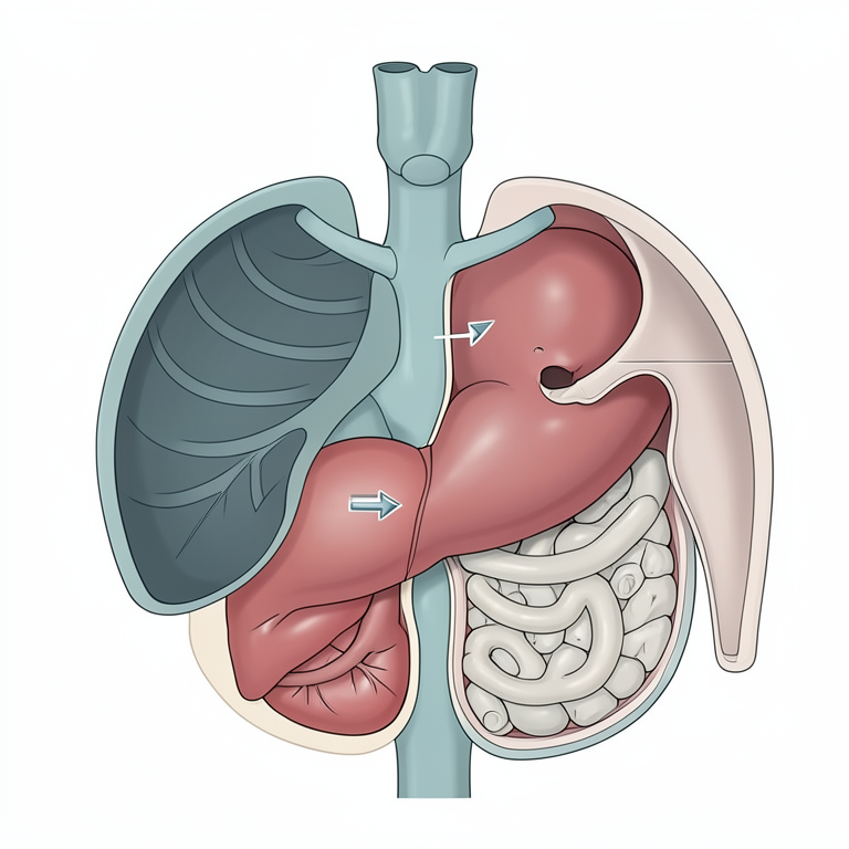
Anatomy of PPDH
AI-generated conceptual illustration (Doberman dog thoracic and abdominal anatomy). It explains the idea and does not show study data.
View source evidence ↗Figures & tables
From the original paper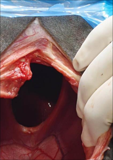 Figure 5
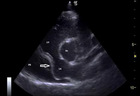 Figure 4
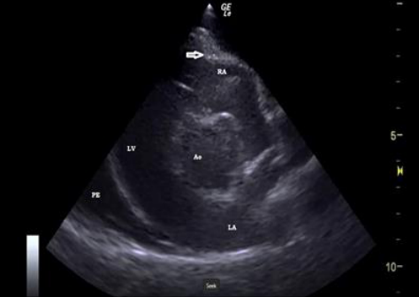 Figure 3
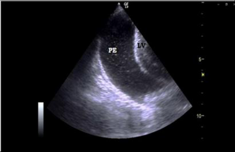 Figure 2
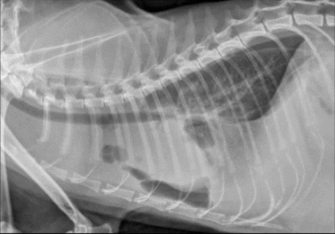 Figure 1
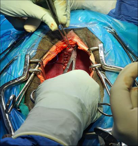 Figure 8
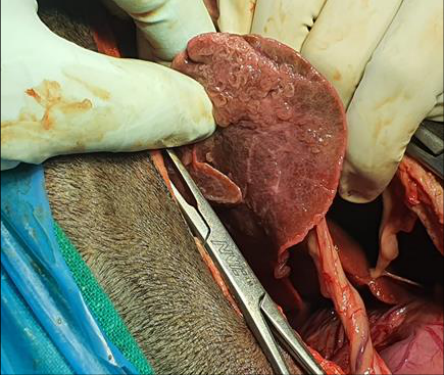 Figure 7
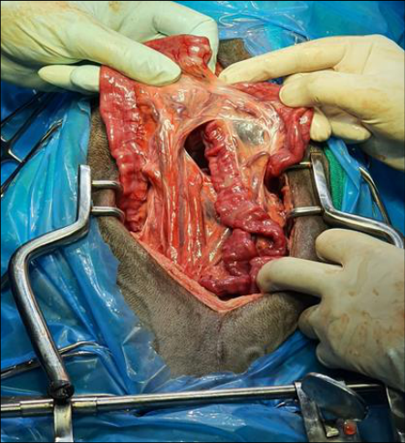 Figure 6
Introduction
Peritoneopericardial diaphragmatic hernia (PPDH) is a congenital anomaly in dogs with a communication between the pericardial sac and the peritoneal cavity that allows displacement of abdominal organs to the pericardial sac (Banz and Gottfried, 2010; Mcclaran, 2013) [1] and [9] . Abdominal organs that can be herniated into the pericardial sac include liver, gallbladder, omentum, intestines, spleen, and pancreas (Banz and Gottfried, 2010; Burns et al. 2013) [1] and [3] . In small animals, PPDH is the most common congenital anomaly involving the pericardium and the diaphragm (Burns et al. 2013; Reimer et al. 2004) [3,13] . Trauma is also one of the causes for acquired diaphragmatic hernias in dogs particularly in very young age (Hoddinott, 2013) [4] . Adhesions between the herniated organs and pericardium or myocardium is the most common associated complications that may result fatal among these species (Banz and Gottfried, 2010) [1] . Malformations and teratogen-induced factors are the most probable causes for PPDH among various species (Rexing and Coolman, 2004) [14] . Most of the dogs remain asymptomatic and many times the PPDH is an incidental finding during routine check-up (Banz and Gottfried, 2010; Hunt and Johnson, 2012) [1,6] or at necropsy (Reza et al. 2014) [15] . An embryonic development defect of the dorsolateral septum transversum that is associated in a structure termed as sterno-costal triangle may result in this developmental abnormality (Berry et al. 1990) [2] . Peritoneopericardial diaphragmatic hernia has been reported in dogs, cats, rabbits, donkey and calf (Margolis et al. 2018; Wright et al. 1987) [8,17] . This paper presents an incidental case of PPDH and its surgical repair in an adult Doberman dog.
Observations and Results
An eight year old male Doberman dog was referred to the Cardiology ward of Veterinary Clinical Complex, CVSc, Rajendranagar, Hyderabad with the history of chronic recurrent vomiting, anorexia, weakness, tachypnoea and going down in condition for a couple of months.
Treatment by a local vet did not completely alleviate the signs and were recurring once the medication was discontinued. Clinical parameters, viz., pulse and heart rate were considerably elevated while the rectal temperature was within the normal range, with slightly pale conjunctival mucous membrane. The dog was partially recumbent and extremely weak. Auscultation of chest revealed low intensity heart beat with muffled sounds, exaggerated respiratory sounds and typical barborygmi sounds of intestines. Physical examination of abdomen didn't reveal any abnormality. Neutrophilic leukocytosis was the CBP abnormality but with normal serum chemistry values. X-ray on left lateral exposure of thorax revealed radiolucent areas within the pericardial space and adjacent soft tissue structures of thorax. Continuity of abdominal soft tissue structures into thorax along with gas filled intestinal loops, through the diaphragm was also noticed on radiograph (Fig. 1). B-mode echocardiographic examination revealed huge amounts of anechoeic area with swirling hyperechoeic crystals within the pericardial space. Occasionally collapse of left ventricle free wall and right atrial wall was also noticed (Fig. 2 to 4). Sample aspirated on ultrasound guided thoracocentesis revealed alkaline pH of 8.5 and thus confirming PPDH as herniation of abdominal viscera through diaphragm. Surgical repair of the PPDH was performed using routine protocol, under inhalation anaesthesia and positive pressure ventilator. The hernia was approached through a midline celiotomy (Fig. 5 to 7) and the diaphragm was closed from dorsal to ventral direction, by opposing the margins in two layers using simple continuous pattern with polydioxanone suture material (Fig. 8). Despite every effort, the dog died on day 7 post-operative. [3,11] . If the diaphragmatic hernia is large, the displaced abdominal organs into the pericardial sac may result in adhesion to the pericardium, organ entrapment or strangulation leading to gastrointestinal, respiratory or cardiac disorders. However, small defects that do not allow organ protrusion may remain asymptomatic (Mcclaran, 2013 and Litman, 2001) [9,7] . The most common signs include intermittent vomiting, lack of appetite, weight loss and diarrhea that might be due to movement of the stomach or intestines into the pericardial sac. Some dogs are also presented with the signs of respiratory system viz., dyspnea, cough, and wheeze, due to restricted movement of lungs associated with herniated organs (Banz and Gottfried, 2010 and Reed, 1988) [1,12] . Unusual manifestations of PPDH include, abdominal distension, shock and collapse (Nikiphorou et al. 2016 and Reed, 1988) [11,12] . Though the signs associated with PPDH are noticed at any stage of life, most commonly recorded later years rather than first few years and this is partly due to the fact that traumatic events, even late in patient's life, may cause abdominal contents to move into pericardial cavity giving rise to acute clinical signs (Evans and Biery, 1980) [4] The most common findings on physical examination of PPDH dogs and cats include muffled heart sounds, heart murmurs, decreased lung sounds and barborygmi on auscultation or an empty abdomen on palpation (Burns et al. 2013; Reimer et al. 2004 and Neiger, 1996) [3,13,10] . The diagnosis of PPDH was an incidental finding in 46% of the dogs (Burns et al. 2013) [3] . PPDH is diagnosed by taking chest and abdominal x-rays (preferably contrast) and identifying stomach, intestines or liver in the pericardial sac. Thoracic radiograph may reveal an enlarged cardiac silhouette, presence of abdominal organs within the pericardial sac or loss of distinction between the heart and the diaphragm (Reimer et al. 2004 and Neiger, 1996) [13,10] . The pathognomic radiographic findings include the evidence of gas filled herniated intestinal loops in pericardial sac (Rexing and Coolman, 2004) [14] . Echocardiography may also be done to assess the amount and nature of fluid in the pericardial sac and the effects of pericardial fluid and organs on the heart (Neiger, 1996; Reed, 1988 and Wallace et al. 1992) [10,12,16] . Surgical procedure followed in the present case is in accordance with (Banz and Gottfried, 2010 and Burns et al.
2013) [1,3] , who reported a ventral midline celiotomy approach for correcting PPDH among dogs and cats. The authors (Burns et al. 2013) [3] also opined that a caudal median sternotomy is required when adhesions are present between the herniated viscera and partial pericardectomy when adhesions of the herniated organs to the pericardium is present (Banz and Gottfried, 2010) [1] . Whereas, (Mcclaran, 2013) [9] suggested reconstruction of diaphragm with pericardial grafts in cases of large defects or in agenesis of the diaphragm. Prognosis following surgical repair of PPDH is usually good with 81-87.5% of the dogs recovering within 10-15 days postoperatively with a short-term postoperative mortality of 5-12.5% among dogs (Banz and Gottfried, 2010) [1] . Common postoperative complications include incisional dehiscence, regurgitation, pericardial steatitis or pneumothorax (Wallace et al. 1992) [16] and whereas, intraoperative complications such as, adhesions of the liver to the pericardium, hypotension, respiratory acidosis, hypoventilation and hypoxia are most commonly observed in (Reimer et al. 2004) [13] . We conclude that, though the occurrence of the peritoneopericardial diaphragmatic hernia in dogs is rare, it should also be considered as one of the differentials when specific signs are present and should be corrected.
Acknowledgement
AcknowledgementsThe authors thank the university authorities for providing the facilities and Dr. Laxmi Srinivasan, Chief Veterinarian, the Animal Care Clinic, Hyderabad, for surgical support.
Figures and tables from the paper
References
17 in the original paper- Banz AC, Gottfried SD. Peritoneopericardial diaphragmatic hernia: a retrospective study of 31 cats and eight dogs. Journal of Americal Animal Hospital Association 2010;46(6):390-404.
- Berry CR, Koblik PD, Ticer JW. Dorsal peritoneopericardial mesothelial remnant as an aid to the diagnosis of feline congenital peritoneopericardial diaphragmatic hernia. Veterinary Radiology 1990;31(5):239-245.
- Burns CG, Bergh MS, Mcloughlin MA. Surgical and non-surgical treatment of peritoneopericardial diaphragmatic hernia in dogsand cats: 58 cases (19992008). Journal of American Veterinary Medical Association 2013;242:643-650.
- Evans SM, Biery DN. Congenital peritoneopericardial diaphragmatic hernia in the dog and cat: A literature review and 17 additional case histories. Veterinary Radiology 1980;21(3):108-116.
- Hoddinott K. Traumatic diaphragmatic hernia in a 5month-old boxer dog. Canadian Veterinary Journal 2013;54:507-509.
- Hunt GB, Johnson K. Diaphragmatic hernias. In: Tobias KM, Johnston SA, eds. Veterinary Surgery of Small Animal. St. Louis: Elsevier 2012, 1380-1390.
- Litman LM. Traumatic diaphragmatic hernia in a clinically normal dog. Canadian Veterinary Journal 2001;42:564-566.
- Margolis C, Maja ZP, Jolie D, Matan O, Paula H, Margret LC. Congenital peritoneopericardial diaphragmatic hernia in a family of Persian cats. Journal of Feline Medicine and Surgery 2018; Open Reports 1-5. DOI: 10.1177/2055116918804305.
- Mcclaran LK. Diaphragmatic and peritoneopericardial diaphragmatichernias. In: (ed: Monnet E) Small Animal Soft Tissue Surgery. Willey-Blackwell, Ames 2013, 278-285.
- Neiger R. Peritoneopericardial diaphragmatic hernia in cats. Compendium continued Education for Practicing Veterinarians 1996;18:461-479.
- Nikiphorou X, Chioti R, Patsikas MN, Papazoglou LG. Peritoneopericardial diaphragmatic hernia in the dog and cat. Journal of Hellenic Veterinary Medical Society 2016;67(3);117-122.
- Reed JR. Pericardial diseases. In: Miller MS, Tilley LP, Fox RP (Eds.). Canine and feline cardiology. New York, USA: Churchill Livingstone 1988, 495.
- Reimer SB. Kyles AE. Fillipowitz DE, Gregory CR. Long term outcome of cats treated conservatively or surgically for peritoneal pericardial diaphragmatic hernia; 66 cases (1987-2002). Journal of American Veterinary Medical Association 2004;224:728-732.
- Rexing J, Coolman B. A peritoneopericardial diaphragmatic hernia in a cat. Veterinary Medicine 2004;99(4):314-319.
- Reza K, Mehdi S, Dariush V, Nasrin A. Congenital peritoneopericardial diaphragmatic hernia in a terrier dog. Veterinary Research Forum 2014;5(2):153-155.
- Wallace J, Mullen HS, Lesser MB. A Technique for surgical correction of peritoneal pericardial diaphragmatic hernia in dogs and cats. Journal of American Animal Hospital Association 1992;28:503-510.
- Wright RP, Wright R, Scott R. Surgical repair of a congenital pericardial diaphragmatic hernia. Veterinary Medicine 1987;82(6):618-624.
Cited by 1
From OpenAlex, OpenCitations and reference lists read by Scinovex
- Repair of a Large Congenital Peritoneopericardial Diaphragmatic Hernia with Canine Small Intestinal Submucosa Patch Reinforcement in a Dog: A Case Report ↗
Taeseong Moon, Sung-Yeon Moon, Su-Han Woo, Eun-Ji Jeong, Changmin Park, A-Jin Lee et al. · Animals · 2026
Peritoneopericardial diaphragmatic hernia (PPDH) is a congenital anomaly allowing communication between the peritoneal and pericardial cavities. Surgical repair of large diaphragmatic defects can be challenging due to insufficient tissue for primary closure. A 2-year-old intact female White Miniature Schnauzer weighing 6.23 kg presented with a 1-month history of panting and exercise intolerance. Thoracic radiography, ultrasonography, echocardiography, and CT confirmed PPDH…
❝ 0PDF