
Research article · 2021 · Full text
International Journal of Veterinary Sciences and Animal HusbandryArt and science: The importance of scientific illustration in veterinary medicine
Volume 6(3) · Pages 30–33 · DOI: 10.22271/veterinary.2021.v6.i3a.357 ↗
Research areasVeterinary Practice and Education StudiesEmpathy and Medical Education
Abstract
Author-writtenThe importance of illustration in veterinary is usually overshadowed by its use in human medicine and forget. Nonetheless, it is important to recognize the importance of illustration in the development of veterinarian, as this is also a profession based on observation. Through history there are several examples of how illustration help to increase and share the knowledge in veterinary sciences. There is no doubt that illustration is an important tool in learning. That makes scientific illustration an important and irreplaceable tool, since they have the ability of takes scientific concepts, from the simplest to the complex, and bring it to life in an attractive and simplified way.
Summary
Source-checkedPlain-language reading of the full paper, prepared with AI and checked against the text. Each point links to the authors' own words.
Why it matters
- Biological illustration, including medical illustration, serves as a critical method for recording and sharing medical knowledge in fields such as virology and anatomy. View evidence ↗
- The role of illustration in veterinary medicine is often overlooked or overshadowed by its more prominent application in human medicine. View evidence ↗
- Historically, animal dissection and vivisection were used as models for human anatomy and physiology because human dissection was often forbidden. View evidence ↗
The question
- The author aims to highlight the importance of scientific illustration in the development of veterinary medicine, a profession fundamentally based on observation. View evidence ↗
What they did
- The author examines historical examples of illustrations to demonstrate how they have increased and shared knowledge within veterinary sciences. View evidence ↗
What they found
- One of the earliest recorded anatomical illustrations dates back to 15,000 BCE in Spain's El Pindal cave, depicting a mammoth with a heart-like area. View evidence ↗
- The Kahun Veterinary Papyrus from the Twelfth Dynasty of Egypt (2025–1700 BCE) represents the first extensive record of veterinary practice. View evidence ↗
- The Shalihotra Samhita from India is noted as one of the most complete and illustrated early treatises, covering surgery, anatomy, and diseases of elephants and horses. View evidence ↗
- Carlo Ruini's 1598 publication, “Anatomia del Cavallo,” is identified as the first great treatise on animal anatomy. View evidence ↗
- The author cites multiple studies indicating that the inclusion of illustrations in medical texts aids the learning process. View evidence ↗
- Scientists prefer drawings over photographs for working models because they can simplify reality, create abstract images, provide more depth than schematics, and share information with non-scientists. View evidence ↗
What it means
- The author concludes that artists play a vital role in transmitting scientific knowledge to both veterinary professionals and the general public. View evidence ↗
- Scientific illustration is described as an irreplaceable tool capable of simplifying complex scientific concepts and making them accessible and attractive. View evidence ↗
Visual summary
1 visualsIllustrations explain the paper; they do not show study data.
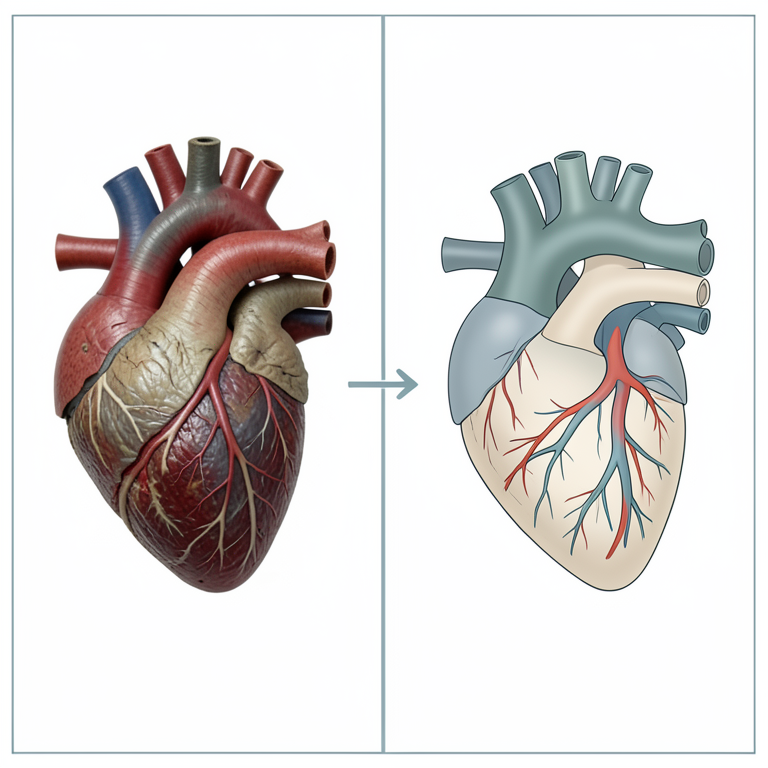
Advantages of Scientific Illustration
AI-generated conceptual illustration (canine heart anatomy). It explains the idea and does not show study data.
View source evidence ↗Figures & tables
From the original paper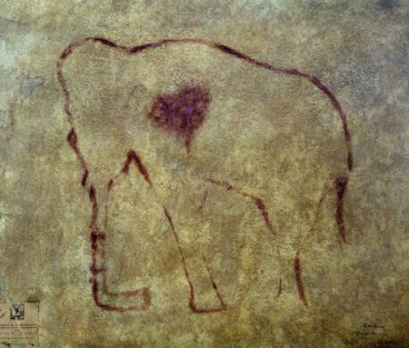 Figure 1
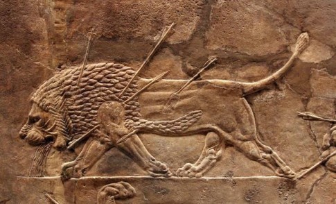 Figure 2
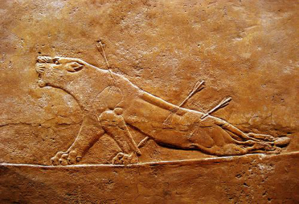 Figure 2
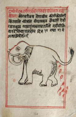 Figure 3
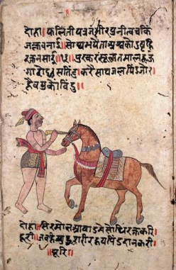 Figure 3
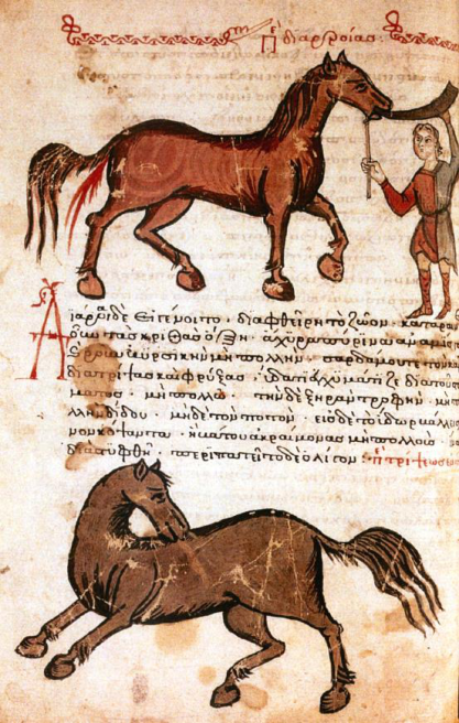 Figure 4
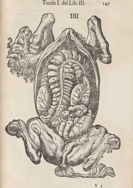 Figure 5
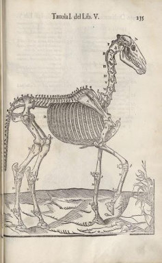 Figure 5
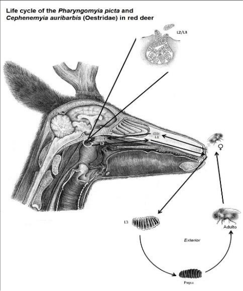 Figure 6
Introduction
Biological illustration, has many branches being one of the medical illustrations. It is a form of illustration that helps to record and disseminate knowledge regarding medicine (e.g., anatomy, virology) [1,2] . Usually, medical illustration is associated with human medicine, with the great anatomical illustrations of Da Vinci and Andreas Vesalius coming to mind [3,4] , but illustration also has an important role in veterinary medicine. Maybe, illustration has been used longer in veterinary than in human medicine but never is referenced its importance [3] . The explanations behind this circumstance are many [5][6][7] . Animals always run through the human visual culture that it's almost impossible to know where to begin, since we always lived in close contact with them, even before we could walk in two limbs. In the past they were hunted to provide food and shelter, later some were worshipped as gods and associated with magic practices and later domesticated to our use [8] . They were accessible and knowledge about them could be easily obtained. Since remote times, the vivisection and dissection of animals were used for teaching and experimentation of anatomy and physiology, and as a model for the human bodies [4,5] . This because, knowledge regarding human anatomy and physiology was hard to obtain because dissection of the human body was forbidden in many cultures thought history [3,4,9] . Many civilizations continued the study of anatomy and medicine in animals, although many times as a replacement to human medicine, but those studies allowed the development of veterinary medicine, with a great source of works existing till nowadays [5,8] .
The contribution of illustration along with Veterinary Medicine History
Archaeologically it is hard to determine when veterinary medicine first start, being the first record of what could be veterinary practice skulls of Bos taurus and Sus scrofa in France with signs of cranial surgery (trepanation) from the Mesolithic period (3400-3000 BCE) [10] . In prehistoric times before the written words exist, some pictograms were left in cave walls and stone all over the world with representations of fauna and flora. The first recorded anatomical illustration was in El Pindal cave in Spain, where is represented a mammoth with a leaf-shaped dark area where the heart should be (15000 BCE) (Fig. 1) [3] . The earlier civilizations produced simple biological and medical drawings which were made principally as ornaments or portraiture on vases, columns, walls, and tablets. For example, in a wall of a Babylonian temple, around 75,000 years ago to 3,000 B.C, is a carving of a wounded lion with arrows lodged in his spine and lungs, with the hind limbs dragging stick-like and blood dripping from his wounds and nose (Fig. 2). These simple drawings give us information that one of the arrows perforated the lung, the consequences of that trauma and the first representation of animal pain [4] . The first extensive record of veterinary medicine practice was provided by the Egyptian Papyrus of Kahun (Twelfth Dynasty of Egypt): the Kahun Veterinary Papyrus (2025-1700 BCE) [11] . In the Mauryan Empire, between 268 BCE to 232 BCE, the edicts of Asoka provide some information regarding veterinary practices, but without any relevant illustration [12] . During the same period, in India (Sravasti), the Shalihotra Samhita appear as one of the most completed and illustrated treaters on veterinary medicine, that described anatomy, physiology, surgery and diseases in equine and elephants (Fig. 3) [13] . In China, during this period some works regarding the treatment of animals start to appear. Later, between 500 ADE to 600 ADE, during the Byzantine empire appears a compilation from unknown authors, dedicated to the care and healing of the horse called Hippiatrica (Fig 4) [14,15] . Entering the Dark Ages, medicine was based on the knowledge of ancient civilization (Rome and Greece, particularly on the works of Galen (131-201 ad)) [5,16] and folk medicine. No new advances or studies were made during this period, with the monks only copied classical texts. Before the 10 th century, the illustrations in the manuscripts were rare and crude. Only, after the 11 th century forwards there is an increase in the number of miniatures and an occasional fullpage illustration began to appear, but nothing very complex or scientifically accurate (e.g., bestiaries) [7,17] . With the Renaissance, physicians and artists start to have a new interest in this field and new advancements were made. This period is marked by great artists like Leonardo Da Vinci (1452-1519), Berengario da Carpi (1460-1530), Albrecht Durer (1471-1528) and Vesalius (1514-1564) [5] . In veterinary many treats regarding these fields start to appear, some richly illustrated. The appearance of printing (15 th Century, Gutenberg) and the Age of Discovery help to bring back the interest in biological illustration and make it accessible to a greater public [5] . In this period biological illustrations could be found in anatomy textbooks, nature guides, scientific magazines and journals, botanical gardens, museums and others. Étienne de Flacourt (1600) was the first to documented his travels to Madagascar, and illustrated the unique fauna there, setting a model for the future explorers (as Charles Darwin or Ernst Haeckel) to document the natural world and improve the fields of medicine [3,5,18] . The first great treaty on animal anatomy was Carlo Ruini's book "Anatomia del Cavallo", published in 1598 (Fig. 5). It was the first complete comprehensive treatise on the anatomy of a non-human species [8,15,18,19] . This was the start, and afterword's numerous publications on anatomy and veterinary procedures appeared, not only in diverse species of animals but in different fields of veterinary, from anatomy to microbiology [15,18] .
The importance of illustration on Veterinary
There is no doubt that illustration is an important tool in learning. Several studies have shown that illustration in a medical text aids in the learning process, as many studies have shown [20][21][22] . In the last century, medical illustration has specialized and became a profession on its own, and develop in an infinite variety of techniques, from charcoal to digital painting. On one side the scientist can help the illustrator in the solution of an artistic problem and by other and the illustration helps the scientist to expose the information. Art always helped to solve communication dilemmas in science, including veterinary. [3,4,6] .
The illustration techniques used to represent medical knowledge have been evolving at the de throughout history along with the artists and technology present, from drawing, engraving, photography, animation to digital processing. The development of each new technique has built on previous experience to improve medical knowledge and make it more accessible to the target audience [3] . Although we live in the Era of technology with digital photography, electron micrographs, magnetic resonance imaging and computers, illustration still has an important role. Both, traditional illustration and computerized images are important tools in veterinary education and transmission of knowledge [3] . Scientists still prefer to use drawings to illustrate their working models (fig. 6), because 1) represent simplified descriptions of the reality; 2) enables the creation of images that are more abstract than photographs; 3) more depth and accurate than schematics; 4) present spatial visualizations that could never be photographed or easily schematized; 4) integrate contextual information obtained from different fields or experiments; 5) possibility of share scientific information to the public without a scientific background [23] . With the development of the new methods in which information is distributed, e.g., internet, the scientific knowledge is available faster and more accessible to everyone, in comparison to what happened in the past. That makes scientific illustration an important and irreplaceable tool, since they have the ability of takes scientific concepts, from the simplest to the complex, and bring it to life in an attractive and simplified way [1,3,6] .
Conclusions
Animal health has always had a very important role, and even more nowadays days with the new concept of "One Health".
Artist has an important role in the transmission of knowledge, not only to professionals in this area but also to the general population. The future of medical illustration, particularly in the veterinary field, are wide and fascinating with the rise and expansion of veterinary sciences. The importance of illustration in veterinary is usually overshadowed by its use in human medicine and forget. Nonetheless, it is important to recognize the importance of illustration in the development of veterinarian, as this is also a profession based on observation.
References
22 in the original paper- Tsafrir J, Ohry A. Medical illustration: from caves to cyberspace ‡. Health Information & Libraries Journal [Internet] 2001 [cited 2021;18(2):99-109. Available from: http://doi.wiley.com/10.1046/j.14711842.2001.d01-16.x
- Mikota SK, Sargent ELee, Ranglack GS. Medical management of the elephant [Internet]. Indira Pub. House, 1994 [cited 2018 Mar 27]. Available from: http://wildpro.twycrosszoo.org/S/00Ref/BooksContents/b 450.htm
- Hajar R. Medical illustration: Art in medical education. Heart Views 2011;12(2):83.
- Loechel WE. The history of medical illustration. Bulletin of the Medical Library Association 1960;48:168-71.
- Donald G. The history of medical illustration. Journal of Visual Communication in Medicine 1986;9(2):44-9.
- Porter CM. Essay review: The history of scientific illustration. Journal of the History of Biology 1995;28(3):545-50.
- Animals | Illustration Chronicles [Internet]. [cited 2021 May 30]; Available from: https://illustrationchronicles.com/animals
- History of Vivisection and Dissection: Dying to Learn : AAVS [Internet]. [cited 2021 May 30]; Available from: http://www.dyingtolearn.org/animalUseHistory.html
- Ramirez Rozzi F, Froment A. Earliest Animal Cranial Surgery: from Cow to Man in the Neolithic. 2018 [cited 2021;8:5536. Available from: www.nature.com/scientificreports/
- Kahun Papyrus. First Writing On Veterinary Medicine In 1895 BC Africa [cited 2021 May 31]; Available from: https://thinkafrica.net/kahun-papyrus-first-writing-onveterinary-medicine-in-1895-bc-africa/
- Romila Thapar. Asoka and the Decline of the Mauryas. Oxford University Press 1997 [cited 2021 May 31]; Available from: http://projects.mcah.columbia.edu/indianart/pdf/asoka_th apar.pdf
- Shalihotra Samhita by Ancient India's Dr Dolittle - Sarmaya [cited 2021 Jun 1]; Available from: https://sarmaya.in/spotlight/shalihotra-samhita-byancient-indias-dr-dolittle/
- A Byzantine Encyclopaedia of Horse Medicine -Anne McCabe [cited 2021 Jun 1]; Available from: https://books.google.pt/books?id=lrqpFCBfmbgC&redir_ esc=y
- Robert H. Dunlop, David J. Williams. Veterinary Medicine -An Illustrated History. St. Louis (USA): Mosby-Year Book Inc 1996.
- Dahan S, Shoenfeld Y. A picture is worth a thousand words: Art and medicine. Israel Medical Association Journal 2017;19(12):772-6.
- History I. History Impressed 1957;33(5).
- Smith DF. Lessons of history in veterinary medicine. Journal of Veterinary Medical Education 2013;40(1):211.
- Evans HE. A History of Anatomy at Cornell. Cornell University 2013. [cited 2021 Jun 1]; Available from: file:///C:/Users/Andreia/Downloads/History_of_Anatomy _at_Cornell.pdf
- Balemans MCM, Kooloos JGM, Donders ART, van der Zee CEEM. Actual drawing of histological images improves knowledge retention. Anatomical Sciences Education 2016;9(1):60-70.
- Alsaid B, Bertrand M. Students' memorization of anatomy, influence of drawing. Morphologie 2016;100(328):2-6. Available from: http://dx.doi.org/10.1016/j.morpho.2015.11.001
- Laakkonen J. Drawing in Veterinary Anatomy Education: What Do Students Use It For? Anatomical Sciences Education 2020.
- Chabrier R, Janke C. The comeback of hand drawing in modern life sciences. Nature Reviews Molecular Cell Biology [Internet] 2018;19(3):137-8. Available from: http://dx.doi.org/10.1038/nrm.2017.126
Cited by 10
From OpenAlex, OpenCitations and reference lists read by Scinovex
- SCIENTIFIC ILLUSTRATION: A BRIEF REVIEW AND THE EMERGENCE OF 3D ILLUSTRATION ↗
Márcia Venâncio · ENVIRONMENTAL SMOKE · 2022
Scientific illustration can be defined as a realistic representation of an object that the illustrator or the scientist observes, pointing out features that they want to show. Over the years, illustration has fulfilled different objectives, becoming an indispensable tool in the science service, being extremely important to understanding the natural world and recognized as a significant factor in the emergence…
❝ 7PDF - Picturing Benjamin Franklin’s Kite Experiment in the Nineteenth Century ↗
Breno Arsioli Moura · Science & Education · 2023
❝ 2 - İllüstrasyon: Bir Görsel İletişim Sanatı ↗
Mesut Tanrıkulu, Emine Jessica McKIE, Barış Hasırcı, Eda Uzun, Elif Ecem COŞKUN, Seda Nur Atasoy et al. · Özgür Yayınları eBooks · 2024
Basitçe, bir iletinin, görsel olarak aktarılması, bir metnin açıklanması amacıyla yapılan resimleme çalışması anlamına gelen “illüstrasyon” sözcüğünün kökeni, Latince “aydınlatmak” anlamına gelen “illustrare” sözcüğüdür. İllüstrasyon olgusu eski çağlarda var olmuş olsa da sözcüğe kaynaklık eden eserler Orta Çağda ortaya çıkan inanç odaklı el yazması kitaplardaki altın boya ve parlak renklerle çalışılmış resim ve süsleme çalışmalarıdır. Bu eserlerin yansımaları Osmanlı minyatürlerinde…
❝ 1PDF - Recent perspectives on impact of Ethnobotany in India: A review ↗
Suresh Kumar Channaveeraiah, Keshamma Entoori · Journal of Medicinal Plants Studies · 2022
Medicinal and aromatic plants constitute most significant group of plants in the welfare of living beings. Most of these plants are found growing wild and collected by different ethnic and rural people of our country. They used them freely for the treatment of various human and animal ailments, on the basis of their ancestral knowledge and beliefs. They know medicinal…
❝ 1PDF - Dijital Oyunlarda İllüstrasyonun Yeri ve Önemi ↗
Mustafa Uygur Çevik, Murat Çalış · Özgür Yayınları eBooks · 2024
Dijital oyunlar, yalnızca eğlence amacıyla değil, aynı zamanda sanatsal ifade ve etkileşim alanı olarak dikkat çeken bir medya formuna dönüşmüştür. Bu bağlamda illüstrasyon, oyunların görsel kimliğini belirlemede ve oyuncularla duygusal bir bağ kurmada kritik bir role sahiptir. İllüstrasyon; oyun tasarımında hikâye anlatımını destekleme, atmosfer yaratma ve oyuncu deneyimini derinleştirme gibi işlevlerle öne çıkmaktadır. Bu çalışma, dijital oyunlarda illüstrasyonun estetik ve…
❝ 0PDF - Bilimin Görsel Dili: Bilimsel İllüstrasyonlar ↗
Seda Nur Atasoy · Özgür Yayınları eBooks · 2024
Bu bölümde, bilimsel illüstrasyonların tarihsel, epistemolojik ve disiplinlerarası önemi incelenmektedir. Bilimsel illüstrasyonlar, bilimsel bilgiyi görsel olarak temsil ederek, anlaşılması zor ve detaylı kavramları daha anlaşılır hale getiren bir iletişim aracıdır. Bölümde, illüstrasyonların anatomi, botanik, zooloji, mikroskobik yapılarının incelenmesi gibi çeşitli alanlarda kullanım örnekleri sunulmuştur. Tarihsel olarak, Eski Mısır ve Yunan döneminden Rönesans’a, modern dönemden dijital çağın yeniliklerine kadar gelişimi ele…
❝ 0PDF - Görsel Öyküleme Süreçlerinde Konsept Tasarım ve İllüstrasyon ↗
Mesut Tanrıkulu · Özgür Yayınları eBooks · 2024
Görsel öyküleme süreçlerinde konsept tasarım ve illüstrasyon, yaratıcı projelerde hikâyenin görsel temellerini oluşturarak anlatının duygusal ve estetik boyutlarını derinleştirmektedir. Konsept tasarım, hikâye dünyasının çerçevesini çizerken, illüstrasyon bu çerçeveyi detaylandırarak izleyiciyi anlatıya bağlayan görsel bir deneyim sunmaktadır. Bu bağlamda, konsept tasarım ve illüstrasyon ilişkisi yaratıcı projelerde görsel bütünlüğü ve tutarlılığı sağlayan önemli bir bağlayıcıdır. Görsel öyküleme, yalnızca hikâyeyi göstermek değil, aynı…
❝ 0PDF - Süpergrafiklerin Dönüştürücü Gücü ↗
Eda Uzun · Özgür Yayınları eBooks · 2024
Süpergrafikler 1960’lardan sonra yaygınlaşan, uygulandığı alanı iç veya dış mekân sınırlaması olmaksızın değiştirerek bu alanın yeni bir kimlik kazanmasına yardımcı olan bir sanat alanıdır. Barbara Stauffacher Solomon ve Jean-Philippe Lenclos gibi öncüler tarafından 20. yüzyılın ortalarında popüler hale getirilen bu yöntem, görsel ifadeyi mekânın fiziksel özellikleriyle harmanlayarak kullanıcılar üzerinde güçlü estetik ve duygusal etkiler yaratmaktadır. Mekânın nasıl algılandığını değiştirmek, ortamların…
❝ 0PDF - Fantastik İllüstrasyon ile Peri Ülkesine Yolculuk: Yüzüklerin Efendisi İllüstrasyonları ve Ötesi ↗
Barış Hasırcı · Özgür Yayınları eBooks · 2024
Fantastik edebiyat ve fantastik illüstrasyon illüstrasyonun önde gelen sanatçılarından olan John Howe’ın, J. R. R. Tolkien’in “Yüzüklerin Efendisi” kitapları ve öykünün içinde geçtiği yer olan Orta Dünya’yla ilişkili olarak yaratmış olduğu illüstrasyonlar, okurların içinde gezmeyi arzuladığı büyülü düş dünyasını 1980’lerden beri kitap kapağı illüstrasyonları, harita çizimleri, posterler ve benzeri ortamlar yoluyla gözler önüne sermektedir. Howe’ın, Tolkien’in kitaplarının film ve dizi…
❝ 0PDF - Effect of garlic extract loaded on zinc oxide nanoparticles against Sarcocystis in vitro ↗
Sawsan Darwish Jari, Sura Waleed Yasir, Saja Alaa Razzaq · International Journal of Forensic Medicine · 2022
The present study aimed to investigate the effect of garlic extract loaded on zinc oxide nanoparticles on the viability of Sarcocystis in vitro Samples of the parasite in the study was divided into three group inside to control group each one consists of 20 parasite in petri dish (5 petri dish) for each concentration contain the Phosphate Buffer Saline(PBS). The…