
Research article · 2020 · Full text
International Journal of Veterinary Sciences and Animal HusbandryPrevalence, economic losses and epidemiology of fascioliasis in cattle presented at slaughter slabs of Juba County, Central Equatoria State, South Sudan
Volume 5(1) · Pages 115–118
Research areasHelminth infection and controlInsect Pest Control StrategiesCoccidia and coccidiosis research
Abstract
Author-writtenA cross sectional epidemiological study was conducted from July to August of 2018 with the aim of assessing the prevalence, economic losses and identify some epidemiological factors associated with bovine fascioliasis in cattle presented at the slaughter slabs of Juba County, Central Equatoria State (CES), and South Sudan. In total, 571 cattle were investigated, of which 109 were positive and the overall prevalence of bovine fascioliasis was found to be 19.1% (109/571). Based on source of cattle, the prevalence of bovine fascioliasis was found to be 37.0%, 35.0%, 28.0% and 25.0% from cattle brought from Maridi, Jebel Ladu, Terekeka and Pibor, respectively. The prevalence was higher in animals slaughtered at Gudele slaughter slab (33.3%) compared to Gumbo slaughter slabs (13.0%). Based on the current local price of liver, the economic loss incurred from totally and partially condemned livers per month amounted for 549,000 SSP which is equivalent to 2,889 USD. Fascioliasis remains prevalent and is associated with significant economic losses in cattle slaughtered at the slaughter slabs of Juba County, CES. Therefore, the study recommend for further epidemiological study to assess the prevalence and economic losses of fascioliasis in small ruminants and urged for immediate control programme of the disease.
Summary
Source-checkedPlain-language reading of the full paper, prepared with AI and checked against the text. Each point links to the authors' own words.
Why it matters
- Fascioliasis is a significant parasitic disease caused by Fasciola hepatica and Fasciola gigantica, commonly known as liver fluke. View evidence ↗
- In livestock, the disease causes reduced growth, poor meat and milk production, anemia, mortality, and the condemnation of livers. View evidence ↗
- The regular condemnation of infected meat at slaughter slabs makes fascioliasis a major economic problem. View evidence ↗
The question
- The study aimed to assess the prevalence, economic losses, and epidemiological factors of bovine fascioliasis in cattle at slaughter slabs in Juba County, South Sudan. View evidence ↗
What they did
- A cross-sectional epidemiological survey was conducted from July to August 2018 at randomly selected slaughter slabs in Juba County, South Sudan. View evidence ↗
- The study investigated a total of 571 cattle. View evidence ↗
- Researchers used descriptive statistics and the Pearson Chi Square test with a 5% significance level to analyze associations between prevalence and variables like age, sex, and breed. View evidence ↗
What they found
- The overall prevalence of bovine fascioliasis was 19.1%, with 109 out of 571 investigated cattle testing positive. View evidence ↗
- Prevalence varied by location, being higher at the Gudele slaughter slab (33.3%) than at the Gumbo slaughter slab (13.0%). View evidence ↗
- Prevalence differed by the source of the cattle, with the highest rates in animals from Maridi (37.0%), followed by Jebel Ladu (35.0%), Terekeka (28.0%), and Pibor (25.0%). View evidence ↗
- There was a significant association between sex and prevalence (p≤0.000), with females showing a higher prevalence than males. View evidence ↗
- A significant association (p≤0.014) was found between age and prevalence, with the disease tending to decrease as animals got older. View evidence ↗
- Monthly economic losses from totally and partially condemned livers were estimated at 549,000 SSP (approximately 2,889 USD). View evidence ↗
What it means
- The authors concluded that fascioliasis is an important disease causing significant revenue loss through liver condemnation and reduced carcass weight in Juba County. View evidence ↗
- The authors recommend introducing a trace-back system to monitor infection sources, encouraging regular de-worming, and conducting further studies on small ruminants. View evidence ↗
Visual summary
3 visualsIllustrations explain the paper; charts are replotted from the paper's own table values.
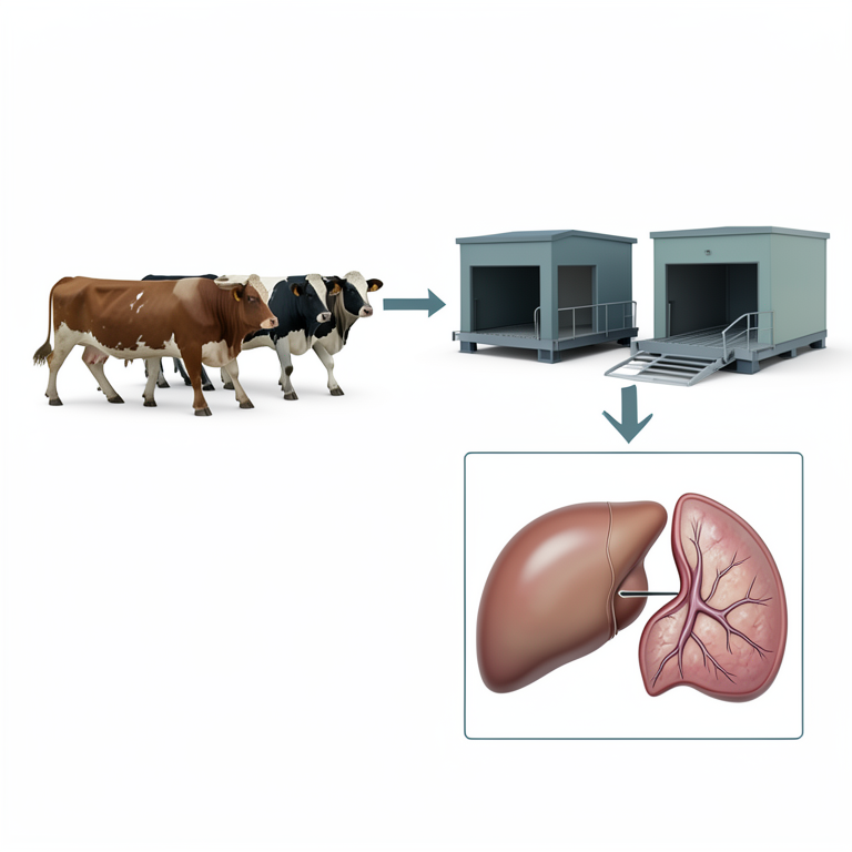
Epidemiological Flow of Bovine Fascioliasis
AI-generated conceptual illustration (cattle and bovine livers). It explains the idea and does not show study data.
View source evidence ↗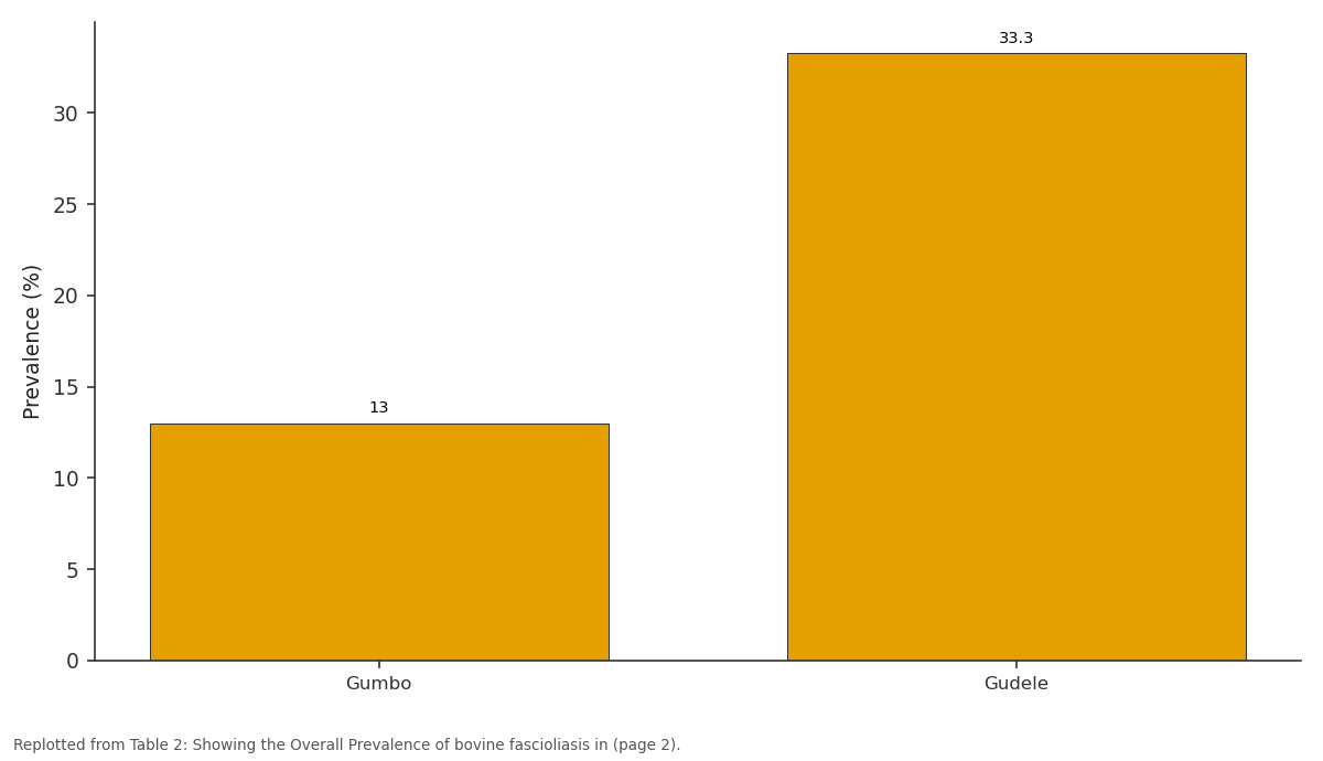
Prevalence (%): Gumbo vs Gudele
Replotted from Table 2 (page 2) of the original paper. Every bar is the paper's own table value.
View source evidence ↗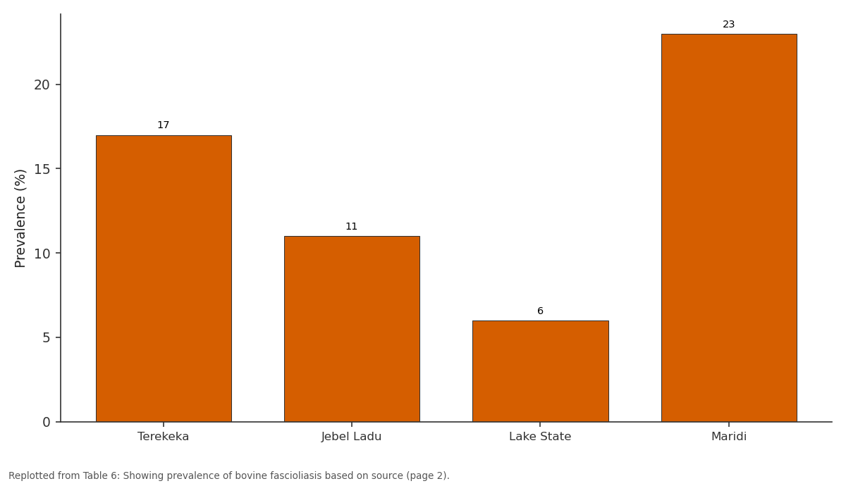
Positive animals: Terekeka vs Jebel Ladu vs Lake State vs Maridi
Replotted from Table 6 (page 2) of the original paper. Every bar is the paper's own table value.
View source evidence ↗Figures & tables
From the original paper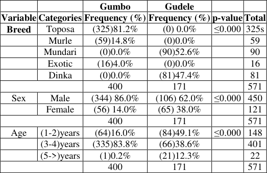 Table 1
 Table 2
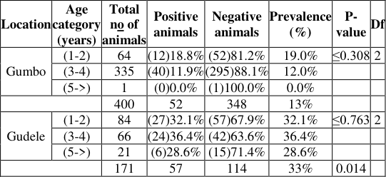 Table 3
 Table 4
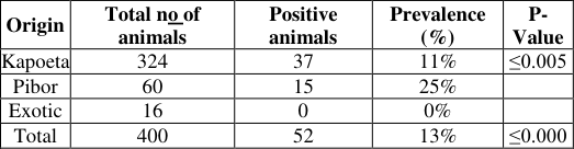 Table 5
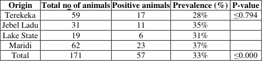 Table 6
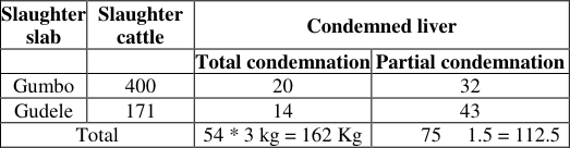 Table 7
Introduction
Fascioliasis, also known as "the common liver fluke" or the "the sheep fluke" [1] , is an important parasitic disease caused by two species of the genus Fasciola: F hepatica and F. gigantica. The development of liver fluke requires an intermediate host, Lymnae, a fresh water snail and various species of ruminants as the final definitive host for its occurrence. Fascioliasis is predominantly found in sheep, goats, cattle and buffalo, as well as other domestic ruminants and human [2] . The transmission of bovine fascioliasis is associated with livestock drinking from snail infected watering places and with contaminated grazing pastures in wetland. Transmission in human is through ingestion of contaminated improperly cooked liver [3] . Infection in livestock usually leads to reduced growth, poor production of meat and milk and other complications such as reduced fertility, abortion in late stages of pregnancy, anemia and mortality, in dairy cattle, reduction in milk yield, and liver condemnation [4] . Prevalence of fascioliasis in domestic animals is much higher in developing continents. In Nigeria, a prevalence of 80.8% has been reported in cattle slaughtered at Maiduguri abattoir, [5] . Additionally, in Jimma Municipal Abattoir in Ethiopia, a prevalence of (53.48%) has been reported by [6] and [7] reported 17% prevalence in Juba Slaughter house in South Sudan. Fascioliasis also causes a substantial economic loss. It is an important parasitic disease affecting cattle in Horro District in Western Ethiopia [8] . Meat infected with this parasites are regularly condemned at inspection in slaughter slabs and thus, constitute a major economic problem [9] . In South Sudan, limited studies have been done to investigate the prevalence, economic losses and epidemiology of bovine fascioliasis. Hence, this study aimed to identifying prevalence, epidemiology and economic losses posed by fascioliasis and update information on bovine Fascioliasis in Juba, Jubek State, and South Sudan
Materials and Methods
Materials and Methods
Study Area
This study was conducted in randomly selected slaughter slabs in Juba County, CES, and South Sudan. Central Equatoria State is one of the 10 States of South Sudan and is geographically located between 4.85° latitude and 31.58° longitude and it is situated at elevation 518 meters above the sea. The state has a rainfall of 600mm-700mm per annum and a minimum and maximum temperature of 24.7 °C and 38 °C, respectively.
Study Design and Animals
This study was a cross-sectional epidemiological survey targeting cattle of both sexes, different age groups and breeds. The cattle presented at the slaughter slabs of Juba County were mainly brought from Terekeka County in CES, Pibor in Jonglei State and Amadi in Eastern Equatoria State.
Sample size Determination
The sample size for this study was calculated based on the predetermined parameters of 95% level of confidence and 5% desired level of precision according to [10] . An expected prevalence of 17% derived from [7] was used to assess the prevalence of bovine fascioliasis. Thus, the following formula was used:-Where; P exp= expected prevalence; d = desired absolute precision n = sample size. Based on the above formula a total of 217 cattle were to be investigated. However, 571 cattle were investigated with the aim of increasing the study precision.
Data Management and Statistical Analysis
The data collected was first, cleaned and checked for errors then entered into SPSS (version 20) directly. Then, cleaning was conducted to check for missing values before the analysis. Descriptive statistic was computed and Pearson Chi Square (X 2 ) test was used to determine associations between the prevalence of bovine fascioliasis and other variables such as ages, sexes, breeds etc. 5% significance level was used to determine whether there are significant differences between the variables. Results were then displayed on tables for clear understanding and presentation.
Ethical Statement
An introduction letter was obtained from the School of Natural Resources and Environmental Studies which was then taken to the State Ministry of Animal Resources and Fisheries, CES for further approval and permission to conduct the study in Juba County.
Results
Results
Prevalence of Bovine Fascioliasis
Prevalence of Bovine Fascioliasis
Economic losses caused by bovine fascioliasis between July and August
The estimated economic loss due to partially and totally condemned livers per month based on the current local market price of 1 kg of liver at 2,000 South Sudanese Pounds (SSP) was 549,000 SSP which is equivalent to 2,889 USD. Therefore, the annual financial loss assuming constant rate of condemnation and market price would be 6,588,000 SSP which is equivalent to 34,674 USD at an exchange rate of 190 SSP per a dollar.
Discussion
In the two slaughter slabs, the Toposa cattle from Eastern Equatoria State constitutes the majority of slaughtered animals compared to the exotic cattle which were the least cattle being slaughtered with a significance of p ≤ 0.001. The possible explanation to this observation is that, the Toposa cattle were cheaper compared to exotic cattle which were mainly brought from Uganda. Additionally, famers/pastoralists locally do not sell their cattle and were not business oriented as such, the prevalence of their cattle is unknown. Higher numbers of bulls were slaughtered compared to cows, which indicates that females are left for reproduction in farms and herds and are kept in farms for breeding purpose with significance (p ≤0.000).This is in agreement with the studies in Nigeria by [11] and [12] , who recorded higher number of bulls being slaughtered compared to cows. Cattle aged between 3-4 years old constitutes majority of the slaughtered cattle compared to 5->years and above as the least of the slaughtered cattle. The reason behind this is that cattle aged 3-4 years old have good body condition unlike the 5-> and above with poor body condition. The overall prevalence of bovine fascioliasis determined in this study was 19.1% which appears to be much higher than the prevalence reported by [7] who reported a prevalence of 17.0% in and around Juba (South Sudan), 16.3% recorded by [13] in Tanzania and 5.13% reported by [14] in Yola Modern Abattoir in Nigeria. This might be attributed to the lack of proper control strategies in the control of fascioliasis and inadequate veterinary services. On the other hand, the overall prevalence was lower compared to studies reported by [15] in Zimbabwe, who reported 37.1%, [16] recorded prevalence of 44.8% in Nigeria and [17] reported 74.0% in and around Woreta of North Western Ethiopia. The lower value of prevalence rate compared to the higher value earlier obtained can be traced probably to the progressive intensification of veterinary and extension services through regular antihelmintic treatments of cattle. There was statistical significant association (≤0.014) of bovine fascioliasis in cattle with different age groups in the present study although the prevalence showed a tendency to decrease with increase in age. This study is in agreement with the findings of Mohammadnur, [18] who recorded a significantly association of prevalence of bovine fascioliasis with the age on which young animals were more affected than adult animals. Additionally, [19] recorded higher prevalence in younger animals compared to adult animals. The reason behind this observation might be due to the fact that younger animals are more susceptible to infections than adults. Adult animals may acquire immunity to parasites through frequent challenges and oust the ingested parasite before they establish infection. Cattle tend to display self-cure in relation to age for instance, cattle display self-cure between 9-26 months after infection. This means fascioliasis tend to decrease with increase in age. Based on this study, sex has a significant association (p≤0.000) on the prevalence of bovine fascioliasis. The prevalence of bovine fascioliasis was observed higher in females than males. This result is in line with the findings [20] and [21] who observed higher prevalence of bovine fasciolosis in females than male. The higher prevalence observed in females than males can be explained that hormonal influence and stress leading to immune-suppression might be associated with this phenomenon. Statistical significant association (p≤0.000) of bovine fascioliasis varies with the place of source of the animal. Higher prevalence was recorded in cattle brought from Maridi in Western Equatoria State (WES), (37%), followed by Jebel Ladu in CES (35%), Lakes State (31), Terekeka in CES (28%), Pibor in Jonglei State (25%), Kapoeta in EES (11%) and Uganda (0%). This could be explained by the difference in the presence of intermediate hosts in the swampy grazing areas and variation in climate such as altitude, rainfall, temperature and management systems of control measures through de-worming [11] and [22] . The total economic loss incurred due to totally and partially condemned of livers during the study period has amounted to 549,000 SSP which was equivalent to 2,889 USD per month. This finding is less compared to the findings reported by [7] , who revealed the sum of monetary losses of 45,180 SSP (15,227.5 USD) and 17,560 SSP (5,918.4 USD) in total and partial liver condemnations, respectively. This dissimilarities could be due to differences on the number of sampled cattle, duration of the study and the current devaluation of South Sudanese pounds.
Conclusion and Recommendations
In conclusion, the present study confirmed that fascioliasis is an important disease causing considerable loss of revenue due to condemnation of affected liver and carcass weight reduction at Juba County slaughter slabs. Hence, demanding the attention of all the stakeholders to mitigate the huge financial losses incurred due to the disease. Therefore, based on the aforementioned conclusion, the study recommends that, the Trace-back system should be introduced in order to monitor the source of the infection and to control the infection at source; regular de-worming of animals should be encouraged and further studies should be conducted on the prevalence of fascioliasis in small ruminants.
Figures and tables from the paper
| — | — | Gumbo | Gudele | — | — |
|---|---|---|---|---|---|
| Variable | Categories | Frequency (%) | Frequency (%) | p-value | Total |
| Breed | Toposa | (325)81.2% | (0) 0.0% | ≤0.000 | 325s |
| Murle | (59)14.8% | (0)0.0% | 59 | ||
| Mundari | (0)0.0% | (90)52.6% | 90 | ||
| Exotic | (16)4.0% | (0)0.0% | 16 | ||
| Dinka | (0)0.0% | (81)47.4% | 81 | ||
| 400 | 171 | 571 | |||
| Sex | Male | (344) 86.0% | (106) 62.0% | ≤0.000 | 450 |
| Female | (56) 14.0% | (65) 38.0% | 121 | ||
| 400 | 171 | 571 | |||
| Age | (1-2)years | (64)16.0% | (84)49.1% | ≤0.000 | 148 |
| (3-4)years | (335)83.8% | (66)38.6% | 401 | ||
| (5->)years | (1)0.2% | (21)12.3% | 22 | ||
| 400 | 171 | 571 |
Table text extracted from the original PDF; superscript letters are shown inline. The original table image is below.
View original table image
| Location | Total no. animal | Positive animals | Prevalence (%) |
|---|---|---|---|
| Gumbo | 400 | 52 | 13.0% |
| Gudele | 171 | 57 | 33.3% |
| Overall total | 571 | 109 | 19.1% |
Table text extracted from the original PDF; superscript letters are shown inline. The original table image is below.
View original table image
| Location | Age category (years) | Total no of animals | Positive animals | Negative animals | Prevalence (%) | P- value | Df |
|---|---|---|---|---|---|---|---|
| Gumbo | (1-2) | 64 | (12)18.8% | (52)81.2% | 19.0% | ≤0.308 | 2 |
| (3-4) | 335 | (40)11.9% | (295)88.1% | 12.0% | |||
| (5->) | 1 | (0)0.0% | (1)100.0% | 0.0% | |||
| 400 | 52 | 348 | 13% | ||||
| Gudele | (1-2) | 84 | (27)32.1% | (57)67.9% | 32.1% | ≤0.763 | 2 |
| (3-4) | 66 | (24)36.4% | (42)63.6% | 36.4% | |||
| (5->) | 21 | (6)28.6% | (15)71.4% | 28.6% | |||
| 171 | 57 | 114 | 33% | 0.014 |
Table text extracted from the original PDF; superscript letters are shown inline. The original table image is below.
View original table image
| Location | Sex | Total no of animals | Positive animals | Negative animals | Prevalence (%) | P-value | 95 CI | Df |
|---|---|---|---|---|---|---|---|---|
| Gumbo | Male | 344 | (37)10.8% | (307)89.2% | 10% | ≤0.001 | (≤0.002-≤0.002) | 1 |
| Female | 56 | (15)26.8% | (41)73.2% | 26% | ||||
| Gudele | Male | 106 | (34)32.1% | (72)67.9% | 32% | ≤0.656 | (≤0.739-≤0.389) | 1 |
| Female | 65 | (23)35.4% | (42)64.6% | 35% | ||||
| Total | 571 | 109 | 462 | 19.1% | ≤0.000 | ≤0.000-≤0.000 |
Table text extracted from the original PDF; superscript letters are shown inline. The original table image is below.
View original table image
| Origin | Total no of animals | Positive animals | Prevalence (%) | P- Value |
|---|---|---|---|---|
| Kapoeta | 324 | 37 | 11% | ≤0.005 |
| Pibor | 60 | 15 | 25% | |
| Exotic | 16 | 0 | 0% | |
| Total | 400 | 52 | 13% | ≤0.000 |
Table text extracted from the original PDF; superscript letters are shown inline. The original table image is below.
View original table image
| Origin | Total no of animals | Positive animals | Prevalence (%) | P-value |
|---|---|---|---|---|
| Terekeka | 59 | 17 | 28% | ≤0.794 |
| Jebel Ladu | 31 | 11 | 35% | |
| Lake State | 19 | 6 | 31% | |
| Maridi | 62 | 23 | 37% | |
| Total | 171 | 57 | 33% | ≤0.000 |
Table text extracted from the original PDF; superscript letters are shown inline. The original table image is below.
View original table image
References
22 in the original paper- Center of Disease Control, CDC. https://www.cdc/parasites/fasciola/index.html, 2018.
- Saleha AA. Liver fluke disease (Fascioliasis): Epidemiology, economic impact and public health significance. The Southeast Asian Journal of Tropical Medicine and Public Health, 1991. PMID 1822928
- Mas-Coma MS, Esteban JG, Bargues MD. Epidemiology of human Fascioliasis: a review and proposed new classification. Bulletin of the World Health Organization. 1999; 77(4)340-346.
- Bekele M, Tesfay H, Getachew Y. Prevalence of Bovine Fasciolosis and Its Economic Loss Due to Liver Condemnation at Adwa Municipal Abattoir North Ethiopia. EJAST. 2010; 1(1):39-47.
- Biu AA, Ahmed MI, Mshelia SS. Economic assessment of losses due to parasitic diseases common at the Maiduguri abattoir, Nigeria. African Scientist. 2006; 7:143-145.
- Terefe D, Wondimu A, Gachen DF. Prevalence, gross pathological lesions and economic losses of bovine fasciolosis at Jimma Municipal Abattoir, Ethiopia. Journal of Veterinary Medicine and Animal Health. 2012; 4(1):P6-11.
- Amal A, Mousa, Khitma H, El Malik, Erneo B. Ochi. Prevalence and Monetary Loss due to Bovine Fasciolosis in Juba Slaughter House, South Sudan. Nature and Science. 2013; 11(11):145-148.
- Tulu D, Gebeyehu Surra. Study of Prevalence and Associated Risk Factors of Bovine fasciolosis in Jimma Horro District of Kellem Wollegha Zone, Western Ethiopia. Arch Vet Sci Medicine. 2018; 1(1):009-018.
- Magaji AA, Kabir Ibrahim, Salihu MD, Saulawa MA, Mohammed AA, Musawa AI. Prevalence of Fascioliasis in cattle slaughtered in Sokoto Metropolitan Abattoir, Sokoto, Nigeria. Advances in Epidemiology, 2014; http://dx.doi.org/10.1155/2014/247258.
- Thrusfield M. Veterinary Epidemiology. Second edition, University of Edinburgh, Black well science, 2005, 180188.
- Karshima NS, Bata SI, Bobbo AA. Prevalence, Risk Factors and Economic Losses Associated with Fasciolosis in slaughter cattle in Bauchi, North-Eastern Nigeria. Alexandria Journal of Veterinary Sciences. 2016; 50(1):87-93.
- Oladele-Bukola MO, Odetokun IA. Prevalence of bovine fasciolosis at the Ibadan Municipal abattoir, Nigeria. African Journal of Food, Agriculture, Nutrition and development, 2014, 14. ISSN 1684 5374.
- Mellau LSB, Nonga HE, Karimuribo ED. A slaughterhouse survey of liver lesions in slaughtered cattle, sheep and goats at Arusha, Tanzania. Research Journal of Veterinary Sciences, 2010; ISSN 1819 -1908.
- Damwesh SD, Ardo MB. Epidemiological studies of Bovine Fasciolosis in Yola modern Abattoir, Adamawa State, Nigeria. Vom Journal of Veterinary Science. 2012; 9:54-60.
- Pfukenyi DM, Mukaratirwa S. A retrospective of the prevalence and seasonal variation of Fasciola gigantica in cattle slaughtered in the major Abattoirs of Zimbabwe between 1990 and 1999. Onderstepoort Journal of Veterinary Research. 2004; 71:181-187.
- Abraham JT, Jude IB. Fascioliasis in cattle and goat slaughtered at Calabar Abattoirs. Journal of Biology, Agriculture and Healthcare. 2014; 4(18):34-41.
- Abebe N, Kumar N, Mekonnen, M, Yohanes T. Study on prevalence of bovine fasciolosis and its economic impact at Sheno Municipal Abattoir, North Shewa, Ethiopia. Ethiopian Journal of Veterinary Science and Animal Production (EJVSAP). 2017; 1(1):35-45.
- Mohammadnur M, Geleta M. Prevalence of Bovine Fasciolosis and its Economic Significance at Robe Municipal Abattoir. Global Journal of Computer Science and Technology, 2018, 18(1). ISSN: 0975-4172.
- Ashebir KW, Tamirat HS. Coprological Prevalence of Bovine Fasciolosis and Associated Risk Factors in Gewata District of Kaffa Zone, Southern Ethiopia. Acta Parasitologica Globalis. 2017; 8(2):68-72.
- Bayu Y, Zewde A, Wondimu A. Prevalence of bovine fasciolosis and its economic loss due to liver condemnation at Wolaita Sodo Municipal abattoir, Ethiopia. Hindawi Veterinary Medicine International, 2019; Article ID 9572373.
- Bhutto B, Arijo A, Phullan MS, Rind R. Prevalence of Fascioliasis in buffaloes under different agro-climatic areas of Sindh Province of Pakistan. International journal of agriculture & biology. 2012; 14:241-245.
- Geneti D, Abdisa M, Desalegn J, Negasa F, Indaleema, Tsega KA. Abattoire survey on prevalence of bovine fasciolosis in Guduru and Abay Chomaan districts. World Journal of Agricultural Sciences. 2016; 12(2):111-118.