
Research article · 2016 · Full text
International Journal of Veterinary Sciences and Animal HusbandryStudy on the prevalence and production impact of Bovine lung worm in and around Woreta town, Western part of Amhara region and Northern part of Ethiopia
Volume 1(6) · Pages 18–23
Research areasHelminth infection and control
Abstract
Author-writtenPrevalence study of lung worm in bovine was conducted in and around Woreta town from October 2017 to April 2018. The study was carried out to determining the prevalence of lungworm infection in bovine through laboratorial examination in and around Woreta town. Likewise, the study was assessed the association factors of different parameters and the burden of larval output during the study time. A total of 352 Bovine were examined during the study period. The output of the study indicated the overall prevalence of lungworm infection is 30.1% based on coprological examination. The prevalence of lungworm in infected bovine with different species were 41.5 % Dictyocaulus filaria, 17.9% Protostrongylus rufescens, 16.0% Muellerius capillaris and 4.7% mixed infections with D. filaria and M. capillaris, 5.7% D.filaria and P.rufescens, 9.4% M. capillaries and P. rufescens, and 4.7% D. filaria, M. capillaries and P. rufescens. Prevalence in age group and body condition score showed statistically significant variation (P0.05). As stated above the prevalence of lung worm in bovine was high and also true in three lung worm species in and around Woreta town. The Prevalence of lungworm diseases main in the area requires attention to minimize the impact of the problems on production. Strategic deworming should be applied on time to reduce the disease on there.
Summary
Source-checkedPlain-language reading of the full paper, prepared with AI and checked against the text. Each point links to the authors' own words.
Why it matters
- Lungworms are among the most economically significant endoparasites and a primary cause of illness and death for livestock in the Ethiopian highlands. View evidence ↗
- Infections of the lower respiratory tract by lungworm nematodes can lead to pneumonia, bronchitis, or both. View evidence ↗
What they did
- The researchers examined a total of 352 cattle, varying by age, sex, body condition, and management system. View evidence ↗
- Fecal samples were collected from the rectum of all 352 animals, preserved in 10% formalin, and analyzed using the Baermannization technique to extract larvae. View evidence ↗
- The study used Chi-square tests to analyze the association between various risk factors and the presence of lungworm infection. View evidence ↗
What they found
- Dictyocaulus filaria was the most prevalent species at 41.51%, followed by Protostrongylus rufescens (17.92%) and Muellerius capillaris (16.04%). View evidence ↗
- While females had a higher prevalence (33.47%) than males (23.28%), this difference was not statistically significant. View evidence ↗
- Age significantly influenced prevalence (P<0.05), with the highest rate found in cattle under 1 year (40.74%) and the lowest in those between 1 and 3 years (20.90%). View evidence ↗
- Body condition score was significantly associated with infection (P<0.05), with poor condition cattle showing the highest prevalence at 34.04%. View evidence ↗
- Prevalence was slightly higher in extensive systems (31.42%) than semi-intensive systems (27.78%), but this difference was not statistically significant. View evidence ↗
What it means
- The authors conclude that lungworm is a major helminthosis in the area, particularly affecting young animals and those in poor body condition. View evidence ↗
- The authors recommend strategic deworming, supplementing feed to improve body condition, and isolating young cattle from adults during the end of the summer grazing season. View evidence ↗
Visual summary
3 visualsIllustrations explain the paper; charts are replotted from the paper's own table values.
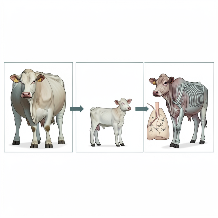
Risk Factor Correlation
AI-generated conceptual illustration (cattle (Bos taurus)). It explains the idea and does not show study data.
View source evidence ↗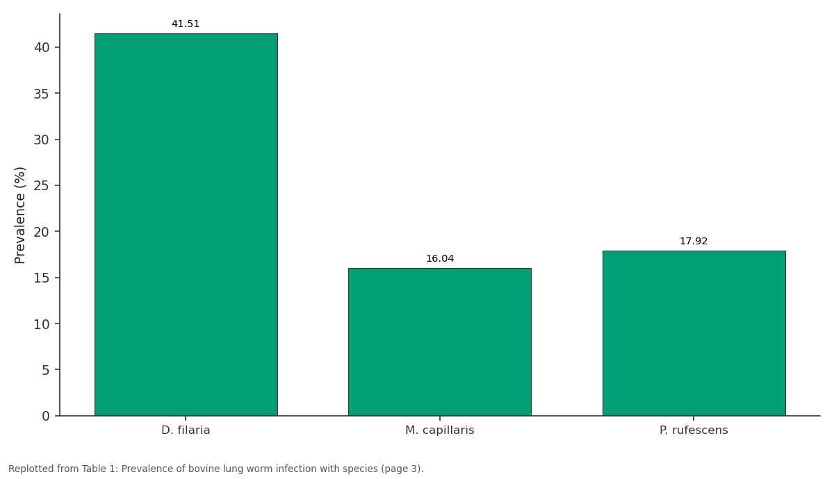
Prevalence (%): D. filaria vs M. capillaris vs P. rufescens
Replotted from Table 1 (page 3) of the original paper. Every bar is the paper's own table value.
View source evidence ↗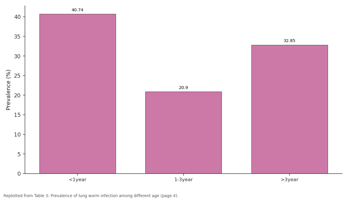
Prevalence (%): <1year vs 1-3year vs >3year
Replotted from Table 3 (page 4) of the original paper. Every bar is the paper's own table value.
View source evidence ↗Figures & tables
From the original paper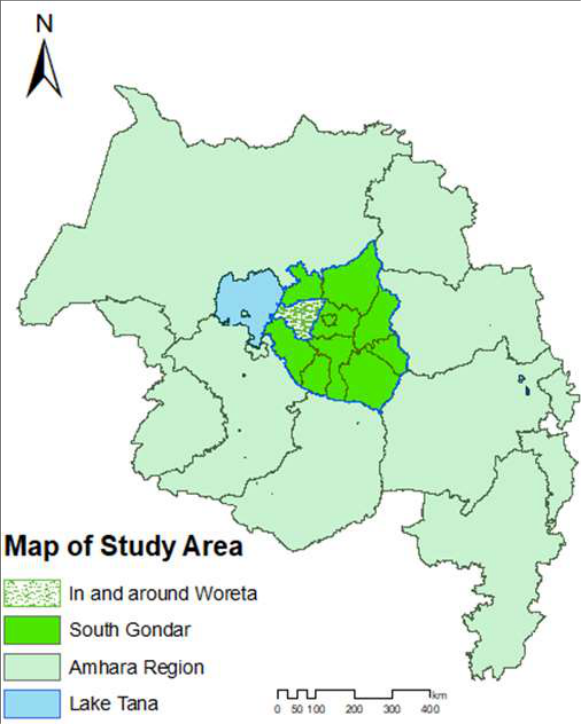 Figure 1
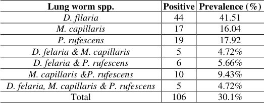 Table 1
 Table 2
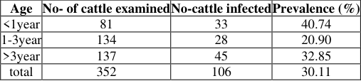 Table 3
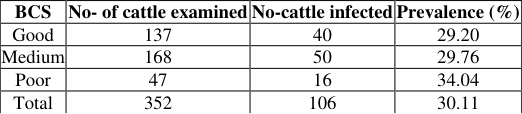 Table 6
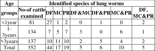 Table 4
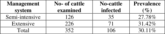 Table 5
Introduction
In Ethiopia, livestock production contributes about 30-33% of agricultural gross domestic prod uct (GDP) and more than 85% of the farm cash income is mainly from meat, milk, eggs, wool, hides and skins [24] . Small ruminants are important contributors to food production in Ethiopia providing 40% of cash income, 33% meat consumption and 14% milk consumption [3,36] . Eventhough sheep are important componens of the Ethiopian farming system their contribution to food production, rural income and export income are far below than expected potential. This is because sheep production is constrained by the compound effect of disease, poor feed quality and poor management [13] . Among the many constrains that limit productivity in livestock populations are parasites. Endo parasites are problem forsheep farmers throughout the world [24] . Among endoparasites the most common and economical significant ones are lungworm. Lung worms are widely distributed throughout the world but are particularly common in countries with temperate climates, and in the highlands of tropical and sub tropical countries. The species of importance in ruminants belongs to two different families; the Trichostrong Lisa Ann the Metastrongyliodae. The Dictyocaulidae include Dictyocaulus viviparus in cattle and buffaloes, and Dictyocaulus filaria in sheep and goats. These worms are 5-10 cm long and live in the trachea and bronchi. The Metastrongylidae are represented by at least two species in small ruminants. Protostrongylus rufescens, a small worm (1.5-3.5 cm) found in the bronchioles and Muellerius capillaris (1.2-2.5 cm) which are located in the alveoli. An infection of the lower respiratory tract by any of these nematode species may result in bronchitis or pneumonia, or both [32] .
Dictyocaulidae and/or certain Metastrongylidae are known to exist in east Africa (Ethiopia, Kenya, Tanzania and South Africa) [32] . Endoparasites, lungworms, are the major cause of mortality and morbidity in the Ethiopian highlands. Up to half of all sheep mortality on farms in Ethiopia highlands are caused by pneumonic and endo-parasites [17] . The effect of lungworms depends on their location within the respiratory tract, the number of infective larvae ingested and the immune system of the animals [11] . Prevention and control of these parasites is, therefore, critical to enhance the economic benefit from these species of livestock. However the incidence of respiratory helminthosis varies greatly from place to place depending on the relative importance of factors [2] . Baseline data on the type, distribution and seasonal incidence of lungworms is important to devise and implement strategic and technical control measures. However, information on lungworm in the study area is scarce. Therefore, the objectives of this study were:-To determine the prevalence of lung worm infection in Cattle, to recognize the species of lung worm involved in study area and to assess possible risk factors of lung worm infection in the study area.
Materials and Methods
Materials and Methods
Study Area
The study was conducted in and around Woreta town. which is located at 11°29'N latitude, 37°29'E longitude around 630km distance North-West of Addis Abeba. The area has midland altitude ranging from 1800 -2500 meters above sea level which is called "Woyenadega". The area receives a bimodal rainfall with an average annual rain fall ranging from 1200-1600mm. The mean annual temperature of the study area is 23°C. The presence of the largest Lake Tana and River Abay influence the climatic condition of the study area. The area has a mixed farming practice with crop and livestock production. According a survey by [7] .
Study Population
Study population included all cattle that were found in and around Woreta town. A total of 352 cattle of different sex, age, body condition within different management system were selected. When I was added some amount of population in the study to increase the accuracy (precisions). Of these, 236 were female and 116 were male. Of which, 236 were selected from extensive farms while the rest 116 cattle were selected from semi-intensive farms, which were selected randomly by their percentage proportion from the total population.
Study design and sampling techniques
Simple random sampling technique was used to determine the prevalence of bovine lung worm infection in and around Woreta town. A total of 352 cattle were randomly selected in and around study area and aslo coprological fecal examinations
was used to identify the species of the parasite from fecal sample.
Sample size determination
The sample size required for this study was determined based on the expected prevalence of lung worm invasion and the desired absolute precision. The sample size was computed using the formula given by [31] as follows: N = (1.96) 2 Pexp (1-Pexp) d 2 Where N = required sample size; Pexp = expected prevalence; d= desired absolute precision. Previous studies that were conducted on prevalence of lung worm in cattle in the study area show 18.16% of prevalence rate. Therefore, an expected prevalence of 18.16% was used to estimate the sample size. Using desired 95% confidence interval, 5% precision and 18.16% expected prevalence, the number of sample used to determine the prevalence of lung worm in the study area was 228 cattle. Alternatively, to increase the level of precision the number of sample was increased by 124 and so a sum total of 352 cattle were used.
Study methodology and laboratory examination Sample collection and laboratory technique
Total of 352 fecal samples were collected directly from the rectum of all selected animals and stored in universal bottles containing 10% formalin and transported to the Bahir Dar animal health investigation and diagnostic laboratory for laboratory examination. In the laboratory, the collected fecal samples were processed according to conventional methods for lungworm larvae (Baermanization) [14] . During sample collection, sex, age, date of sampling, body condition and management was recorded. In the laboratory, 25 g of feces was weighed from each sample for the extraction of larvae using modified Baermann technique [14] . The feces were enclosed in gauze fixed on the string rod and submerged in a clean glass tube filled with warm water. The whole apparatus was left for overnight. The larva leaves the feces and migrates through the gauze and settles at the bottom of the glass. After siphoning off the supernatant, the sediment was examined under the low power of the microscope to the larvae of lungworm [12,34] . Finally a drop of 1% iodine solution was added to the slide to immobilize the larvae when the larvae are detected under microscope to identify the species of the larvae. If have not a chance to see the larvae under microscope, the examined samples were registered as negative for lung worm infection. In both cases, the result that was obtained for each sample was indicated to their corresponding specific animal. All the area under the cover slip was thoroughly and uniformly searched for the presence of lungworm larvae [16] . The age of the animal was gathered from dentitions [32] .
Data management and analysis
The data were entered and managed in MS -Excel spreadsheet. All the data analysis was done by Statistical Package for Social Science (SPSS) software version18. Descriptive statistics such as percentages and frequency distributions were used to describe the nature and the characteristics of data. The prevalence of lung worm infection was analyzed using percentages. The association of different risk factors with the disease was computed by Chi -square (x 2 ) test.
Result Overall prevalence of lungworm
Of a total352 fecal sample of cattle that were examined, 106(30.1%) were found to be infected with either one or more of the lungworm species.
Species identification
The prevalence rate of species of lung worm in cattle at study area was different and mixed infestation of two or more species of lungworm were observed. Therefore, the prevalence rate of
Prevalence of bovine lungworm in different sex
An attempt was made to see if there was difference in prevalence rate of lung worm between sex groups. The result indicated that there was difference in isolation rate of lung worm between sex groups with prevalence rate of 33.47% in female and 23.28% in male cattle. However, the difference in prevalence rate of lungworm infection did not show statistically significant association between sexes of the animals (Table 2 Contrary to sex, the influence and association of age on the prevalence rate of lung worm infection was determined and it was found that there was difference in the prevalence rate of lung worm infection between age groups. It was 40.74% less than 1year, 20.90%% between 1&3 years and 32.85% greater than 3 years. There was statistically high significant difference (P<0.05) between the prevalence rate of lung worm and age groups (Table 3). The prevalence rate of lung worm species that were isolated from different age groups was different within the age groups and it was statistically significant (P< 0.05).Generally, the prevalence of M. capillaris and P. rufescens increases with age of the animal, whereas the prevalence of D. filaria was high in young age groups of the animals. Mixed infection was common in old age group of the animals (Table 4).
| Age | Identified species of lung worms | — | — | — | — | — | — | — |
|---|---|---|---|---|---|---|---|---|
| groups | No-of cattle examined | DF | MC | PR | DF&MC | DF&PR | MC&PR | DF, MC&PR |
| <1year | 81 | 27 | 1 | 2 | 0 | 1 | 0 | 2 |
| 1- 3years | 134 | 7 | 5 | 7 | 3 | 0 | 6 | 1 |
| >3years | 137 | 10 | 11 | 10 | 2 | 5 | 4 | 2 |
| Total | 352 | 44 | 17 | 19 | 5 | 6 | 10 | 5 |
Table text extracted from the original PDF; superscript letters are shown inline. The original table image is below.
View original table image
| Age | No- of cattle examined | No-cattle infected | Prevalence (%) |
|---|---|---|---|
| <1year | 81 | 33 | 40.74 |
| 1-3year | 134 | 28 | 20.90 |
| >3year | 137 | 45 | 32.85 |
| total | 352 | 106 | 30.11 |
Table text extracted from the original PDF; superscript letters are shown inline. The original table image is below.
View original table image
| Sex | No-of cattle examined | No-of cattle infected | Prevalence (%) |
|---|---|---|---|
| Female | 236 | 79 | 33.47 |
| Male | 116 | 27 | 23.28 |
| Total | 352 | 106 | 30.11 |
Table text extracted from the original PDF; superscript letters are shown inline. The original table image is below.
View original table image
below).
below).
Prevalence of ovine lungworm under different management systems
In this study the prevalence of lung worm was determined in cattle that were reared in two different management systems (extensive and semi -intensive management systems) and the prevalence was found to be higher in cattle in the extensive management system (31.42%) as compared to the semiintensive management system (27.78%). However, there was not statistically significant association within the management systems (χ 2 = 7.452, P > 0.05) (Table 5).
| Management system | No- of cattle examined | No-cattle infected | Prevalence (%) |
|---|---|---|---|
| Semi-intensive | 126 | 35 | 27.78% |
| Extensive | 226 | 71 | 31.42% |
| Total | 352 | 106 | 30.11% |
Table text extracted from the original PDF; superscript letters are shown inline. The original table image is below.
View original table image
Prevalence of Bovine lungworm infection in different body conditions of animals
All cattle that were examined for lung worm infection were categorized in to three groups as poor, medium and good body condition. It was observed that the prevalence of lung worm infection was 34.04%, 29.76% and 29.20% in cattle with poor, medium and good body conditions, respectively. There was statistically significant difference (P<0.05) in lungworm infection rate among the different body condition scores (BCS) (Table 6).
| BCS | No- of cattle examined | No-cattle infected | Prevalence (%) |
|---|---|---|---|
| Good | 137 | 40 | 29.20 |
| Medium | 168 | 50 | 29.76 |
| Poor | 47 | 16 | 34.04 |
| Total | 352 | 106 | 30.11 |
Table text extracted from the original PDF; superscript letters are shown inline. The original table image is below.
View original table image
Discussion
Lung worm infection is a chronic and prolonged nematodosis that affects the lungs of cattle and goats and other animals. This disease results in substantial economic losses due to the reduction of growth rate, morbidity and mortality as the disease exposed the animals to secondary bacterial infections. It also causes an economic loss due to organ condemnation and medication costs.
In the present study, coproscopic results made known an overall lungworm infection prevalence of 30.11% in cattle in and around Woreta. Of the total infected animals, 41.51%, 16.04%, 17.92%, 4.72%, 5.66%, 9.43% and 4.72% prevalence was due D. filaria, M. capillaris, P. rufescens, D.filaria & M. capillaries, D. filaria & P. rufescens and D. filaria, M. capillaris, P. rufescens species of lungworm, respectively. The overall prevalence result almost coincides with the previous researcher reports in Ethiopia, like with that of [33] in Gaint awraje (32.4%), Addis and his colleagues [1] in Gondar (33.83%), Weldesenebet and Mohamed [35] in Jimma (26.7%), Dawit and Abdu (2011)in Jimma (25.6% ), Teffera(1993) in Dessie and Kombolcha (27.8%), Brook and co-workers [5] in and around Bahir Dar (27.8%). However, a relative higher and lower prevalence were also reported in different parts of the country including 40.4% in Dessie and Kombolcha districts, Northern Ethopia, by Regassa [28] 22.7% in and around Bahir Dar, by Tewdrose (2010), 21.57%in Tigray, by Mengstom [20] and 20.2% in and around Bahir Dar, by Kassa and Abdu [18] . But, the present study is higher than the observation made from the same area by Muluken [24] , 18.16%, and Sisay [29] , 13%. Contrarily, the finding is lower than the prevalence of lung worms that were reported by Netsanet [25] in Debrebirhan, Alemu [2] in North West Ethiopia, Mezgebu [21] in Addis Ababa and Mihreteab and Aman [22] in Tiyo with the prevalence of 73.25%, 53.6%, 48% and 57.1%, respectively. The possible explanation for such infection rate variation could be attributed to variation in season of examination on the respective study areas, which may have good turn or bad turn to the survival of parasite larvae [9] . In addition to that, the snail which is the intermediate host of P. rufescens and M. capillaris in encouraging seasons may be considered to be creating the difference. At the end of summer, the parasites multiplication increases as a result of wet season, which is favors multiplication of the parasite. And the intermediate host also obtains complimentary state to survive and multiply. Therefore, the increase in prevalence rate might be due the fact the data was collected in November and December. Moreover, the occurrence of lungworms is associated with nutritional status, level of immunity, and management practice of the animal [4] which is at different levels among the different parts of the country. With regard to the species of lungworms, it was observed that D. filaria was the predominant species in the area followed by P. rufescens, whereas M. capillaris was the least prevalent. This is in total agreement with the work by Mihreteab and Aman [22] . In the current study higher level of prevalence was observed in female (33.47 %) animals compared to that of male animals (23.3%). However, sex dependent variation was not significantly associated; hence both sexes are believed to have almost equal susceptibility to infection with lungworms. This was coinciding with research study reported by Nibret [26] and Hasen [15] , but disagree with report of Alemu [2,22] . These may be due to the fact that improper distribution of sample selection between the two sexes [27] ; or else most of the sampled females were not in preparturient period and early lactation during the study time. These preparturient period and early lactation are relaxation of resistance result in the female's inability to expel adult worms [34,6] . Which cause higher level of larvae detection. In comparison to age groups, significant difference (χ 2 = 7.04, P < 0.05) in the prevalence rate of lungworm infection was observed. Among lungworms identified, high prevalence rate of D. fillaria (41.51%) followed by P. rufescens (17.92%) was recorded. The prevalence of M. capillaris and P. rufescens increases with age of the animal, whereas the prevalence of D. filaria was high in young age groups of the animals. This could be due to not well develop immunity against the infection. However, a high infection prevalence of overall lungworm was recorded in young than adults. This is in agreement with the observations of Soulsby [30] and Mireteab and Aman [22] This has been partly explained by the analysis given as; infection with M. capilleris and P.rufescens did not show development of acquired immunity. The acquired immunity developed by older cattle due to previous exposure of D. filaria and that recovered from the infection have better immunity against reinfection [30,6] . Mixed infection was common in old age group of the animals. An infection rate of bovine lungworm was statistically analyzed on the basis of body condition to study the impact of the disease in debilitating/emaciating infected animals. The result indicated that an infection rate of 34.04%, 29.76% and 29.2% in poor, medium and good body condition scores, respectively; with a significant variation (P<0.05). This signifies that poor body conditioned animals are more susceptible to an infection. The possible explanation for this observation could be due to immuno-suppression in cattle with poor body conditions, concurrent infection by other parasites including GIT helminthes and/or malnutrition [10] . The reason for this could be partly due to the fact that poorly nourished animals appear to be less competent in getting ride-off lungworm infection although it is unusual for well-fed animals to submit to the disease provided the right environmental conditions are made available. Kassai [19] has also reported that well nourishment and watering lead to less risk of helminthes infection. In this study, an attempt was conducted to know if there is difference in prevalence of bovine lung worm infection under different management systems. The overall prevalence of bovine lungworm infection was 31.42 % and 27.78% in extensive and semi-intensive management systems, respectively. However, the difference was not statistically significant (P > 0.05); it could be due to the fact that animals under semi-intensive management system have a chance of grazing in the field, being exposed to the intermediate host (snail or slugs) or directly can be infected with lung worm while they are grazing in the grazing field. This study agrees with the finding that was reported by Kassa and Abdu [18] in and Bahir Dar; with the prevalence of 20.9% and 9.7%in extensive and semi -intensive farms, respectively. In contrary, it is not in agreement with the finding of Dawit [8] in Tse-Ada-Emba; with the prevalence of 30.43% and 25% in extensive and semi -intensive sytems, respectively. This could be due to the difference in agro-ecology and season of the two study areas; sampling and sampling techniques that are used and it may also be due to the differences in breed and the biology of animals.
Conclusion and recommendations
The study on lungworm infection of cattle using fecal examination in and around Woreta town revealed prevalence of 30.1%. The present study result indicated that lungworm is one of the most important helminthosis of cattle in the study area and the prevalence of infection was higher in young age groups, and poor body conditioned cattle. Young animals are mostly affected by the most pathogenic species of lungworm, D. filaria, than adults, on the contrary which are highly affected by M. capillaries and P. rufescens. Based on the above conclusions the following recommendations are forwarded:-Isolation of most susceptible age groups (young) from adult at summer end season of grazing, Supplementation of additional feed to cattle to ensure animals are wellnourished and became good body condition, Emphasis strategic deworming were provided well to be reduced its prevalence.
Acknowledgement
AcknowledgementThe grateful thanks go to Fogera woreda veterinary clinic for provision of materials what I need and University of Gondar, Faculty of Veterinary Medicine for material and ideally supports, and their cooperation to bring this research to completion.
Figures and tables from the paper
| Lung worm spp. | Positive | Prevalence (%) |
|---|---|---|
| D. filaria | 44 | 41.51 |
| M. capillaris | 17 | 16.04 |
| P. rufescens | 19 | 17.92 |
| D. felaria & M. capillaris | 5 | 4.72% |
| D. felaria & P. rufescens | 6 | 5.66% |
| M. capillaris &P. rufescens | 10 | 9.43% |
| D. felaria, M. capillaris & P. rufescens | 5 | 4.72% |
| Total | 106 | 30.1% |
Table text extracted from the original PDF; superscript letters are shown inline. The original table image is below.
View original table image
References
36 in the original paper- Addis M, Fromsa A, Ebuy Y. Study on the Prevalence of Lung worm Infection in Small ruminantsin Gondar Town, Ethiopia. Vet. Res 2011;4(3):85-89.
- Alemu S, Esayas G, Galegay A, Aschalew Z. Study on small ruminant the lung worm in Northern Ethiopia. Veterinary Parasitolgy 2006;142:330-335.
- Asfaw W. Country report: Ethiopia. Proceedings of the Seminar on Livestock Development Policies in Eastern and Southern Africa, Swaziland, 1997, 398-398.
- Bradford PS. Large Animal Internal Medicine: Disease of horses, cattle, sheep and goats, 3 ed . Mosby, Inc 2002;51415:1452-1455.
- Brook L, Fesseha G, Shibru T. The seasonal occurrence of D.filaria (Rudolph: in four selected sites of Ethiopia; Sci 1986;9(1):25-38.
- Practitioner, Western Veterinary Conference, Las Vegas 1998.
- CSA. Summery and report of the (2007) population and housing census. F.D.R.E. population census commission. December (2008). Addis Abeba, Ethiopia, 2008.
- Dawit M. Prevalence of ovine lungworm in and around Tse-Ada-Emba. DVM Thesis Jimma University, Jimma, Ethiopia, 2009.
- Eyob E, Matios L. The prevalence and risk factors associated with ovine lungworm infestation in the Asella province, Central Ethiopia Journal of Parasitology and Vector Biology 2009;5(8):116-121.
- Kimberling CV. Jensen and Swift's diseases of sheep, 3 rd ed. Lea and Febiger, Philadelphia, Pa, USA 1998, 99-101.
- FAO. The epidemiology, diagnosis and control of helminthe parasites of ruminants. Printed by the ILCA, Addis Ababa, Ethiopia, 1994, 25-28.
- Fraser CM. The Merck veterinary manual, a hand book of diagnosis therapy and disease prevention and control for the veterinarians. 7 th ed. Merk and Co., Inc. Rahway, Nif, USA, 1991, 714:817.
- Getachew MR. Parasite of small ruminants. In: Gray GD, Uilengerg G (eds), Parasitological Research in Africa, 1995.
- Hansen J, Perry B. The Epidemiology, Diagnosis and Control of Helminth parasites of ruminants. ILRAD, Nairobi, Kenya 1994, 38.
- Hasen A, Takele S, Simenew K. Ovine lungworm infestation rate on fecal larvae recovery basis. Acta Parasitol Globalis 2013;4(1):29-33.
- Hendrix C. Diagnostic of veterinary parasitology, 2 nd ed. Mosby, London 1998, 49.
- ILCA. Annual report. ILCA (International Livestock Center for Africa), Addis Ababa, Ethiopia, 1990.
- Kassa T, Abdu M. Prevalence of Ovine Lung Worm in and Around Bahir Dar, East Africa, DVM Thesis, Jimma University, Jimma, Ethiopia, 2011.
- Kassai T. Veterinary Helminthology. Butter worth helminthology. Hungary; Budagest, 1999.
- Mengestom G. Preliminary survey on prevalence of ovine lung worm infection in Atsbi. DVM Thesis, JimmaUniversity, Jimma, Ethiopia, 2008.
- Mezgebu M. Study on bovine Fasciolosis and lung worm infection in Addis Ababa and the surrounding highland areas. DVM Thesis, Faculty of Veterinary Medicine, Addis Ababa University, Debre-Zeit, Ethiopia, 1995.
- Mihreteab B, Aman A. Ovine Lungworms in Tiyo District, South-East Ethiopia: Prevalence, Effect of Altitude and Major Host Related Risk Factors. Global Vet 2011;7(3):219-225.
- MoARD. Estimation of weight and age of sheep and goats (ESGPIP) 2009. [Available at file:///C:/Users/user.DELLPC/Downloads/age%20determi ned%20by%20the%20pair%20of%20teeth%20growth].
- Muluken B. Preliminary study on prevalence of ovine lung worm infection in Bahir Dar. DVM Thesis, Jimma University, Jimma, Ethiopia, 2009.
- Natsanet B. Study on prevalence and control of lungworm in local Ethiopian highland sheep in and around Debere Birhan, DVM thesis, Faculty of Veterinary Medicine Addis Ababa University Debreziet 1992, 42.
- Nibret M, Basaznew B, Mersha C. Dictyocaulus filaria and Muellerius capillaris are Important Lungworm Parasites of Sheep in Wogera District, Northern Ethiopia. Int. J. Anim. Vet. Adv 2011;3(6):465-468.
- Paulos A. Importance and seasonal dynamics of lungworminfection of small ruminants in Chilalo area, Arsizone, DVM thesis, Faculty of Veterinary Medicine, Addis Ababa University Debreziet. 2000.
- Regassa A, Toyeb M, Abebe R, Megersa B, Mekibib B, Mekuria S. Lungworm infection in small ruminants: Prevaence and associated risk factors in Dessie and Kombolcha districts, northeastern Ethiopia. Vet. Parasitol. 2010;169(1-2):144-148.
- Sissay A. Preliminary study on the prevalence of study on the prevalence of ovine lungworm infestation in and around Bahir Dar. DVM Thesis, AAU-FVM, Debrezeit Ethiopia, 1996.
- Soulsby EJL. Helminths, Arthropods and Protozoa of Domesticated Animals. 6 th ed, Bailliere Tindall, London 1982, 56-58.
- Thrusfield M. Survey in Veterinary Epidemilogy 2 nd ed. Blackwell Science Ltd., Cambridge, USA, 2005, 178-198.
- Torncy PM. Mannual of Tropical Veterinary. Parasitology. Helminthes of Livestock and poultry in Tropical Africa. The International Technical Center for agricultural and rural cooperation, C, A.B International, 1989, 81-85.
- Tsegaye S. Prevalence of Dictyocaulus filarial in Gayint Awraja, DVM thesis, Faculty of Veterinary Medicine, Addis Ababa University, Debre Zeit, Ethiopia, 1985.
- Urquhart GM, Rmour JL, Duncan AM, Dunn, Jennings FW. Veterinary Parasitology. 2 nd .ed, Black Well Science Ltd., London, UK, 1996, 307.
- Weldesenebet D, Mohamed A. Prevalence of small ruminant lung worm infection in Jimma town. Glob Vet. 2012;8(2):153-159.
- Zelalem A, Fletcher IC. Small ruminant productivity in the central Ethiopia Mixed farming system. In: Proceeding of the 4 th National Livestock Improvement Conference, IAR, and Addis Ababa, Ethiopia, 1993.