
Research article · 2020 · Full text
International Journal of Veterinary Sciences and Animal HusbandryA review on canine ehrlichiosis and its zoonotic implications
Volume 5(3) · Pages 36–42
Research areasVector-borne infectious diseasesViral Infections and VectorsRabies epidemiology and control
Abstract
Author-writtenCanine ehrlichiosis is an infectious vector borne disease of dogs caused by different Ehrlichia species, which are intracellular rickettsial organism belonging to the family Rickettsiaceae; Included in order Rickettsiales, the obligate intracellular location of these organisms makes an effective host immunologic response difficult. It is typically a disease of leukocyte and platelet manifested by an acute reduction in cellular blood elements, most often Pancytopaenia, thrombocytopenia. The disease is widespread in dogs in tropical and subtropical and geographic distribution of tick vectors the brown dog tick (Rhipicephalus sanguineous), lone star tick (Amblyoma americanum). Canine ehrlichiosis has been a subject of increasing interest from veterinary and public health perspectives over the last few decades with identified zoonotic important species, from which Ehrlichia chaffeensis, the etiologic agent of human monocytotropic ehrlichiosis (HME) is an emerging zoonosis. Traditional diagnostic techniques including hematology, cytology, serology and isolation are valuable diagnostic tools for canine ehrlichiosis; however, a definitive diagnosis of Ehrlichial species infection requires molecular techniques. This disease can be prevented through avoidance of tick bites. Tetracycline-related antibiotics have been the treatment of choice for Ehrlichia infections for years. Therefore this paper will focus on canine ehrlichiosis and its public health importance.
Summary
Source-checkedPlain-language reading of the full paper, prepared with AI and checked against the text. Each point links to the authors' own words.
Why it matters
- Canine ehrlichiosis is an infectious disease in dogs caused by various species of Ehrlichia, which are intracellular rickettsial organisms. View evidence ↗
- The disease has gained veterinary and public health interest because some species, such as Ehrlichia chaffeensis, are emerging zoonoses that can infect humans. View evidence ↗
- Because these organisms live inside host cells, it is difficult for the host's immune system to mount an effective response. View evidence ↗
What they did
- Diagnosis typically involves hematology, cytology, serology, and isolation, though molecular techniques are required for definitive species identification. View evidence ↗
- A reliable diagnosis is reached by combining patient history, clinical signs, blood tests, serology, and molecular confirmation. View evidence ↗
What they found
- The disease primarily affects leukocytes and platelets, typically resulting in thrombocytopenia and pancytopenia. View evidence ↗
- The primary mode of transmission is through the bite of infected ticks, specifically Rhipicephalus sanguineus for E. canis. View evidence ↗
- The acute phase of the disease occurs after an incubation period of 8 to 20 days and typically lasts for 2 to 4 weeks. View evidence ↗
- Recommended treatment includes tetracycline (22 mg/kg every 8 hours) or doxycycline (5 mg/kg every 12 hours) for at least six to eight weeks. View evidence ↗
- As of September 2002, over 1,363 cases of E. chaffeensis infection were reported to the CDC in the United States, though this is considered an underestimate. View evidence ↗
- Human monocytic ehrlichiosis (HME) is most frequently diagnosed in adults over the age of 50. View evidence ↗
What it means
- The authors conclude that the disease can be prevented by controlling tick populations and avoiding tick bites. View evidence ↗
- The authors recommend that researchers in Ethiopia conduct studies on the disease, as the vectors and agents are present in neighboring countries but not yet characterized in Ethiopia. View evidence ↗
- A current limitation in prevention is that vaccines for the disease are still in the early stages of development. View evidence ↗
Visual summary
1 visualsIllustrations explain the paper; they do not show study data.
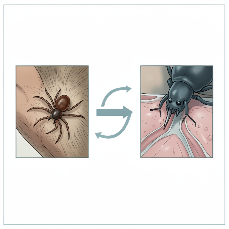
Transmission Cycle
AI-generated conceptual illustration (Rhipicephalus sanguineus ticks and a domestic dog). It explains the idea and does not show study data.
View source evidence ↗Figures & tables
From the original paper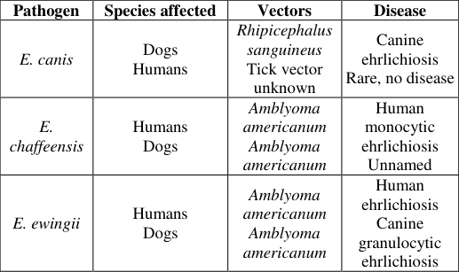 Table 1
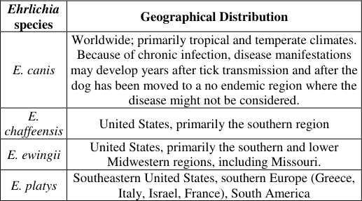 Table 2
Introduction
Canine ehrlichiosis is an infectious vector borne disease of dogs caused by variety of Ehrlichia species such as E. `canis, E. chaffeensis, E. ewingii, and E. platy. Although the clinicopathologic course of disease can vary depending up on the infecting Ehrlichia species, illness is typically characterized by an acute reduction in cellular blood elements, most often, pancytopaenia and thrombocytopenia. [33] The causative agents of canine ehrlichiosis are intracellular rickettsial organisms. In dogs specific Ehrlichia have been identified parasitizing monocyte (E. canis), granulocyte (E. ewingii), and platelets (E. platy). Acute, subacute and chronic syndromes have been described for canine ehrlichiosis. E. chaffeensis is better known as the agent of human monocytotropic ehrlichiosis (HME) in the Southern United States, but also infects dogs. Experimental infections suggest that E. chaffeensis produces relatively mild disease in dogs. However, when co-infection with other Ehrlichial agents is present, dogs may be more severely affected. [32] The distribution of canine ehrlichiosis is related to the distribution of the vector ticks, Rhipicephalus sanguineus, the brown dog tick and vector for E. canis throughout the world as well as Amblyoma americanum, the lone star tick and primary vector for E. chaffeensis and E. ewingii in the United States. It is widespread in dogs in tropical and subtropical regions. [7] Even if, studies in Africa lacks to identify Ehrlichia canis, the causative agent of canine ehrlichiosis or tropical canine Pancytopaenia (TCP) has been identified as stated by Kamijolo [19] in Kenya, Nidp [26] in Cameroon. The canine ehrlichiosis agents are maintained in nature through enzootic ticks and, wild and domestic animals. Because transovarial transmission is inefficient in ticks, animals seem to play a major role in propagation and as reservoir of these pathogens. Dogs are competent reservoir hosts of several zoonotic agents and can serve as a readily available source of nutrition for many blood feeding arthropods. Therefore, the growing medical interest in canine Vector-borne diseases is directly related to public health. Despite being considered rare, human infestation with brown dog tick varies regionally and R. sanguineus feed on humans much more commonly than previously thought. This may imply that the tick is becoming more anthropophilic or that a more human adapted population of R. sanguineous has been introduced. There after the canine ehrlichiosis have been subject to increasing interest from veterinary and public health perspectives. [31] In canine ehrlichiosis the diagnosis is routinely based on serology (indirect fluorescent antibody-IFA, ELISA, Western immunoblotting), since its clinical and clinico-pathological features are largely nonspecific . [39] Therefore, this seminar paper at hand is aimed:
To provide an over view on canine ehrlichiosis and its public health importance
Canine Ehrlichiosis
Canine ehrlichiosis; also known as canine rickettsiosis, canine hemorrhagic fever, canine typhus, dog AIDS and tropical canine Pancytopaenia is a tick-borne disease of dogs. It can be classified in to three forms depending on the Ehrlichial species that are affecting specific blood cells. [32] These are; Canine granulocytic ehrlichiosis, Canine cyclic thrombocytopenia and canine monocytic ehrlichiosis; which caused by E. ewingii, E. platy and E. canis, and affecting neutrophil, platelets and monocyte, respectively.
Etiology Taxonomy and its characteristics
Etiological agents of canine ehrlichiosis are E. canis, E. ewingii, E. platy, E. chaffeensis and E. phagocytophila, from the genus Ehrlichia belonging to the family Rickttsiaceae; included in the order Rickettsiales. They are pleomorphic cocci capable of causing disease in people and in several species of domestic and wild animals. They have distinguished from gram-negative organisms in that they do not cause endotoxemia and they require a vector for transmission. [40] These organisms are found in membranelined vacuoles within the cytoplasm of infected eukaryotic host cells, most often leukocytes. The obligate intracellular location of these organisms makes an effective host immunologic response difficult, and this complicates antimicrobial therapy. [25] The diseases caused by these pathogens have traditionally been categorized by the type of blood cell most commonly infected. For example, E. chaffeensis and E. canis reside primarily in monocytes, and the disease caused by these agents is frequently called monocytic (or monocytotropic) ehrlichiosis and E. ewingii reside primarily in granulocytes, and the disease caused by these agents is often referred to as granulocytic (or granulocytotropic) ehrlichiosis with their respective vectors and hosts as shown below (Table 1). [15] Some of the Ehrlichia organisms have been cultivated in human myeloblastic leukemia cell lines with potential for monocytic or myelocytic differentiation. Canine leukemia cell lines of histolytic origin also have been used to grow E. chaffeensis. Many Ehrlichia remain uncultivable; however. These organisms stain blue with Romanov sky stain. [37] [15] Transmission and Epidemiological Factors The principal mechanism of transmission for the agents' canine Ehrlichia species is via tick bite. Infection with Ehrlichia transmitted through the salivary secretions of an attached tick. Dogs are get infection through the bite of an infected Rhipicephalus sanguineous tick. Transmission, in the tick, occur transstadially, but not transovarialy. Larvae and nymphs become infected while feeding on rickettsemic dogs and transmit the infection to the host after moultig to nymphs and adults, respectively. Ehrlichiosis occurs mainly in the spring and early summer. [37] Ehrlichia species can also be transmitted by blood transfusion and it is recommended to screen for its presence in the blood of donor dogs. The severity of the disease depends on the dog's age (i.e., young dogs are more susceptible), strain of the organism, the presence of concurrent disease, and breed (example, German shepherds) are more likely to be infected. [28] Ehrlichia canis has a worldwide geographic distribution, occurring particularly in tropical and subtropical areas. [11] Geographic distribution of tick vectors has a direct impact on disease prevalence in a given region. For instance; As stated by Hinrichsen [12] in the United States, E. phagocytophila infection is reported most often in the northwestern, upper Midwestern, and northeastern states, the same regions in which Ixodes ticks are most abundant, E. ewingii infection also follows the distribution of its tick vector, being found most often in the southeastern and south-central United States as sown below (Table 2). Several different tick species are capable of horizontal transmission of infection from vector to eukaryotic host. Ehrlichia canis usually is spread by the bite of the brown dog tick (Rhipicephalus sanguineous), which also can transmit infection with E. ewingii and probably E. platys. E ewingii is transmitted predominantly (but not solely) by the lone star tick (Amblyoma americanum). Dogs may serve as a reservoir host for any Ehrlichia species rather than E. canis, which has not been clearly established. Besides deer, rodents, coyotes and other small mammals may serve as major reservoirs for the other Ehrlichia species, with dogs playing only a minor role in the maintenance of the organism in a given geographic location. Dogs serve as a key reservoir host for E. canis and also as the maintenance host for the primary vector tick, Rhipicephalus sanguineous.
Immature stages of R. sanguineous are infected when feeding on a rickettsemic dog and then maintain that infection transstadially, enabling transmission to occur when the tick feeds again as a nymph or an adult. Adult male R. sanguineous have also been shown to be capable of transmitting E. canis in transstadially, a route that may be important in outbreak situations, as male ticks have been shown to readily move between dogs as they intermittently feed and mate. The maintenance cycle of E. canis is particularly pernicious because R. sanguineous populations can establish and survive inside homes and kennels, providing a near-constant source of infection to dogs in an infested environment. [41] Table 2: Geographic distribution of Ehrlichia species
Ehrlichia
species Geographical Distribution
E. canis
Worldwide; primarily tropical and temperate climates.
Because of chronic infection, disease manifestations may develop years after tick transmission and after the dog has been moved to a no endemic region where the disease might not be considered.
E. chaffeensis
United States, primarily the southern region
E. ewingii
United States, primarily the southern and lower Midwestern regions, including Missouri.
E. platys
Southeastern United States, southern Europe (Greece, Italy, Israel, France), South America Source: [27]
Tick-vectors
In canine ehrlichiosis transmission; mainly two important tick species are involved those are Rhipicephalus sanguineus and Amblyoma americanum from the genus Rhipicephalus and Amblyoma respectively even if other are also can transmit. Rhipicephalus sanguineous (Brown Dog Tick): Is a three host tick. The engorged female detaches and lays its eggs in cracks and crevices of buildings or under the dog's bedding. These larvae then detach and moult to the nymphs find another host, engorge, detach and moult to adults usually within a few weeks. The adults then engorge and the cycle continues. All these stages take place around where the dog lives and may be indoors or in a kennel. [5] Rhipicephalus sanguineus is active throughout the year in the tropics and subtropics. On dogs, adult ticks attach on the ears, neck and shoulders, nymphs are also found on the ears and shoulders, and larvae attach particularly to the belly and flanks. Rhipicephalus sanguineus is a potent disease vector for dogs and can transmit Ehrlichia canis, the causative agent of canine ehrlichiosis, and Babesia canis, the causative agent of canine babesia, east coast fever, and Nairobi sheep disease in east Africa. [38] In Ethiopia R. sanguineous is not still identified rather other Rhipicephalus species of ticks are recognized. [2] But according to Jongejan [17] and Ahmed [1] tick Rhipicephalus sanguineous is identified in southern Sudan from the border of Ethiopia.
Life cycle
The life cycle of Ehrlichia species in the vector is not completely understood. But in the host there are three intracellular forms. Initial bodies are small spherical structures (1-2 microns) which are believed to develop into larger multiple units known as morulae. The morula is thought to dissociate into small granules called elementary bodies. The elementary body is the infective stage and enters to the monocyte or other leukocyte types by phagocytosis.
Elementary bodies are individual Ehrlichia about one µm in diameter and usually coccoid or ellipsoid in shape. Once they are captured inside the phagosomes, the pathogens replicate by binary fission, forming clusters of tightly packed elementary bodies termed initial bodies. Additional growth and replication leads to the formation of the morula, the configuration that typifies the genus. Rupture of the host cell releases the elementary bodies to infect new cells. [29] Pathogenic effects After entering to the canine host through the bite of the tick vector, ehrlichial organisms travel through the circulation, invade cells and disseminate to various tissues. Once in tissues, they continue to invade, persist, and replicate in cells. Circulating infected cells may induce vasculitis and subsequent intravascular coagulation, which in combination with an altered cell-mediated immunity; result in the destruction of platelets. Similar destruction of leukocytes and erythrocytes in combination with reduction of erythrocyte production may cause clinical leucopenia and anemia respectively. [37] There are three clinical phases of ehrlichiosis: acute, subclinical, and chronic. The acute phase begins after an incubation period of 8-20 days and lasts 2-4 weeks, during which time the organisms multiply in reticuloendothelial cells, lymphocytes, and monocytes. Infected mononuclear cells marginate in the small vessels or migrate into endothelial tissues and cause vasculitis, which leads to hyperplasia of endothelial cells and localized thrombus formation then obstruction of blood flow, with escape of blood cells in to the surrounding tissue and finally pancytopaenia and thrombocytopenia. [32] Immunologic and inflammatory mechanisms are involved with increased platelet consumption. Polyartheritis may arise from haemarthrosis and immune complex in to the joints and is often accompanied by neutrophilic inflammation. Plateletassociated IgG and antibodies that recognize platelet proteins in dogs with E. canis infection may play a role in the thrombocytopenia. [7] In addition, platelet migration-inhibition factor (PMIF) has been found to exist in dogs with ehrlichiosis and its level is related inversely to the platelet count. The acute phase usually resolves spontaneously. The subclinical phase can persist for years. Immunocompetent dogs may be able to eliminate E. canis; however, the organism persists intracellularly in most dogs, leading to the chronic phase. [23] This phase may be mild to severe. In the mild form, there is vague illness and weight loss. Bone marrow hypoplasia leading to pancytopaenia occurs in the severe chronic form. The severity of the disease depends on the dog's age (i.e., young dogs are more susceptible), strain of the organism, the presence of concurrent disease, and breed (example, German shepherds) are more likely to be infected. [28]
Immunological features
The ability of Ehrlichia to evade the host immune mechanisms, both innate and adaptive immunity appears to be essential for its survival in natural hosts as well as incidental hosts such as humans. Ehrlichia are obligately intracellular rickettsial organisms that exhibit tropism for mononuclear phagocytes and other leucocytes. Recently, a number of studies have demonstrated that antibodies play an essential role in immunity against Ehrlichia pathogens. Furthermore, a small subset of E. chaffeensis and E. canis proteins react strongly with antibodies in sera from infected humans or dogs and thus are considered to be major immunoreactive proteins. [24] Clinical and Pathological features There are three clinical phases of ehrlichiosis: acute, subclinical, and chronic. Clinical findings in dogs with ehrlichiosis vary with the phase of the infection. During the acute phase, nonspecific signs such as fever, oculonasal discharge, anorexia, weight loss, dyspnea, and lymphadenopathy may occur. During the subclinical phase, thrombocytopenia, leucopenia and anemia may continue. Clinical signs commonly seen during the chronic phase include depression, weight loss, pale mucous membranes, abdominal pain, hemorrhage, lymphadenopathy, hyperglobinaemia, spleno-megaly, dyspnea, increased lung sounds, hepatomegaly, arrhythmias, pulse deficits, polyuria, polydypsia, and stiff, swollen, painful joints. Ocular abnormalities such as perivascular retinitis, hyphemia, retinal detachment, anterior or posterior uveitis, and corneal edema may occur. Dogs which are severely affected can die from this disease. [6] Ehrlichiosis is not characterized by specific pathologic findings, but gross lesions may include petechial and ecchymotic hemorrhages on the serosal surfaces of the gastrointestinal and urogenital tracts and kidneys, edematous or hemorrhagic enlargement of most lymph nodes, and edema of the limbs. Dogs are generally emaciated at death and may have signs of epistaxis [21] . Splenomegaly and/or hepatomegaly may be observed. Histopathologic findings include widespread perivascular accumulations of lymphoreticular and plasma cells, particularly in the meninges, kidneys, liver and lymphopoietic tissues. Multiple Kupffer cell hyperplasia and degeneration and acute centrilobular necrosis of the liver may be seen. Lesions of the CNS include hemorrhage and plasma cell accumulations in the meninges and occasionally lymphocytic and plasma cell infiltrations are present in the brain parenchyma. Other microscopic findings may include crescent-shaped perifollicular hemorrhages in the spleen, bone marrow hypoplasia, interstitial pneumonia, and glomerulonephritis [16,13] .
Diagnostic techniques
Diagnosis of canine ehrlichiosis is routinely based on serology (indirect fluorescent antibody-IFA, ELISA, Western immunoblotting), since its clinical and clinico-pathological features are largely non specific. Nevertheless, serology may be diagnostically misleading, especially in the endemic areas of the disease. [14] Hence, observation of Ehrlichia morulae within monocytes and/or lymphocytes, the detection of its DNA in target-tissues by PCR amplification, or in-vitro culture are required for a sound diagnosis. Therefore, there is no single method of diagnosis for this disease; instead, the diagnosis is achieved to varying degrees of certainty through a combination of history, clinical and hematological indicators, serologic evidence, and molecular confirmation [39,27] Residence in or travel to known endemic areas, and a history of tick infestation should be increase the suspecting of infection. In addition, blood tests also show abnormalities in the numbers of red blood cells, white blood cells, and most commonly platelets, if the disease is present [9] . An ELISA test has also been developed to detect antibodies and circulating antigen in dogs with E. canis. Cross-reactivity occurs between several of the Ehrlichia species [20] .
Differential diagnosis
The differential diagnosis for suspect canine ehrlichiosis is extensive and varies according to the organ systems most affected. The nonspecific nature of the clinical presentation mimics interstitial pneumonia, and glomerulonephritis, hepatitis, leptospirosis, gastroenteritis, endocarditis, pneumonia, and meningo-encephalitis. Ehrlichiosis may also resemble non-infectious diseases, such as collagen vascular diseases and hematologic malignancies. But canine ehrlichiosis is accompanied by pancytopaenia and thrombocytopenia [10] . In human; ehrlichiosis is a difficult infectious disease to diagnose because it manifests as an acute, undifferentiated, febrile, RMSF-like illness with few or no physical findings. Most patients who are diagnosed with RMSF without rash probably have ehrlichiosis. Co-infections of various tickborne pathogens transmitted by the same vector are rare, but they do occur. Ehrlichiosis has the same distribution as RMSF and is transmitted by the same tick species (e.g. Amblyoma, Dermacentor). However, RMSF causes physical findings that ehrlichiosis does not, including bilateral periorbital edema, edema of the dorsum of the hands and feet, and conjunctival suffusion. The petechial rash of RMSF is absent in ehrlichiosis. Laboratory findings associated with RMSF and ehrlichiosis are similar (example, thrombocytopenia, relative lymphopenia, increased levels of serum transaminases, atypical lymphocytes). However, neutropenia is more common in ehrlichiosis than in RMSF. Exposure to large Dermacentor ticks would suggest RMSF [18] .
Treatment, Prevention and Control
Supportive care must be provided to animals that have clinical signs. Subcutaneous or intravenous fluids are given to dehydrated animals, and severely anemic dogs may require a blood transfusion. Treatment for ehrlichiosis involves the use of antibiotics such as tetracycline (22 mg/kg given every 8 hours) or doxycycline (5 mg/kg every 12 hours) for a period of at least six to eight weeks; response to the drugs may take one month. In addition, steroids may be indicated in severe cases in which the level of platelets is so low that the condition is life threatening [36] . Treatment must be extended for many months through at least one tick season if the endemic cycle is to be successfully eliminated [18] . For prevention of infection with Ehrlichia, effective tick control is paramount. Several highly efficacious products are available for direct application to the dog, and premises sprays are available to decrease tick populations in the dog's local environment. Other strategies for disease prevention have been considered. The prophylactic use of tetracycline antibiotics (3 mg/kg doxycycline by mouth every 24 hours) in endemic regions during tick season has been advocated for preventing infection [8] . Prophylaxis for travelling dogs involves tick control using fipronil, permethrin or deltamethrin and owners should be encouraged to examine dogs daily to remove ticks prior to attachment [22] . At present, Vaccines are still in early stages of development. According to Rudoler and Baneth [35] the attenuated E. canis strain may serve as an effective and secure future vaccine for canine ehrlichiosis. Prevention of transfusion-associated transmission can be reduced by using seronegative screened blood donors, although new donors with a negative screen cannot be presumed free of infection for several weeks as they may be incubating infection [30] . Prevention in human being can be through avoidance of tick bites. Activity in areas of high tick density should be avoided or minimized, particularly during months when tick abundance is greatest. If exposure to tick habitat is unavoidable, measures such as wearing long-sleeved, lightcolored clothing and application of approved repellants to clothing and skin are effective means of reducing tick attachment. Thorough daily, whole-body examinations for, and prompt removal of attached ticks should reduce one's risk considerably [34] .
Zoonotic implications of canine ehrlichiosis
Ehrlichiosis has been a subject of increasing interest from veterinary and public health perspectives over the last few decades [31] . Because people can be infected with Ehrlichia organisms, the zoonotic potential of these infections must be considered. By virtue of the need for a vector host, there is no evidence that any of these infections are passed directly from animals to people. Because pets are susceptible to infection with some of the same organisms that people are, pets might serve as disease sentinels, or perhaps even as reservoirs of infection [27] . Many factors have contributed to the emergence of E. chaffeensis as a zoonotic pathogen in the United States including increased density of A. americanum ticks, expanded vector geographic distribution, and vertebrate host populations of the tick vector, increase in reservoir host populations for E. chaffeensis. increased human contact with tick vectors through recreational and occupational activities, increased size and immunocompromised status of the human population, and the availability of diagnostic reagents, and improved surveillance. The most important factor in the emergence of E. ewingii appears to be increased immunocompromised populations, because the infection has been observed primarily in human immune deficiency virus (HIV) infected individuals and patients on immunosuppressive therapies [31,35] . According to Ganguly and Mukhopadhayay [10] more than 20 years have elapsed since the first case of HME was reported in United States in 1987, presented to medical attention. During this interval, much has been learnt about the pathogen, the disease, and the multiple ecological elements involved in the maintenance of this zoonosis. In United States of America more than 1363 cases of infection caused by E. chaffeensis had been reported to the center for Disease control and prevention (CDC) as of September 2002. However, since HME is not a reportable disease in most states, this figure is a gross under estimate [3] .
Disease in Man
Human ehrlichiosis is an emergent tick-borne infection increasingly reported from the world. It is later shown to be caused by a new species, Ehrlichia chaffeensis. And then, a 400 base pair fragment of the 16s rRNA gene which is 100% identical with E. canis ''Venezuelan human Ehrlichia'' strain is detected in 6 of 20 patients presenting to the central hospital in Barquisimeta-Venezuela with human monocytic ehrlichiosis [3] . But the most common disease in human may occur in two forms; these are; Human Monocytic Ehrlichiosis (HME) and Human Granulocytic Ehrlichiosis (HGE) [30] . Human monocytic ehrlichiosis and Human granulocytic ehrlichiosis Human monocytic ehrlichiosis: Is caused by E. chaffeensis; Appears as an undifferentiated febrile illness. The main clinical signs include fever, headache, myalgias, vomiting, petecchiae, macular, maculopapular or diffuse erythema, cough, neurologic findings and mental status changes. Malaise as well as various manifestations including lymphadenopathy, gastrointestinal symptoms, pharyngitis or less frequently, conjunctivitis, dysuria, and peripheral edema may occur. Leucopenia, thrombocytopenia, and elevated hepatic transaminases levels are the most common laboratory findings. HME is most commonly diagnosed in adults more than 50 years old. Severe or fatal HME has also been reported in immunocompromised patients. Human granulocytic ehrlichiosis: Is Caused by E. ewingii and E. phagocytophila; presented with the main clinical signs included fever, malaise and myalgia, headache, nausea and vomiting, leucopenia, thrombopenia and anemia. Other laboratory findings included an elevated aspirate aminotransferase level, elevated alanine aminotransferase level, and hyponatremia [31] .
Persons at risk
The frequency of reported cases of ehrlichiosis is highest among males and people over 50 years of age. A compromised immune system (such as may occur through cancer treatments, advanced human immunodeficiency virus infection, prior organ transplants, or some medications) may increase the risk of severe outcome. Individuals who reside near or spend time in wooded areas or areas with high grass may be at increased risk for infection. Although cases of ehrlichiosis can occur during any month of the year, the majority of cases reported have an illness onset during the summer months and a peak in cases typically occurs in the months of June and July. This period is the season for increased numbers of adult and nymphal lone star ticks, which are the primary life stages of ticks that bite humans and if infected can transmit disease [4] .
Conclusion and recommendations
It is concluded that canine ehrlichiosis is a disease that has been used to describe infection of dog with the Ehrlichia species such as E. canis, E. ewingii and E. chaffeensis, which are intracellular, vector-borne pathogens. So the disease is transmitted to mammals by way of vector, mainly Rhipicephalus sanguineus and Amblyoma americanum. Deer, rodents, coyotes and other small mammals are serving as a reservoir hosts. Canine ehrlichiosis have been known for a long time in veterinary medicine but now a day this disease is considered as emerging diseases in human medicine because of their relatively recent description and their steady increase in incidence in several parts of the world. Individuals who reside near or spend time in wooded areas and have compromised immune system are a group at risk. It can be prevented through avoidance of tick bites, control of tick as well. Tetracycline-related antibiotics have been the treatment of choice. In Ethiopia, canine ehrlichiosis and its vector still yet not identified but Rhipicephalus sanguineus in southern Sudan, Ethiopia border and etiological agent in Kenya. Therefore, based on the above conclusion the following recommendations are forwarded: Because the complex cycles of microbial pathogens, vector ticks, environment, and mammalian hosts evolve continually and can lead to the emergence of canine ehrlichiosis in previously non-endemic areas, veterinarians should consult their local or state departments of public health for the most current information on canine ehrlichiosis and the tick-vector in their community.
Since tick-vectors R. sanguineous and A. americanum play pivotal role in transmission of canine ehrlichiosis in dog and human, they should prevented by using fipronil, permethrin or deltamethrin and owners should be encouraged to examine dogs daily to remove ticks prior to attachment. Public education should be given and human settlement around forest area should be given attention to avoid their exposure.
Avoiding importation of affected dogs from endemic area.
In Ethiopia canine ehrlichiosis and its tick-vector are not yet still identified and characterized. So researcher should conduct study on this vector-borne zoonotic infectious disease since the vector and its etiological agent are there in neighboring countries.
In human, activities in areas of high tick density should be minimized, particularly during months when tick abundance is greatest.
Owners and especially those in the group at risk must be informed about the potential risk and possible routes of infection, including the life cycles of vectors and agents.
Eradication of reservoir dogs and other (rodents, coyotes, and deer and other small mammals) reservoir animal in non-endemic areas must also be considered.
References
41 in the original paper- Ahmed BM, El Hussein AM, El Khider AO. Some observations on ticks (Acari: Ixodidae) in River Nile. Onderstepoort Journal of Veterinary research. 2005; 72: 239-243.
- Belew T, Mekonnen A. Distribution of Ixodid Ticks on cattle in and Around Holeta, Ethiopia. Global Veterinaria. 2011; 7:527-531.
- Beran GW. Hand book of Zoonoses. Section A: Bacterial, Rickettsial, Chlamydial, and Mycotic. 2 nd ed. USA: CR Cpuev LLC, 1994, 447-454.
- Center for disease prevention and control (CDC). United States of Ameica Statistics and Epidemiology Annual Cases of Ehrlichiosis in the United States, Atlanta, 2010.
- Dantas-Torres F. Biology and ecology of the brown dog tick, Rhipicephalus sanguineus. Parasites and Vectors. 2010; 3:26.
- Dessiris A, Koutinas AF, Ofri R. Ocular manifestations of natural canine monocytic ehrlichiosis (Ehrlichia canis): a retrospective study of 90 cases. Veterinary ophthalmology. 2007; 10:137-142.
- Ettinger SJ, Feldman EC. Veterinary Internal Medicine, a Text book of Disease of the dog and cat.6 th ed. California: Elsevier Saunders, 2005, 632-633.
- Ettinger SJ, Feldman EC. Veterinary Internal Medicine, a Text book of Disease of dog and cat. 4 th ed.California: Elsevier Saunders. 1995, 6795-7216.
- Forbes BA, Sahm DF, Weissfeld AS. Diagnostic Microbiology.11 th ed. China: Mosby, Inc, 2002, 580-582.
- Ganguly S, Mukhopadhayay SK. Tick-borne ehrlichiosis infection in human beings. Journal of Vector Borne Disease. 2008; 45:273-280.
- Harrus S, Waner T. Diagnosis of canine monocytotropic ehrlichiosis (Ehrlichia canis): an overview. Veterinary Journal. 2011; 187:292-296.
- Hinrichsen VL, Whitworth UG, Breitschwerdt EB. Assessing the Association between the geographic distribution of deer ticks and seropositivity rates to various tick-transmitted disease organisms in dog. Journal of American Veterinary Medical Association. 2001; 218:1092-1097.
- Hirsh DC, Maclechlan NJ, Walker RL. Veterinary Microbiology. 2 nd ed. Oxford: Blackwell publishing ltd, 2004, 253-256.
- Ijdo JW, Wu C, Magnarelli LA, Stafford KC, Anderson JF, Fikrig E. Detection of Ehrlichia chaffeensis DNA in Amblyoma americanum ticks in Connecticut and Rhode Island, Journal of Clinical Microbiology. 2000; 38:456465.
- Jennifer H, Mcquiston CL, William L, Nicholson. Zoonoses update Ehrlichiosis and related infections. Journal of the American Veterinary Medical Association. 2003; 223:750-753.
- Jones TC, Hunt RD, King NW. Veterinary Pathology.6 th ed. USA, Lippincatt William and Wilikins, 1996, 386.
- Jongejan F, Zivkovic D, Pegran RG, Tat RJ, Fison T, Latif AA, et al. Ticks (Acari: Ixodidae) of the blue and white Nile ecosystems in the Sudan with particular reference to the Rhipicephalus sanguineous group. Experimental and applied Acarology. 1987; 3:331-346.
- Kahn CM. The Merck Veterinary Manual.9 th ed. Philadelphia, National publishing Inc, 2005, 638-641.
- Kaminijolo JS, Nyindo MB, Sayer PD, Rurangirwa F, Johnson LW, Hird SF. et al. Identification of ehrlichia canis in East Africa. The journal of British veterinary association. 1976; 99:434-435.
- Kasper DH, Fauci AS, Longo DL, Braunwald E, Hauser SL, Jameson JL. Harrison's Principlersof Internal Medicine.16 th ed. USA: McGraw Hillmedical publishing division, 2005, 1005-1006.
- Little SE. Ehrlichiosis and Anaplasmosis in Dogs and Cats. Journal of Small Animal. 2010; 40:121-140.
- Mark G, Susan S. Exotic diseases of dogs and cats at risk of importation to Ireland. Irish Veterinary Journal. 2005; 58:271-277.
- Mayer DJ, Harvey JW. Veterinary Laboratory Medicine: Interpretation and Diagnosis.3 rd ed, USA, Saunders, 2004, 98-99.
- McBride JW, Doyle CK, Zhang X, Cardenas AM, Popov VL, Nethery KA, et al. Identification of a glycosylated Ehrlichia canis 19-kilodalton major immunoreactive protein with a species-specific serine-rich glycopeptides epitope. Infection and Immunity. 2007; 75:74-82.
- Murphy PR, Rosenthal KS, Kobayeshi JP, Pfaller MA. Medical Microbiology. 4 th ed. USA: Mosby, Inc., 2002, 400-410
- Ndip LM, Dickmu SN, Walker EB. Ehrlichia infection in Cameronianes by ehrlichia canis and ehrlichia ewingii. Journal of Veterinary Microbiology. 2005; 111:59-66.
- Neer TM, Breitschwerdt EB, Green RT. Consensus statement on ehrlichial disease of small animals from the infectious disease study group of the ACVIM. Journal of Veterinary Internal Medicine. 2002; 16:309-15.
- Nelson RW, Couto CG. Small Animal Internal Medicine.3 rd ed, china, 2003, 1267-1270.
- Nicholson WL, Allen KE, MincQuiston JH. The increasing recognition of rickettsial pathogens in dogs and people. Trends in Parasitology. 2010; 26:205-212.
- Olano JP, Walker DH. Human ehrlichiosis. Tick-borne diseases. 2002; 86:375-388
- Paddock CD, Childs JE. Ehrlichia chaffeensis: Atypical emerging pathogen. Clinical Microbiology. 2003; 16:3764.
- Quinn PJ, Markey BK, Carter EN, Domelly WJ, Leonard FC. Veterinary microbiology and microbialdisease.1 st ed, Blackwell science, 2002, 206-207.
- Ramsy I. Manual of Canine and Feline Infectious Diseases. British Small Animal Veterinary Association, 2001, 86.
- Rohrbach BW, Harkess JR, Ewing SA. Epidemiologic and clinical characteristics of persons with serologic evidence of E. canis infection. American Journal of Public Health. 1990; 80:442-445.
- Rudoler N, Baneth G. Evaluation of an attenuated strain of Ehrlichia canis as a vaccine for canine monocytic ehrlichiosis. Vaccine. 2012; 31:226-33.
- Sainz A, Tesouro MA, Amusetequi I. Prospective study of 3 treatment protocols using doxy cycline or imidocarb dipropione in dogs with naturally occurring ehrlichiosis. Journal of Veterinary Internal Medicine. 2000; 14:134139.
- Taylor MA, Coop RL, Wall RL. Veterinary Parasitology.3 rd ed.UK: Black well science ltd, 2007, 421424.
- Wall R Shearer. Veterinary Ectoparasites; Biology, Pathology, and control. 2 nd ed. Berlin, Black well Science, 2001,75-78
- Waner T, Jongejan F, Bark H, Keysary A, Cornellissen AWCA. Significance of serological testing for ehrlichial diseases with special emphasis on the diagnosis of canine monocytic ehrlichiosis caused by Ehrlichia canis. Veterinary Parasitology. 2001; 95:1-15.
- Williams KP, Sobrol BW, Dickerman AW. Evolution and diversity of Rickettsia bacteria. Journal of Bacteriology. 2007; 189:4578-4586.
- Yabsley MJ. Natural history of Ehrlichia chaffeensis; Vertebrate hosts and tick vectors from the United States and evidence for endemic transmission in other countries. Journal of Veterinary Parasitology. 2010; 167:136-149.
Cited by 5
From OpenAlex, OpenCitations and reference lists read by Scinovex
- Pet and Stray Dogs’ Contribution to Zoonotic Transmission Pathways: A Bibliometric Review ↗
Thibaut Langlois, Sylvie Hurtrez‐Boussès, Michel De Garine-Wichatitsky · Transboundary and Emerging Diseases · 2025
) emerge as the most frequently cited host species in the context of zoonoses, being mentioned in at least 10% of publications for nearly a quarter of the pathogens recognized as zoonotic to humans. This review examines the contributions of pet and stray dogs to various zoonotic transmission pathways, highlighting some mismatches between research focus and actual epidemiological risks. Among…
❝ 12PDF - Study of Haematological and Biochemical Alterations in Clinical Cases of Canine Ehrlichiosis to Understand the Clinical Picture of the Disease ↗
Sonika Verma, Mukesh Srivastava, Jitendra Tiwari, Ashish Srivastava, Rupam Sachan, Shruti Bhatt et al. · Journal of Advances in Microbiology · 2024
Aims: To study the effect of Ehrlichia canis infection on various haematological and biochemical parameters as compared to the normal healthy animals in order to understand the disease in dogs. Study Design: Dogs who comes to the veterinary hospital with symptoms of ehrlichiosis were included in the study. Haematological and biochemical parameters of dogs found positive for ehrlichiosis were analysed.…
❝ 2PDF - Immune Mediated Haemolytic Anaemia Associated with Haemoparasitic Infections in Dogs ↗
Elone Lucy, J. B. Rajesh, Ankita Debnath, Payel Kar, S K Behera, Gollahalli Eregowda Chethan et al. · Journal of Scientific Research and Reports · 2024
A haematological condition known as Immune-Mediated Haemolytic Anaemia (IMHA) affects dogs and is caused by antibodies directly destroying red blood cells in the bloodstream. It can be primary (idiopathic) or secondary (caused by neoplastic or infectious illnesses). An attack on circulating red blood cells by the immune system is the hallmark of IMHA, a potentially fatal autoimmune disease. Though the…
❝ 1PDF - Immune-mediated haemolytic anaemia in dogs associated with haemoparasitic infections: Pathogenesis, diagnosis, and treatment
S Sinha, SK Maiti, HK Ratre · International Journal of Advanced Biochemistry Research · 2026
Immune-Mediated Haemolytic Anaemia (IMHA) is a hematological disorder that impacts dogs, characterized by antibodies that directly attack and destroy red blood cells present in the bloodstream. This condition can be classified as primary (idiopathic) or secondary (resulting from neoplastic or infectious diseases). The defining feature of IMHA, a potentially life-threatening autoimmune disorder, is the immune system's assault on circulating red blood cells. Although this disease is observed naturally in both humans and dogs, it is notably more common in the canine population. The purpose of thi
- ALTERAÇÕES OFTÁLMICAS SECUNDÁRIAS À ERLIQUIOSE MONOCÍTICA CANINA ↗
Júlia Saito, Fernanda Alves Vieira · Enciclopédia Biosfera · 2023
A Erliquiose monocítica canina (EMC) é uma hemoparasitose com distribuição mundial causada por Ehrlichia canis que parasita principalmente células do sistema fagocítico mononuclear.Sua transmissão ocorre através do carrapato Rhipicephalus sanguineus durante o repasto sanguíneo.A EMC é uma doença multissistêmica que pode afetar o sistema ocular, especialmente o trato uveal.As alterações oftálmicas secundárias à EMC são diversas e podem ocorrer isoladamente…
❝ 0PDF