
Research article · 2021 · Full text
International Journal of Advanced Biochemistry ResearchA prospective study of biochemical markers in diagnosing carcinoma breast
Volume 5(1) · Pages 24–29 · DOI: 10.33545/26174693.2021.v5.i1a.61 ↗
Research areasFree Radicals and AntioxidantsGlutathione Transferases and Polymorphisms
Abstract
Author-writtenIntroduction: Various biochemical tumour markers have been studied singly or in combination, in breast malignancies which help in diagnosing carcinoma of breast cancer.Aim: To assess the levels of enzymes Lactate dehydrogenase (LDH), Gamma glutamyl trans peptidase (GGT), and superoxide dismutase (SOD) in patients with newly diagnosed carcinoma breast and study if there is any clinical correlation of the levels of these enzymes with the presence of early untreated breast carcinoma.Materials and Method: This is prospective study done during the period February 2018 to Jan 2019.In Government Pudukkottai medical college. It was carried out in two groups, namely apparently healthy 27 females controls and 50 females with newly diagnosed carcinoma breast.Results: The imbalance between free radicals and the anti-oxidants in the causation of breast cancer is being researched. Free radicals has been implicated in initiation and promotion of carcinogenesis. This study was undertaken to evaluate if the levels of biochemical markers LDH and GGT, and the level of antioxidant enzyme SOD are altered in cases with carcinoma breast, when compared to the levels in apparently normal controls.In humans, three forms of superoxide dismutase are present. SOD1 is located in the cytoplasm, SOD2 in mitochondria and SOD3 in extracellular matrix. SOD causes detoxifies super oxide to hydrogen peroxide. LDH is a glycolytic enzyme with five iso enzymes. The level of LDH is increased in various malignancies. GGT helps in transportation of amino acids in the cells.Conclusion: The levels of LDH and GGT were found to be significantly elevated in the cases, whereas the levels of SOD were found to be significantly lowered in the cases. The SOD levels were lower in cases with stage-3 carcinoma breast than in cases with stage-2 carcinoma breast.
Summary
Source-checkedPlain-language reading of the full paper, prepared with AI and checked against the text. Each point links to the authors' own words.
Why it matters
- Biochemical tumor markers are used as indicators to help diagnose breast cancer malignancies. View evidence ↗
- The initiation and promotion of cancer are believed to be linked to the presence of free radicals. View evidence ↗
- Research is investigating whether an imbalance between antioxidants and free radicals contributes to the cause of breast cancer. View evidence ↗
The question
- The study aimed to assess LDH, GGT, and SOD enzyme levels in newly diagnosed breast cancer patients to find correlations with early untreated carcinoma. View evidence ↗
What they did
- This prospective study was conducted at the Government Pudukkottai Medical College from February 2018 to January 2019. View evidence ↗
- The study compared two groups: 50 females with newly diagnosed breast carcinoma and 27 apparently healthy female controls. View evidence ↗
- Biochemical parameters were measured using an ERBA CHEM- 5 plus V2 semi-auto analyzer. View evidence ↗
What they found
- Lactate dehydrogenase (LDH) levels were significantly higher in breast cancer cases (423±198 IU/L) than in controls (213±112 IU/L). View evidence ↗
What it means
- The authors conclude that these biochemical markers could potentially serve as adjuvants for breast cancer screening. View evidence ↗
- The authors state that the link between these markers and the clinical course of the disease is not yet established, and further study is needed for nonmalignant breast tumors. View evidence ↗
Visual summary
3 visualsIllustrations explain the paper; charts are replotted from the paper's own table values.
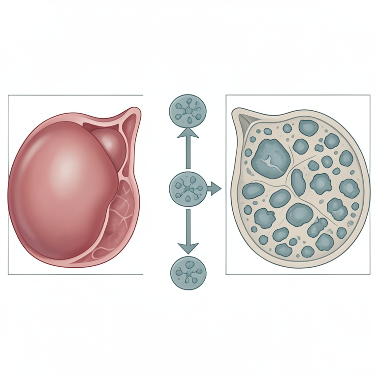
Biochemical Marker Trends in Breast Cancer
AI-generated conceptual illustration (human breast tissue). It explains the idea and does not show study data.
View source evidence ↗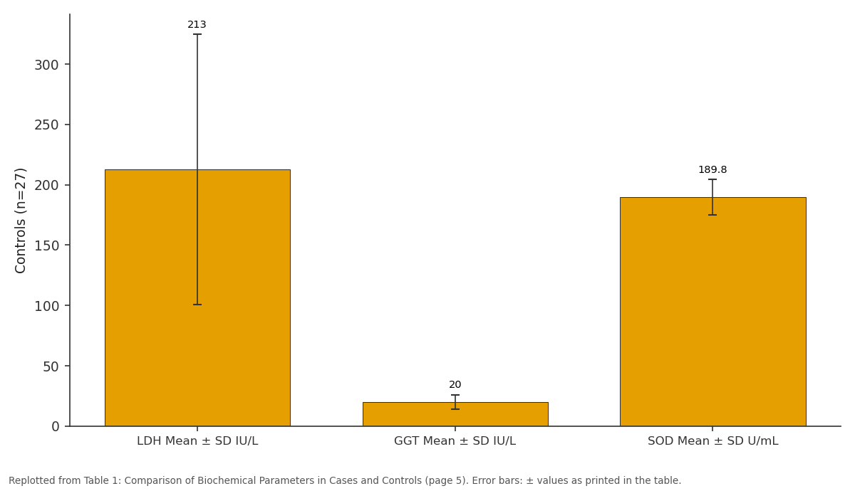
Controls (n=27): LDH Mean ± SD IU/L vs GGT Mean ± SD IU/L vs SOD Mean ± SD U/mL
Replotted from Table 1 (page 5) of the original paper. Every bar is the paper's own table value.
View source evidence ↗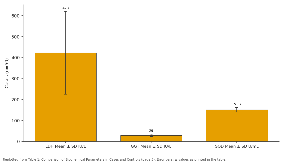
Cases (n=50): LDH Mean ± SD IU/L vs GGT Mean ± SD IU/L vs SOD Mean ± SD U/mL
Replotted from Table 1 (page 5) of the original paper. Every bar is the paper's own table value.
View source evidence ↗Figures & tables
From the original paper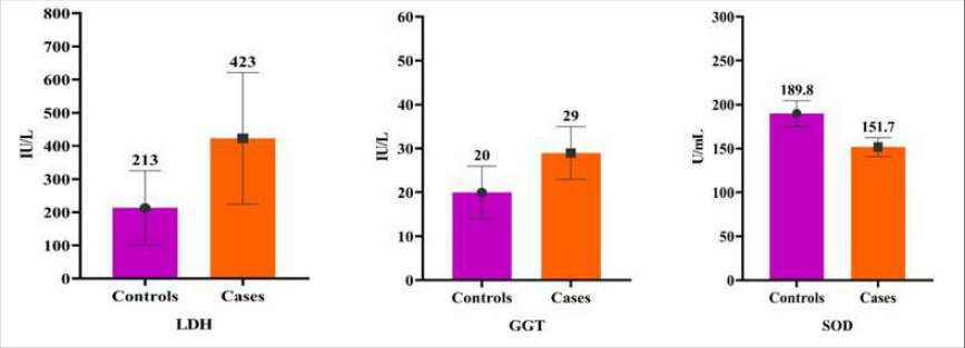 Chart 1
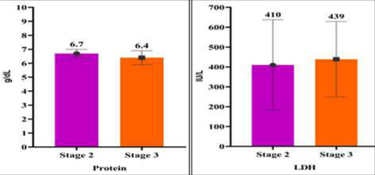 Chart 1
 Table 1
 Table 2
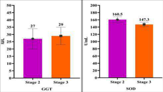 Chart 2
Introduction
Tumour markers are biochemical indicators of the presence of tumour. Various tumour markers have been studied singly or in combination, in breast malignancies. In this study, the level of three biochemical parameters, namely SOD, LDH and GGT was assessed and analyzed for possible correlation to breast cancer.
Aim of the study
On reviewing the role of reactive oxygen species in the causation of carcinogenesis and the changes in biochemical parameters in serum of patients with carcinoma breast, this study has been taken up with keen interest to establish the following aims. To determine the reference ranges for the following biochemical parameters in female individuals of age between 35 and 75-serum lactate dehydrogenase, gamma glutamyl transpeptidase, total protein, albumin, albumin-globulin ratio, and blood superoxide dismutase.
Control Group
The group comprised of 27 apparently healthy female subjects with no significant medical illness, and they were selected from the patients attending the general outpatient department of the Pudukkottai Govt Medical College.
Test Group
This group comprised of 50 females recently diagnosed with carcinoma breast through tissue diagnosis, being managed under the general surgery wards of the Pudukkottai Govt Medical College.
Inclusion criteria
Females with a confirmed diagnosis of carcinoma breast through tissue diagnosis, any stage of carcinoma, of any age group.
Exclusion Criteria
1. Individuals who have undergone any surgical intervention for the carcinoma breast other than tissue biopsy 2. Individuals who have received chemotherapy or radiotherapy for carcinoma breast. 3. Individuals diagnosed to have diabetes mellitus, liver disease, or pancreatic diseases 4. Individuals with a history of recent acute myocardial infarction.
Sample Collection
5ml of peripheral venous blood was withdrawn under sterile conditions with disposable syringes from all the 77 subjects of the study. 1.5 ml of blood was transferred into a test tube containing EDTA, for superoxide dismutase estimation. After thorough mixing, the contents were transferred into a 2 ml Eppendorf tube and labelled properly. These Eppendorf tubes were then stored at a temperature of -20 °C till the samples were analyzed. The remaining blood of 3.5 ml from each subject was transferred to another test tube without any anti-coagulant. Serum separated from this tube was pipetted into a centrifuge tube and was centrifuged at 2000 rpm for 5 minutes to obtain clear fluid without cells. The clear serum was then analyzed for the estimation of serum lactate dehydrogenase, serum gamma glutamyl trans peptidase, serum total proteins, and serum albumin. The biochemical parameters undertaken for the study were determined by semi-auto analyzer ERBA CHEM-5 plus V2 using the following methodology:
Estimation of Superoxide Dismutase in Whole Blood Principle
The role of superoxide dismutase (SOD) is to accelerate the dismutation of the toxic superoxide radical (O2•), produced during oxidative energy processes to hydrogen peroxide and molecular oxygen. This method employs xanthine and xanthine oxidase (XOD) to generate superoxide radicals which react with 2-(4iodophenyl)-3-(4-nitrophenol)-5 phenyl tetrazolium chloride (I.N.T) to form a red formazan dye. The superoxide dismutase activity is then measured by the degree of inhibition of this reaction. One unit of SOD is that which causes a 50% inhibition of the rate of reduction of I.N.T. under the conditions of the assay.
Sample Preparation
EDTA whole blood samples were used. 0.5 ml of whole blood was centrifuged for 10 minutes at 3000 rpm and then plasma discarded. The erythrocytes were washed with 3 ml of 0.9% NaCl solution and centrifuged for 10 minutes at 3000 rpm. The supernatant was discarded. This NaCl wash and centrifugation was repeated four times. The washed centrifuged erythrocytes was made up to 2 ml with cold redistilled water mixed and left to stand at 4 ℃ for 15 minutes. The lysate was diluted with 0.01 mol/L phosphate buffer pH 7 so that the percentage inhibition falls between 30% and 60%. The semi-auto analyzer was calibrated to the above mentioned system parameters. 1 ml of the reconstituted reagent and 100 µL of the sample were dispensed into a test tube, mixed and analyzed in the semi-auto analyzer immediately.
Reagent Composition
Reagent Composition
Total protein
Method: BIURET method Kit used: Bayer AUTOPAK
Principle
Peptide bonds of protein form a blue violet colored complex with cupric ions in an alkaline medium. The intensity of colour is proportional to the amount of peptide bonds and the colour is read at 540 nm (530 to 570 nm). The final colour is stable for 8 hours. Sample used: Serum.
Reagents
Reagents
Reagent reconstitution
The reagents were allowed to attain the room temperature. 41 ml of distilled water was added to one bottle of reagent 1 and then the contents of one bottle of reagent 1A were added, mixed gently to avoid foaming.
Procedure
The reconstituted reagent was brought to room temperature prior to use. In comparing the levels of the biochemical markers in Table 2 among the subjects with stage-2 and stage-3 breast cancers, only SOD levels were lowered which is significant (p-value 0.00)-mean blood level of 147.3 IU/mL in stage 3 compared to the mean blood level of 160.5IU/mL in stage-2. This could be indicative of further radical-induced inhibition of SOD activity in advanced stages of breast cancer.
| Sl. No. | Study group | Protein Mean ± SD g/dL | LDH Mean ± SD IU/L | GGT Mean ± SD IU/L | SOD Mean ± SD U/mL |
|---|---|---|---|---|---|
| 1 | Stage 2 | 6.7±0.3 | 410±228 | 27±7 | 160.5±7.9 |
| 2 | Stage 3 | 6.4±0.5 | 439±190 | 29±6 | 147.3±6.3 |
| p-value | 0.05 | 0.18 | 0.28 | 0.01 | |
| Level of significance* | Significant | Significant |
Table text extracted from the original PDF; superscript letters are shown inline. The original table image is below.
View original table image
Conclusion
The levels of biochemical markers LDH and GGT have been found to be significantly elevated. The levels of antioxidant enzyme SOD significantly lowered in cases with carcinoma breast. The SOD levels were lower in cases with stage-3 carcinoma breast than in cases with stage-2carcinomabreast. Definite association of these markers with carcinoma breast might open up the scope for use of these biochemical markers as adjuvant in breast cancer screening. The association of the levels of these biochemical parameters with the clinical course of the carcinoma breast, is yet to be established. Further, the levels of these biochemical markers in nonmalignant breast tumours need to be studied. Definite association of these markers with carcinoma breast might open up the scope for use of these biochemical markers as adjuvant in breast cancer screening. This also would warrant further studies of these biochemical markers in patients post-surgery and/or chemotherapy and/or radiotherapy, which could open up possibilities of these biochemical markers to be used in estimation of these in the diagnosis, management and follow-up of individuals at risk of and those diagnosed to have carcinoma breast which will considerably help in reducing morbidity and mortality associated with cancer breast.
Figures and tables from the paper
| — | Study group | LDH Mean ± SD IU/L | GGT Mean ± SD IU/L | SOD Mean ± SD U/mL |
|---|---|---|---|---|
| 1 | Controls (n=27) | 213±112 | 20±6 | 189.8±14.8 |
| 2 | Cases (n=50) | 423±198 | 29±6 | 151.7±10.7 |
| p-value | 0.01 | 0.01 | 0.01 | |
| Level of significance* | Significant | significant | significant |
Table text extracted from the original PDF; superscript letters are shown inline. The original table image is below.
View original table image
References
10 in the original paper- Estimates of the worldwide incidence of 25 major cancers in 1999. Int J Cancer Esteva FJ, Hortobagyi GN. Surgical clinics N. America. R.A. Eeles, 1999;80(6):827-41.
- Eeles RA. Future possibilities in the prevention of breast cancer: intervention strategies in BRCA1 and BRCA2 mutation carriers. Breast Cancer Research. 2000;2(4):1-8.
- Yang JH, et al. Cancer statistics CA Cancer J Clin. 1994;44(1):7-26.
- Duffy MJ. Effect of axillary node status on the long term survival following mastectomy for breast carcinoma. Nodal metastases may not always suggest systemic disease. J Surg Oncol. 1987;36(4):243-8.
- Andreasen PA, et al. Proteases as prognostic markers in cancer Clin Cancer Res. 1996;2(4):613-8.
- Duffy MJ, et al. The urokinase-type plasminogen activator in cancer metastasis: A review. Int J Cancer. 1997;72(1):1-22.
- Prechtl A, et al. Urokinase plasminogen activator: A prognostic marker in multiple types of cancer. J Surg Oncol. 1999;71(2):130-5.
- Risk-adapted adjuvant chemotherapy in node-negative breast cancer patients guided by prognostic factors uPA and PAI-1: First interim analysis of a randomized trial. Proc Am Assoc Cancer Res. 2001;34(5):347-352.
- Cheung K, et al. Tumour marker measurements in the diagnosis and monitoring of breast cancer. Cancer treat rev. 2000;26(2):91-102.
- Elston CW, et al. Pathological prognostic factors in breast cancer. Crit Rev Oncol Haematol. 1991;19(5):403-10.