
Research article · 2020 · Full text
International Journal of Veterinary Sciences and Animal HusbandryEstimation of normal kidney length in clinically healthy adult mongrel dogs using ultrasonography
Volume 5(1) · Pages 119–123
Research areasRenal and Vascular PathologiesVeterinary Medicine and SurgeryPediatric Urology and Nephrology Studies
Abstract
Author-writtenRenal size is an important parameter to be considered for renal disease. The normal kidney size varies depending on the breed of dogs and body conformation so the study is required to estimate the breed wise findings. Thus, the use of ratio comparing renal length and aorta diameter was investigated in thirty mongrel dogs including nineteen females and eleven males with bodyweight ranged from ten kilograms to twenty seven kilograms. The micro convex curved array probe of 4.5-8 MHz was used. In this study, the length of both kidneys displayed significant positive with aorta diameter and bodyweight. However, no significant difference was observed between length of right and left kidney. The mean kidney length: Aorta diameter was found to be ranged from-7.23. In conclusion, the ratio of kidney length to aorta diameter can be considered as a useful parameter for assessing the length of kidneys in healthy dogs.
Summary
Source-checkedPlain-language reading of the full paper, prepared with AI and checked against the text. Each point links to the authors' own words.
Why it matters
- Monitoring kidney size is a critical factor when assessing renal disease in dogs. View evidence ↗
- Because normal kidney size varies by breed and body conformation, breed-specific estimates are necessary. View evidence ↗
- Ultrasonography is a vital diagnostic tool for determining the size and shape of the kidneys. View evidence ↗
The question
- The study aimed to establish a reference interval for normal kidney length in healthy mongrel dogs and determine its relationship with body weight and aorta diameter. View evidence ↗
What they did
- Real-time images were captured using a 4.5-8 MHz micro convex curved array electronic transducer. View evidence ↗
- Maximal kidney length and aortic luminal diameter were each measured three times to ensure accuracy. View evidence ↗
- Linear correlation analysis was used to assess the relationships between aorta diameter, kidney length, and body weight. View evidence ↗
What they found
- There was a significant positive linear correlation between aorta diameter and the length of both the left (r=0.41, P=0.023) and right (r=0.41, P=0.024) kidneys. View evidence ↗
- Body weight showed a significant positive correlation with aorta diameter (r=0.65, P<0.01), left kidney length (r=0.47, P<0.01), and right kidney length (r=0.45, P=0.01). View evidence ↗
- No statistically significant difference was found between the lengths of the left and right kidneys (P=0.4469). View evidence ↗
- The mean ratio of kidney length to aorta diameter was 6.22, with a range of 5.17 to 7.23. View evidence ↗
What it means
- The authors conclude that the kidney length to aorta diameter ratio is a useful and reliable method for evaluating canine kidney size. View evidence ↗
- The sample size was too small to analyze potential differences based on sex. View evidence ↗
- Future research is needed to determine if the physical development of puppies affects these dimensional relationships, as only adult dogs were studied. View evidence ↗
Visual summary
2 visualsIllustrations explain the paper; charts are replotted from the paper's own table values.
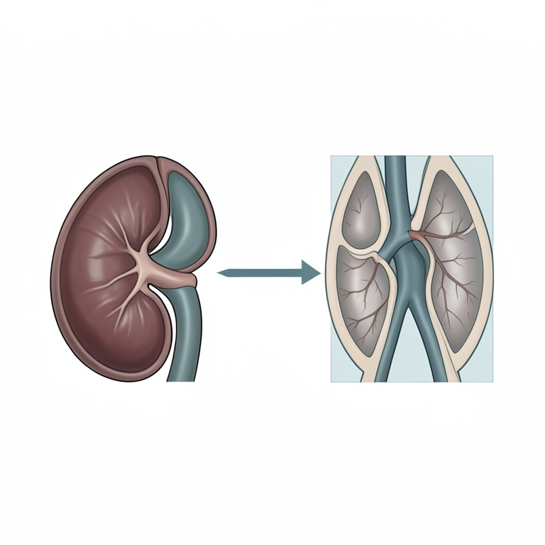
Kidney-to-Aorta Ratio Concept
AI-generated conceptual illustration (adult mongrel dog kidney and abdominal aorta). It explains the idea and does not show study data.
View source evidence ↗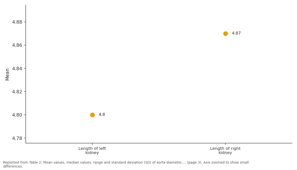
Mean: Length of left kidney vs Length of right kidney
Replotted from Table 2 (page 3) of the original paper. Every bar is the paper's own table value.
View source evidence ↗Figures & tables
From the original paper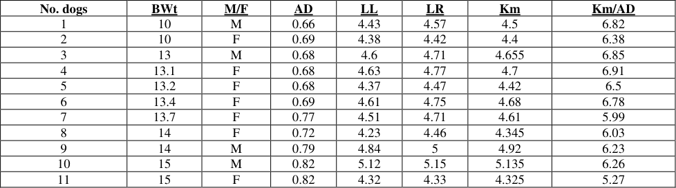 Table 1
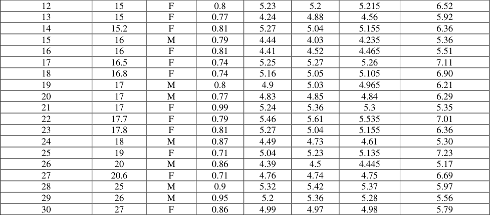 Table 2
 Table 2
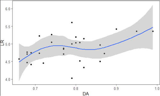 Figure 1
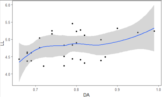 Figure 2
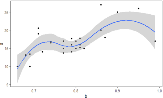 Figure 3
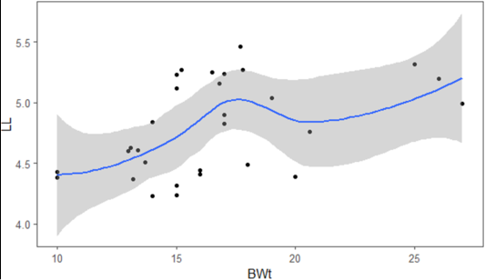 Figure 4
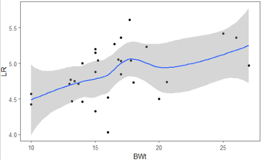 Figure 5
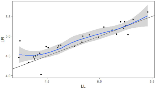 Figure 6
Introduction 1.1 Background
Ultrasound is the examination technique which is safe and painless, and real time pictures are produced of the inside body using sound waves. Small probe (transducer) and ultrasound gel are used directly in the skin through which sound wave is transmitted during ultrasound imaging. The high frequency sound waves get reflected and collected by transducer which are used by a computer to create an image. A mongrel is a mixed breed dog which does not belong to one officially recognized breed and is also not the result of intentional breeding [1] . The total number of mongrel dogs were estimated to be 150 million worldwide [2] . As kidney plays very crucial role in maintaining constant internal environment through homeostasis and in body functioning, it remains vulnerable for various diseases and disease conditions. Ultrasonography can be considered as one of the vital diagnostic tool to determine the shape and size of kidney [3] . Several canine diseases may be associated with changes in renal size. Therefore, accurate estimation of renal size can provide useful clinical information. Radiography can be used in dogs to estimate renal size by comparing renal length on ventro-dorsal projections to the length of second lumbar vertebra4. Furthermore, ultrasonography allows more consistent visualization of the kidneys and does not require the use of intravenous contrast media. Ultrasonographic renal volume measurements have been useful in humans and also in dogs. Overall kidney size in a dog can be assessed on ventro-dorsal position or required lateral recumbency. Because of the variable size of dogs a method using kidney length to aortic diameter can be used. The ratio of the length of the kidney compared to the aortic diameter at the level of the kidney (K/Ao ratio) is considered normal between 5.5 and 9.15. The kidneys should be oval to bean shaped and symmetrical in size. Smooth margins of the kidney and good corticomedullary definition are also the characteristics of healthy kidney. The renal cortex is more echogenic than medulla. The renal cortex is less echoic than the spleen and similar to the liver of dog. Linear echoic lines divide the medulla to give it a lobulated appearance. For convenient and routine use in veterinary medicine, the technique for kidney size examination must be reliable, quick and simple. Thus, the ratio obtained by comparing ratio of renal length with vertebrae in radiographs and in the same way relating renal measurements through ultrasound to some indicator of body size could assist to accomplish these conditions. Aortic diameter has been considered as a reliable landmark for ratio studies. B ultrasound is the most convenient and simplest check for kidney size measurement. What is more, it is cheap and causes no additional injuries to the kidneys, therefore B ultrasound can be done regularly to determine the kidney size, kidney problem, monitor disease progression and assess the curative effects of treatments 1.2 Objective of the study 1.2.1 General objective This study was to find out the normal kidney length and it's relation with diameter of aorta and body weight with the help of ultrasound in clinically healthy mongrel dogs to establish reference interval that could be used in practice.
Specific objectives
To find correlation between aorta diameter and length ok kidneys (right and left). To find relation between body weight of dogs with diameter of aorta and length of kidneys. To compare the size of both left and right kidney length. To calculate the reference ratio value of kidney length and diameter of aorta
Materials and Method 2.1 Time and Site of study
This study was conducted in Animal Medical Centre (AMC), Chuchhepati, Kathmandu, Nepal and some datas were also collected from Sneha's Care centre, Lalitpur, Nepal. The study was done from July 2018 to September 2018
Materials A. Ultrasound machine, Probe, Gel
Ultrasound sound machine (Chison MC6-A) with the micro convex curved array probe of 4.5 MHz-8 MHz and ultrasound gel was used for this study.
B. Animals
Mongrel Dogs visiting AMC for castration or spaying were taken as sample and data were recorded. Seventeen dogs were studied in AMC among which four were male and thirteen were females. We visited Sneha's Care centre for the additional sample where thirteen dogs were further included in the study. Out of which seven were males and six were females. Clinically healthy dogs with no any present disease condition, no any history of renal disease, with body condition Score 3/5 and having BUN and creatinine level in normal range were selected for the study.
Methods
Thirty healthy adult client owned or street mongrel dogs visiting AMC of various body weights of either sex that were scheduled to undergo castration or spaying were used as study population. All the dogs were restrained by hand and kept in the positioner after hair clipping in ventrodorsal position. Hair was clipped just behind the last rib on the left side and abdominal region including over the last two intercostal spaces on the right. The skin was prepared by cleaning the area and an enough quantity of ultrasound gel was applied. Transducer skin contact was achieved using gel. Real time ultrasonographic images were obtained using a 4.5-8 MHz micro convex curved array electronic transducer. Each kidney was evaluated with a subcostal and/ or intercostal ventrolateral approach and the maximal length of each kidney was measured thrice. To prevent slice obliquity and consequent underestimation of maximal renal length, each measure was obtained while smoothly twisting the probe. The aortic luminal diameter was measured thrice in transverse plane just caudal to the origin of the left renal artery. Measurements were recorded from still images acquired at maximum luminal diameter. Measurement cursors were placed at the margins of the lumen, excluding the vessel walls. Average of all the three measurements taken were calculated and then considered as final data to keep in MS-Excel spreadsheet for analysis.
Statistical Analysis
All the measurements were recorded in the Microsoft Excel spreadsheet 2013. Using R-Program descriptive statistics such as mean, median, range (minimum to maximum), SDs were calculated for the aorta diameter, kidney length (right and left) and kidney/aorta ratio (right and left). Relationship between aorta diameter, and kidney length, aorta diameter and bodyweight, bodyweight and kidney length were assessed using linear correlation analysis. The absolute length of the right and left kidneys were finally compared using the paired student's t-test. A probability value (P=0.05) was considered statistically significant.
Result
Thirty adult mongrel dogs included 11 males and 19 females. Body weight ranged from 10 kg to 27kg. Physical examinations and laboratory work from the dogs used in this study were normal. The rest of results obtained ultrasonographically are summarized in Table1. The mean values, median values, range and SDs for kidney length (both left and right), aorta diameter (AD), mean kidneys length (Km), and mean kidney length/aorta diameter ratio (Km/AO) are reported in Table 2. Mean kidneys length: Aorta diameter 6.22 6.27 5.17 Paired t-test revealed that there was no statistically significant difference between the length of left and right kidney (P=0.4469). The mean value± SDs of length of left kidney and length of right kidney are 4.80±0.39 and 4.87±0.37 respectively. This shows there is no such difference in mean values between kidneys length. Therefore, it was possible to consider the lengths of both kidneys right and length. The average of two measurements (mean kidney length, Km) was then analysed to identify its relationship with the aorta diameter. The reference range thus obtained was Km: AD=5.17-7.23 (SDs=±0.6).
| S.N | — | Mean | Median | Range | SD |
|---|---|---|---|---|---|
| 1. | Aorta Diameter | 0.78 | 0.79 | 0.66-0.99 | 0.08 |
| 2. | Length of left kidney | 4.80 | 4.80 | 4.23-5.46 | 0.39 |
| 3. | Length of right kidney | 4.87 | 4.87 | 4.03-5.61 | 0.37 |
| 4. | Mean kidneys length | 4.83 | 4.80 | 4.23-5.53 | 0.37 |
| 5. | Mean kidneys length: Aorta diameter | 6.22 | 6.27 | 5.17-7.23 | 0.6 |
Table text extracted from the original PDF; superscript letters are shown inline. The original table image is below.
View original table image
| 12 | 15 | F | 0.8 | 5.23 | 5.2 | 5.215 | 6.52 |
|---|---|---|---|---|---|---|---|
| 13 | 15 | F | 0.77 | 4.24 | 4.88 | 4.56 | 5.92 |
| 14 | 15.2 | F | 0.81 | 5.27 | 5.04 | 5.155 | 6.36 |
| 15 | 16 | M | 0.79 | 4.44 | 4.03 | 4.235 | 5.36 |
| 16 | 16 | F | 0.81 | 4.41 | 4.52 | 4.465 | 5.51 |
| 17 | 16.5 | F | 0.74 | 5.25 | 5.27 | 5.26 | 7.11 |
| 18 | 16.8 | F | 0.74 | 5.16 | 5.05 | 5.105 | 6.90 |
| 19 | 17 | M | 0.8 | 4.9 | 5.03 | 4.965 | 6.21 |
| 20 | 17 | M | 0.77 | 4.83 | 4.85 | 4.84 | 6.29 |
| 21 | 17 | F | 0.99 | 5.24 | 5.36 | 5.3 | 5.35 |
| 22 | 17.7 | F | 0.79 | 5.46 | 5.61 | 5.535 | 7.01 |
| 23 | 17.8 | F | 0.81 | 5.27 | 5.04 | 5.155 | 6.36 |
| 24 | 18 | M | 0.87 | 4.49 | 4.73 | 4.61 | 5.30 |
| 25 | 19 | F | 0.71 | 5.04 | 5.23 | 5.135 | 7.23 |
| 26 | 20 | M | 0.86 | 4.39 | 4.5 | 4.445 | 5.17 |
| 27 | 20.6 | F | 0.71 | 4.76 | 4.74 | 4.75 | 6.69 |
| 28 | 25 | M | 0.9 | 5.32 | 5.42 | 5.37 | 5.97 |
| 29 | 26 | M | 0.95 | 5.2 | 5.36 | 5.28 | 5.56 |
| 30 | 27 | F | 0.86 | 4.99 | 4.97 | 4.98 | 5.79 |
Table text extracted from the original PDF; superscript letters are shown inline. The original table image is below.
View original table image
| No. dogs | BWt | M/F | AD | LL | LR | Km | Km/AD |
|---|---|---|---|---|---|---|---|
| 1 | 10 | M | 0.66 | 4.43 | 4.57 | 4.5 | 6.82 |
| 2 | 10 | F | 0.69 | 4.38 | 4.42 | 4.4 | 6.38 |
| 3 | 13 | M | 0.68 | 4.6 | 4.71 | 4.655 | 6.85 |
| 4 | 13.1 | F | 0.68 | 4.63 | 4.77 | 4.7 | 6.91 |
| 5 | 13.2 | F | 0.68 | 4.37 | 4.47 | 4.42 | 6.5 |
| 6 | 13.4 | F | 0.69 | 4.61 | 4.75 | 4.68 | 6.78 |
| 7 | 13.7 | F | 0.77 | 4.51 | 4.71 | 4.61 | 5.99 |
| 8 | 14 | F | 0.72 | 4.23 | 4.46 | 4.345 | 6.03 |
| 9 | 14 | M | 0.79 | 4.84 | 5 | 4.92 | 6.23 |
| 10 | 15 | M | 0.82 | 5.12 | 5.15 | 5.135 | 6.26 |
| 11 | 15 | F | 0.82 | 4.32 | 4.33 | 4.325 | 5.27 |
Table text extracted from the original PDF; superscript letters are shown inline. The original table image is below.
View original table image
Discussion
In veterinary ultrasonography, the length of the kidney is compared with that of aorta diameter but during radiological examinations, lumbar vertebrae (L2, L3, L5, L6) are used for comparison. To identify a lumbar vertebra, the transducer is placed over lumbosacral junction and the moved cranially, maintaining contact with the skin surface. It was difficult to maintain scanning continuity all the way to L2 or L3, in part because cranial progression of the transducer was hindered by the presence of costal arch. For this reason, we decided to use the aorta diameter as our references.
During image acquisition, the most frequently encountered problems were related to visualization of the cranial pole of the right kidney which is situated more cranially than left kidney. Because of which, right kidney was not always easily and immediately identifiable. Several scans had to be made to obtain a frame with an accurate measurable image. No problem were encountered acquiring images of the aorta and left kidney. Analysis of the linear correlations between renal length and the aorta diameter revealed significant correlation (P<0.05).
In addition, body weight of healthy dogs with the good body condition score had significant relationship with aorta diameter as well as kidney length. This findings were similar with the previous literature proposed by Mareschal et [5,6,7] . The results of paired t-test showed that there is no significant difference between the lengths of the left kidney and length of right kidney measured ultrasonographically; therefore, the ratios of kidney length to aorta diameter length can be considered reliable for both the right and left kidney.
The reference range we established for the ratios is 5.1-7.3 with standard deviation 0.6. This ratio can be included in the reference ratio proposed by Mareshchal et al., which was 5.5-9.5. Furthermore, it is also near to the value obtained by Seamus et al., which was 7.4. Also Study found no any significant difference between the length of right and left kidney [5,6] . Our sample was not sufficiently large to allow analysis of eventual sex-related differences, but there are no data in the literature indicating that the dimensions of the kidneys or aorta diameter are subject to such differences. As for age, it is important to recall that in this study we examined only adult dogs. In the future, it will be interesting to see whether the rapid physical development that occurs in puppies has any influence the dimensional relationships we have identified.
Conclusion
On the basis of the data obtained in this study, calculating the ratio of kidney length to diameter of aorta appears to be a useful method for evaluating the size of the canine kidney and this can be used with reasonable confidence in dogs. In my opinion, this approach offers several advantages. First, the measurements used to calculate the ratio are sufficiently easy to obtain without complex reference and secondly, ultrasonographic images of the required structure were acquired without any particular difficulty. Therefore, it was concluded that linear kidney ultrasound measurements "in vivo" appear to be sufficiently accurate for clinical use and the reference value resulted with this study will provide more idea in relation to kidney size in different condition in Mongrel dogs
Figures and tables from the paper
References
15 in the original paper- Morris D. "Feral dogs". Dogs: The Ultimate Dictionary of over 1,000 Dog breeds, 2008, 696-697.
- Website: Quick dissolving tablets. http://www.researchgate.net/publication/276440122. 13 September, 2018.
- Sohn J, Yun S, Lee J, Chang D, Choi M, Yoon J. Reestablishment of radiographic kidney size in Miniature Schnauzer dogs. Journal of Veterinary Medical Science. 2016; 78:1805-1810.
- Mareschal A, D'anjou MA, Moreau M, Alexander K, Beauregard G. Ultrasonographic measurement of kidneyto-aorta ratio as a method of estimating renal size in dogs. Vet Radiol Ultrasound. 2007; 48(5):434-8.
- Lobacz MA, Sullivan M, Mellor D, Hammond G, Labruyère J, Dennis R. Effect of breed, age, weight and gender on radiographic renal size in the dog. Veterinary Radiology and Ultrasound. 2012; 53(4):437-441.
- Seamus EH, Brianne LH, Scott JH, Kenneth RW III. Use of computed tomography for measurement of kidneys in dogs without renal disease. Journal of the American Veterinary Medical Association. 2016; 248(3):282-287.
- Barella G, Lodi M, Sabbadin LA, Faverzani S. A new method for ultrasonographic measurement of kidney size in healthy dogs. Journal of Ultrasound. 2012; 15(3):186191.
- Barr FJ, Holt PE, GibbsC. Ultrasonographic measurements of normal renal parameters. J Small Anim. Pract. 1990; 31:180-184.
- Barrera R, Duque J, Ruiz P, Zaragoza C. Accuracy of ultrasonographic measurements of kidney dog for clinical use. Revista Cientifica. 2009; 6:576-583.
- Beregi A, Felkai C, Borzsonvi L, Molnar V. A preliminary study of the ultrasonographic determination of renal size in the guinea pig (Cavia porcellus). 2001; 40(5):50-2.
- Felkai CS, Voros K, Vrabely T, Karsai F. Ultrasonographic determination of renal volume in the dog. Veterinary Radiology Ultrasound. 1992; 33:292-296.
- Finco DR, Stiles NS, Kneller SK, Lewis RE, Barrett RB. Radiologic Estimation of kidney size of the dog. J Am Vet Medic Assoc.1971; 159(8):995-1002.
- Konde LJ, Wrigley RH, Park RD, Lebel JL. Ultrasonographic anatomy of the normal canine kidney. Vet. Radiol. 1984; 25:173-178.
- Nyland TG, Kantrwitz BM, Fisher PE, Oleander HJ, Hornof WJ. Ultrasonic determination of kidney volume in the dog. Vet. Radiol. 1989; 30:174-180.
- Tyson R, Logsdon SA, Werre SR, Daniel GB. Estimation of feline renal volume using computed tomography and ultrasound. Vet. Radiol. Ultrasound. 2013; 54:127-132.
Cited by 2
From OpenAlex, OpenCitations and reference lists read by Scinovex
- Ultrasonographic measurement of the prostatic dimension‐to‐aortic luminal diameter in healthy intact male dogs and dogs with benign prostatic hyperplasia ↗
Suppawiwat Ponglowhapan, K. Pattanaviboon, Y. Maneerattanavichien, P. Phitchayangkoon, P. Lueangprasert, A. Preechatangkit et al. · Journal of Small Animal Practice · 2024
OBJECTIVES: To establish ultrasonographic measurements of the prostatic dimension-to-aortic luminal diameter ratios including the prostatic volume-to-aortic luminal diameter ratio in healthy, intact male dogs and compare these ratios between healthy dogs and dogs affected by benign prostatic hyperplasia. MATERIALS AND METHODS: Healthy dogs (n = 75) and dogs affected by benign prostatic hyperplasia (n = 104) were included in the…
❝ 2 - Quantitative studies on B‐mode ultrasound and point shear wave elastography of kidneys in nonazotemic dogs ↗
Biswadeep Jena, JITENDER MOHINDROO, Kuldip Gupta, Navdeep Singh · Veterinary Radiology & Ultrasound · 2024
Renal diseases in dogs can be diagnosed effectively using B-mode ultrasound. Point shear wave elastography (pSWE) has demonstrated usefulness in diagnosing renal diseases in human medicine. However, its application in veterinary medicine is in its nascent stage. It was hypothesized that establishing pSWE reference values in nonazotemic dogs would prove valuable in differentiating renal diseases. In light of this, a…
❝ 1