
Research article · 2020 · Full text
International Journal of Veterinary Sciences and Animal HusbandryTransmissible spongiform encephalopathies: Emerging threats
Volume 5(6) · Pages 64–71
Research areasPrion Diseases and Protein MisfoldingNeurological diseases and metabolism
Abstract
Author-writtenTransmissible spongiform encephalopathies (TSEs) or prion diseases are a group of fatal neurodegenerative diseases caused by prions (PrPSc). The Prion proteins are devoid of any nucleic acid and have molecular weight of approximately 30 kDa. These exist in two forms: a normal cellular prion protein (PrPC) and a pathogenic misfolded conformer (PrPsc). The disease affects a variety of animal species, including human. Prions derive from a conformational conversion of the normally folded prion protein (PrPC) and acquire pathological and infectious features which is still not well understood. The infectious prion proteins cause mammalian TSEs including Scrapie (sheep), Chronic Wasting Disease (deer and elk), Bovine Spongiform Encephalopathy (BSE; cattle) and Creutzfeldt-Jakob Disease (CJD; human).The prion diseases are generally characterized by a long incubation period, usually in years, a relatively short clinical course and without exception, they cause dementia, loss of motor control, paralysis and ultimately death. The prion accesses the central nervous system (CNS) through autonomic nerves, directly after intracerebral inoculation, or via aerosols through immune-independent pathways. During the course, they first colonize and replicate in secondary lymphoid organs (SLOs). At present, a reliable diagnosis of prion disease is possible only through autopsy since all ante mortem tests are less sensitive. The commonly used diagnostic tests are ELISA, IHC and immunoblotting. A histopathological test is generally performed for confirmation. However, the final confirmation is obtained by a bioassay to assess the infectivity of the pathogen. This is the most sensitive test and infectivity in transgenic mouse to detect pathogenic PrPd. The prevention and control measures adopted for the bovine spongiform encephalopathy are surveillance, culling of sick animals, banning specified risk materials or BSE supplement. The most stringent control measures include a UK program that excludes all animals more than 30 months of age from the human food and animal feed supplies. The program appears to be highly effective. Other control measures include banning the use of mechanically recovered meat from the vertebral column of cattle, sheep and goats for human food and BSE testing of all cattle more than 30 months of age destined for human consumption. All these strategies have worked successfully in controlling the BSE outbreaks in Europe and other part of world.
Summary
Source-checkedPlain-language reading of the full paper, prepared with AI and checked against the text. Each point links to the authors' own words.
Why it matters
- Transmissible spongiform encephalopathies (TSEs), also known as prion diseases, are fatal neurodegenerative conditions caused by infectious proteins called prions. View evidence ↗
- These diseases typically feature long incubation periods lasting years, followed by a short clinical phase resulting in dementia, paralysis, and death. View evidence ↗
- Prions are formed when a normally folded cellular prion protein (PrPC) undergoes a conformational change into a pathogenic form (PrPSc). View evidence ↗
The question
- The paper aims to review the emerging threats and characteristics of various mammalian TSEs, including those affecting sheep, deer, cattle, and humans. View evidence ↗
What they did
- Diagnosis typically involves ELISA, immunohistochemistry (IHC), and immunoblotting, with histopathology and bioassays used for final confirmation. View evidence ↗
- Control strategies for BSE have included banning specified risk materials and excluding cattle over 30 months of age from food and feed supplies. View evidence ↗
What they found
- In hamster brains, the transition from normal PrPC to scrapie-infected PrP involves a shift from 42% α-helix and 3% ß-sheet to 30% α-helix and 43% ß-sheet. View evidence ↗
- Scrapie in sheep has an incubation period of 2 to 5 years, with death occurring between 2 weeks and 6 months after symptoms appear. View evidence ↗
- Bovine Spongiform Encephalopathy (BSE) has an incubation period of 2 to 8 years, most commonly affecting dairy cattle aged 4 to 5 years. View evidence ↗
- Sporadic CJD is the most common human form, accounting for 85% of CJD cases with a global incidence of 1 to 2 cases per million people annually. View evidence ↗
What it means
- The authors conclude that feed bans and the control of specified risk materials (SRM) are the most critical policies for regulating BSE risk. View evidence ↗
- The authors state there is an urgent need to develop improved diagnostic tools and treatments for prion diseases. View evidence ↗
- A significant limitation is that current clinical diagnosis typically occurs only after severe neuronal cell loss has already happened, which cannot be reversed. View evidence ↗
Visual summary
1 visualsIllustrations explain the paper; they do not show study data.
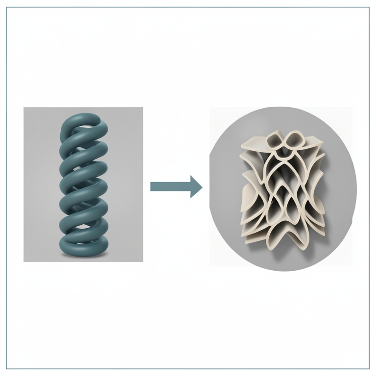
Prion Protein Conformational Change
AI-generated conceptual illustration (Prion proteins in brain tissue). It explains the idea and does not show study data.
View source evidence ↗Figures & tables
From the original paper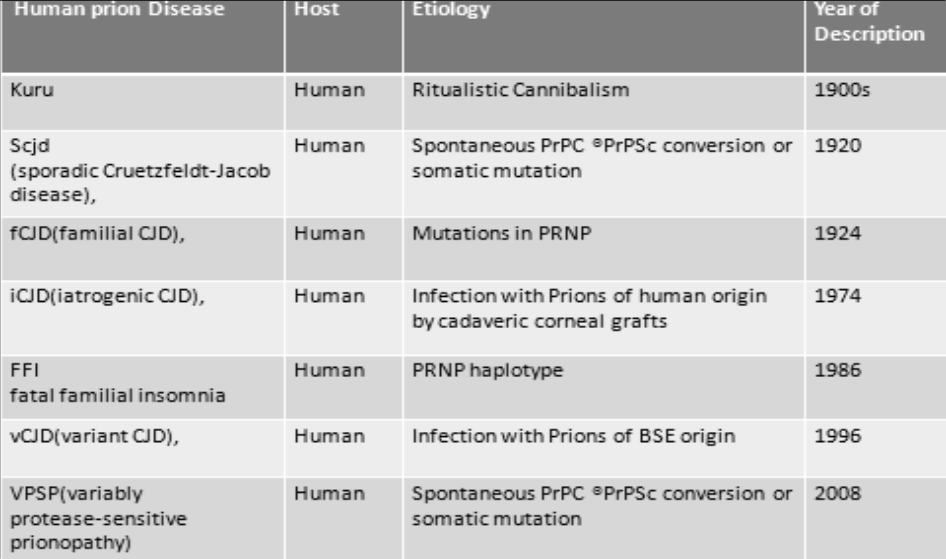 Figure 1
Introduction
The Prion Prions are unprecedented infectious pathogens that cause a group of invariably fatal neurodegenerative diseases mediated by an entirely novel mechanism. Prions are devoid of nucleic acid and seen to be composed exclusively of a modified isoform of PrP designated PrP Sc . The normal, cellular PrP, denoted PrP C , is converted into PrP Sc through a process whereby a portion of its α-helical and coil structure is refolded into ß-sheet. This structural transition is accompanied by profound change in the physicochemical properties of the PrP. (Prusiner, S.B. 1998) [28,29] . The name Prion is derived from its definition as a proteinaceous infectious particle that lacks nucleic acid The Prion was defined as an infectious pathogen that requires a protein for infectivity yet is highly resistant to procedures that modify or destroy nucleic acids. Prions differ from bacteria, viruses, and viroids by their new structure and properties. Experiments designed to uncover participation of a nucleic acid in prion structure or infectivity consistently were given negative results. Human PrP C is a glycoprotein of 253 amino acids before cellular processing. There is an 85-90% homology to prion proteins of other mammalian species. PrP C is a membrane protein expressed mainly in neurons, but also in astrocytes and a number of other cells. It has an N-terminal signal sequence of 22 amino acids, which is cleaved off the translation product. Twenty-three terminal amino acids are removed when glycosylphosphatidylinositol (GPI) is attached to serine residue 230. Mature PrP C is attached to the cell surface by this GPI anchor and undergoes endocytosis and recycling. There are two N-glycosylation sites that are glycosylated differently in different human CJD variants. The N-terminal moiety of the protein contains an octa peptide repeat, (PHGGGW_GQ)x4, which has been suggested to function in copper binding. PrP C purified from hamster brain consisted of 42% α-helical and only 3% ß-sheet structure, whereas PrP purified from scrapie-infected hamster brain is composed of 30% α-helix and 43% ß-sheet (Kretzschmar, H.A. 2002) [22] . The term Prion was proposed to distinguish the infectious pathogen from viruses or viroids (Collinge, J. 2001) [11] . No differences in amino acid sequence between PrP Sc and PrP C have been identified. PrP Sc is known to be derived from PrP C by posttranslational process (Borchelt, et al. 1990) [8] The physiologic roles attributed to PrP C are rather disparate and include: a) function as a membrane receptor; b) regulator of apoptosis; c) carrier or binding protein for copper ions; d) effectors in signal transduction mechanisms; e) regulator of synaptic transmission; and f) transcription factor. The normal function of PrP C remains unknown, although its localization on the cell surface would be consistent with roles in cell adhesion and recognition, ligand uptake, or trans membrane signaling. The normal prion protein, PrP C , is encoded by the prion protein gene (PRNP) on human chromosome 20, with equivalent prion genes in animals. The function is not known but it may have roles in anti-oxidant systems and cellular copper metabolism. In prion diseases, the normal host gene produces the normal host PrP C but there is then an incompletely understood posttranslational conformational change to a disease-related form, PrP Sc . PrP Sc is relatively insoluble and relatively protease-resistant and accumulates in tissues forming amyloid structures. The precise pathogenesis of the neurological illness is not known, but PrP Sc deposition is associated with the neurological changes of neuronal loss, astrocytic gliosis, and spongiform changes. In the acquired prion diseases, material from an affected host infects another. The infective agent (termed the "prion") has not been fully characterized, but PrP Sc is associated with infectivity (Knight, et al. 2007) [21] .
History
An increasing number of animal prion diseases are being recognized, scrapie, a naturally occurring disease of sheep and goat, has been recognized in Europe for over 200 years. Transmissible mink encephalopathy (TME) and chronic wasting disease (CWD) of mule deer and elk were described in captive animals from the 1940s. The appearance in UK cattle in 1986 of BSE, which rapidly evolved into a major epidemic, was widely attributed to transmission of sheep scrapie to cattle via contaminated feed prepared from rendered carcasses (Collinge, J. 2001) [11] . Kuru reached epidemic proportions among a defined population living in the Eastern Highlands of Papua New Guinea, onset of disease ranged from 5 to over 60 years.
Fig 1: Human prion Diseases
Fig 1: Human prion Diseases
Clinical features of prion diseases
Classical (sporadic) CJD is a rapidly progressive, multifocal dementia, usually with myoclonus. Onset usually occurs in the 45-to-75-years age group, manifested as fatigue, insomnia, depression, headaches, ill-defined pain sensation, mental deterioration and myoclonus. (Alpers, M.P. 1987) [2] . The central clinical feature is progressive ataxia; dementia is often absent. vCJD has a clinical presentation in which behavioral and psychiatric disturbances predominate, (Hill, et al.1999) [16] . GSS is an autosomal dominant disorder that present classically as a chronic cerebellar ataxia with pyramidal features, with dementia occurring later. The histologically hallmark is the presence of multi centric PrP-amyloid plaques (Giaccone et al. 1999) [15] . Although first associated with the P102L PRNP mutation (Hsiao, et al. 1989) [17] . GSS is now known as a pathological syndrome associated with several different PRNP mutations and forms a part of the phenotypic spectrum of inherited prion diseases. Sheep scrapie is the protype of the growing group of TSEs. The typical symptoms of scrapie-sick sheep include hyper excitability, pruritus, and myoclonus. The disease is characterized by rapid progression leading to tetra paresis and ultimately to the death of the affected animal. The clinical symptoms of BSE are insidious, and consist of behavioral changes (including aggressive behavior, which is proverbially atypical in cows), and uncoordinated gait. A striking hallmark applying to all TSEs is that the brain is heavily affected in sharp contrast to the body that remains unharmed. The communal lesions are neuronal loss, spongiosis, and astrogliosis, accompanied by an accumulation microglia, and occasionally, the presence of amyloid plaques and various kinds of small deposits immune labeled with anti-PrP antibodies (Aguzzi, A.1996) [1] . Clinical findings vary in the different forms of acquired CJD. In kuru and in diseases caused by inoculation of contaminated growth hormone extracts, cerebellar ataxia is the primary sign. Dementia is less prominent and usually occurs late in the disease course. The incubation time is long ranging from 2 years to greater than 30 years. Interestingly, in diseases following corneal or dural transplants, or use of contaminated neurosurgical instruments (Lasmezas, et al.1996) [23] . dementia is more prominent and the latency is shorter 1-2 years (Chesebro, B. 2003) [10] .
The Prion Diseases and Neurodegeneration
Prion diseases may present as genetic, infectious, or sporadic disorders, all of which involve modification of the prion protein (PrP), a constituent of normal mammalian cells. CJD generally present as progressive dementia, whereas scrapie of sheep and bovine spongiform encephalopathy (BSE) are generally manifest as ataxic illness (Prusiner, S.B. 1998) [28,29] . The brains of patients usually show spongiform degeneration and astrocytic gliosis under the microscope The hallmark of all prion diseases whether sporadic, dominantly inherited, or acquired by infection is that they involve the aberrant metabolism and resulting accumulation of the prion protein. A group of infectious pathogens called prions cause transmissible neurodegenerative diseases in both human and animals. In humans, these diseases are kuru, Creutzfeldt-Jakob Disease (CJD), and Gerstmann-Sträussler-Scheinker Syndrome (GSS), whereas animal prion diseases include scrapie, bovine spongiform encephalopathy (BSE), and transmissible mink encephalopathy (TME). Gerstmann-Sträussler-Scheinker Syndrome (GSS) and familial CJD are unique in that they are inherited and transmissible, these diseases occur in families as an autosomal dominant trait with high penetrance, and extracts of brain tissue from the affected individual can transmit a scrapie-like disease to experimental animals (Stahl, N. and Prusiner, A.B. 1991) [32] . The inherited prion diseases include GSS, familial Creutzfeldt-Jakob Disease (fCJD), and Fatal Familial Insomnia (FFI). These patients present with characteristic clinical and neuropathological findings as early as their third or fourth decade of life and their family histories are compatible with an autosomal dominant pattern of inheritance. Prion diseases are rapidly progressing, invariably fatal, neurodegenerative diseases associated with dementia and neurological deficits such as ataxia, visual disturbances, or myoclonus. Histologically, nerve cell loss, spongiform change, and various forms of prion protein deposits are found in the brain. They are a hetero genius group of diseases that can be acquired, hereditary, or idiopathic. All prion diseases are experimentally transmissible with a relatively long incubation time and a comparatively short clinical duration. The first possible person-to-person transmission of CJD was reported in a recipient of a corneal transplant from a donor with CJD in 1974. The clinical symptoms of BSE are insidious, and consist of behavioral changes (including aggressive behavior, which is proverbially atypical in cows), and uncoordinated gait. A striking hallmark applying to all TSEs is that the brain is heavily affected in sharp contrast to the body that remains unharmed. The communal lesions are neuronal loss, spongiosis, and astrogliosis, accompanied by an accumulation microglia, and occasionally, the presence of amyloid plaques.
Conversion and Aggregation of Prion Protein
The conversion of the PrP C into PrP Sc involves a conformation change, whereby the α-helical content diminishes and the amount of ß-sheet increases. Understanding of how PrP C unfolds and refolds into PrP Sc will be of paramount importance in transferring advances in the prion diseases to studies of other degenerative illnesses. The mechanism by which PrP Sc is formed must involve a templating process whereby existing PrP Sc directs the refolding of PrP C into a nascent PrP Sc with the same conformation. In prion diseases, the normal protein undergoes a number of posttranslational modifications to accumulate within the neutrophil of the central nervous system. This accumulation is accompanied by a change in the protein structure from a predominantly αhelix to a ß-sheet structure. The N-terminus of the protein is highly flexible and undergoes profound conformational change during the conversion to abnormal PrP (Peretz et al. 1997) [27] . The exact mechanism of conversion is poorly understood and may occur by protein dimerization or nucleated seeding. Prusiner has suggested that another protein (Protein X) might be responsible for this conversion as molecular chaperone.
Species Barriers and Strain Diversity of Prions
Transmission of prion diseases between different mammalian species is restricted by a "species barrier". On primary passage of prions from species A to species B, usually not all inoculated animals of species B develop disease. Those that do have much longer and more variable incubation periods than those that are seen with transmission of prions within the same species, where typically all inoculated animals would succumb within a relatively short and markedly consistent incubation period. On second passage of infectivity to further animals of species B, transmission parameters resemble within-species transmissions, with most, if not all, animals developing the disease with short and consistent incubation periods. Species barriers can therefore, be quantitated by measuring the fall in mean incubation period on primary and secondary passage or perhaps more rigorously, by a comparative titration study. Distinct isolates or strains of prions were first described in scrapie-diseased goats, where two dissimilar clinical manifestations ("scratching" and "drowsy") were identified. These strains differ in their incubation times in various inbred mouse lines and by their lesion pattern in the brain. Strikingly, distinct strains of prions can be propagated in an inbred mouse strain that is homozygous with respect to PRNP. A perplexing finding with regard to the protein-only hypothesis meaning that an identical polypeptide chain is able to mediate different strain phenotypes. Both the "refolding" and the "seeding" model propose that each strain is associated with a distinct conformation of PrP Sc and that each of these can convert PrP C of its host into a likeness of itself. In deed, PrP Sc species associated with two hamster-adapted scrapie strains, namely hyper (HY) and drowsy (DY), proved to display characteristic clinical and histopathological properties as well as distinct biochemical patterns with respect to proteinase K digestion (Bessen, R, and Marsh, R. 1994) [7] readily explainable by the presence of different conformations of PrP. Sc Scrapie Scrapie is the ancient form of TSEs. It is known since1732 and has occurred in sheep, goats and moufflons (Jeffrey M, González L.2007) [18] . As is the case with other prion diseases, clinico pathological phenotypes of scrapie vary according to the prion strain and animals' genetic background. Multiple prion strains may exist in a single scrapie isolate and aPrPSc conformer underrepresented in one breed may be selected as dominantly propagating strain in another breed (Thackray et. al. 2011) [35] . Clinical symptoms may include behavioral changes, blindness, ataxia, incoordination, hyperexitability and tremors. Intense pruritus is the most common symptom which usually leads to wool loss by rubbing and scraping, and results in a characteristic nibbling response from animal when the dorsum is scratched or pressure over the base of tail is applied. The incubation period of scrapie is 2-5 years and death occurs within 2weeks to 6 months after clinical onset. Neuropathological signs are spongiform vacuolization and astrogliosis and the deposition of PrPSc amyloid plaques in the central nervous system (CNS). PrPSc has been detected in the nervous system, tonsils, spleen, lymph nodes, nictitating membrane, muscles, placentas, distal ileum and proximal colon. Detection of PrPSc in the third eye lid is used as rapid diagnostic test in live animals. Excretions and secretions have also been found to contain PrPSc infectivity which may transmit between animals horizontally. About 3-5% animals per affected flock may die annually and an increase up to 20% in annual mortality rates has also been noted in some flocks (Maddison et al. 2010) [24] . The abovementioned facts are for classical or typical sheep scrapie. Atypical cases of both sheep and goat scrapie have also been described. In atypical scrapie, the major clinical symptoms are ataxia and incoordination. Pruritus is uncommon. Clinical signs of goat scrapie overlap with those of sheep scrapie and may include irritability, loss of inquisitiveness, unusual alertness, restlessness, impaired vision, hyperesthesia, incoordination, posture abnormalities, tremors, and teeth grinding, salivation, or regurgitation of rumen contents. Several amino acid polymorphisms in both the ovine and caprine PrP encoding genes (PRNP) have been reported to be associated with scrapie susceptibility. Sheep exposed to natural or experimental PrPSc infection have been shown to gain maximum scrapie resistance in the presence of Q171R polymorphism and maximum scrapie susceptibility in the presence of A136V polymorphism. A three codon system based on A136V, R154H and Q171R/H polymorphisms has been developed by using five alleles (ARQ, VRQ, AHQ, ARR and ARH). These five alleles can be combined into 15 genotypes i.e. ARR/ARR or VRQ/ARQ which, in turn, can be classified into five groups (R1-R5) according to the susceptibility they confer to the disease. The most resistant R1 genotype is ARR/ARR and the most susceptible R5 genotypes are VRQ/VRQ, VRQ/ARQ, VRQ/ARH and VRQ/AHQ. The remaining genotypes are of intermediary susceptibility. This risk classification system has helped many European states in selective breeding of sheep against scrapie. The selective breeding of sheep against scrapie caused no considerable negative impacts on production, reproduction and health of animals (Dawson et al. 2008) [12] . The presence of ARR/ARR in either ewe or her fetus would resist the PrPSc accumulation in placenta. Though ovine PRNP polymorphism has been abundantly linked to the scrapie risk, a few studies have been carried out for the analysis of caprine PRNP gene in this regard. This is due to the fact that the prevalence of typical scrapie outbreaks in goat is comparatively much lower than in sheep. However, caprine PrP polymorphismsI142M, H143R, N146S/D, R154H, R211Q and Q222Khave been shown to be associated with low scrapie risk. Atypical scrapie is also influenced by variations in PRNP and has been reported to occur in animals carrying genotypes conferring resistance to typical scrapie (Benestad et al. 2008) [5] .
Transmissible Mink Encephalopathy (TME)
TME is a rare TSE of farmed mink. Mink is a small semiaquatic wild mammal that is raised in several countries for the production of fur. TME was first recognized in Wisconsin and Minnesota in 1947. Subsequently, TME outbreaks in US have occurred in 60s and70s with the most recent outbreak in 1985. TME has also been seen in farmed mink in Canada, Finland, East Germany and the former USSR (Marsh RF, Hadlow WJ (1992) [25] . Although the origin of TME is still unknown, contaminated feed, mainly with the scrapie agent, was presumed to be the main source of infection. Mink inoculated with various strains of the scrapie agent revealed the development of TME. Oral infection of minks with the classical BSE agent also caused TME, but animals exhibited docile rather than aggressive behavior. More recently, the Ltype BSE agent has been described as the most likely candidate for being causative of TME (Sigurdson CJ, Miller MW. 2003) [31] TME is readily transmissible to raccoons by parenteral, intra cerebral and oral routes, and can intra cerebrally be transmitted to striped skunks, ferrets, American sable (pine martens), beech martens, cattle, sheep, goats, hamsters andnon-human primates such as rhesus macaque, stump tailed macaque and squirrel monkey. Non-transgenic mice are not susceptible to TME. TME passaged in cattle has also been transmitted to mink both intracerebrally and orally with incubation periods of only 4-7months. A bifurcation in clinical phenotypes with distinct incubation periods, neuropathological lesions and biochemical profiles was produced in hamsters on inoculation with TME. Depending on clinical symptoms of the disease, one strain was named as "hyper (HY)"while the other as "drowsy (DY)" (Bessen RA, Marsh RF 1992) [6] . On confection of these strains, DY shows a dominant competition for the recruitment of cellular PrPC into oligomers, and may reduce the incubation time or even block the ability of HY to cause the disease. When housed in the same cage, minks may acquire the infection through cannibalism or biting of each other. However, at least in one outbreak, kits housed with their dams did not develop the disease. Environmental exposure and vertical transmission have not been found to cause the spread of the infection. TME has been detected only in adult mink, and may cause60-90% or even 100% mortality during an outbreak [http://www.cfsph.iastate.edu]. The clinical manifestation of the disease includes behavioral changes such as increased aggressiveness and hyperesthesia, depression, restlessness, and neglect in parental care and coat grooming. The affected minks often soil the nest or scatter feces in the cage.
At the earlier disease stages, they may also exhibit difficulty in eating and swallowing. Later, symptoms such as abnormal gait, ataxia, incoordination, occasional tremors, clenching of the jaw, curved tail like those of squirrels, and compulsive biting or mutilation of objects or of the self, particularly of the tail, may be noted. Near the end of the disease course, convulsions may occur, and minks become somnolent and unresponsive, and can be seen to press their heads against the cage for hours. Incubation time in naturally occurring TME may range from 6to 12 months, and death usually occurs within 2 to 8weeks.Neuropathological features of TME include extensive spongiform degeneration in the neutrophil of the brain. Astrocytosis also occurs. Spongiform changes are intense in the cerebral cortex, particularly in the frontal cortex, as well as the corpus striatum, thalamus and hypothalamus, but are less severe in the midbrain, pons and medulla, and usually, are not evident in the cerebellum and spinal cord. In addition to CNS, PrP deposits of the TME agent, but not amyloid plaques, have been detected in spleen, intestine, the mesenteric lymphnode, thymus, kidney, liver and salivary glands of experimentally infected mink.
Chronic Wasting Disease (CWD)
CWD is a TSE of captive as well as free-ranging members of the family cervidae. From 1967 to now, CWD has been seen in 14 USA states, 2 Canadian provinces and in imported animals in South Korea. However, surveillance for CWD has been at minimum around the globe. The affected species include mule deer, white tailed deer, black-tailed deer, Rocky Mountain elk, and Shira's moose. The origin of CWD is still unknown, although intra cerebral transmission of the scrapie agent has been shown to induce the disease in elk. More than1 PrPCWD strains may exist among the affected populations (Tamgüney et al. 2009) [33] . Epidemiological and experimental data provide evidence that horizontal transmission of CWD can efficiently occur by contact with affected animals or through environmental exposure Until now, the natural transmission of CWD has not been evident in humans who have been exposed for long to the affected area and consumed venison, and also not in domestic bovids such as sheep and cattle co sharing the habitat with the affected cervids. Moreover, transgenic mice expressing either the human, ovine or bovine PrPCcoding frames did not develop the disease when inoculated with the CWD agent (Tamgüney et al. 2006) [34] . However, other cervids such as red deer and reindeer/caribou are also susceptible to CWD through intracerebral and oral routes and may be contributive to sustaining the ongoing CWD epidemic in North America. CWD is also intracerebrally transmissible to cattle, sheep, goats, ferrets, hamsters, bank voles, mink, raccoons and squirrel monkeys. The PrPCWD infectivity can be detected in the nervous system, the lymphoreticular system, the hematopoietic system, skeletal and cardiac muscles and salivary glands of naturally and/or experimentally infected animals. Virtually, no tissue in infected cervids should be considered free of the CWD agent. Susceptible animals may acquire the infectivity from their habitats during feeding on grasses or by drinking water contaminated with PrPCWD which affected cervids excrete or secrete or deposit into the environment, even in the asymptomatic carrier state, in the form of feces, urine, saliva, blood, placenta and carcasses. Acquisition of the infectivity may be enhanced further by oral abrasions and nasal exposure to PrPCWD-containing droplets and aerosols (Denkers et al. 2010) [13] . An important point is that the TSE agents can bind to soil particles, persist there for years still retaining the infectivity and transmit the disease via oral route with even more efficiency [36][37][38] . These data provide a plausible explanation for the high incidence of CWD and efficient transmission of the infectivity among cervids. Prevalence of the disease in affected herds may range from 0.1 to 50% or even 100%, sometimes. CWD has also been detected in USA in areas far from the original endemic area and raised several questions: Whether the infectivity has been transported to these areas illegally in the form of tainted materials or infected animals or by some another way, whether the scrapie agent has adapted to cause CWD by repeated natural passages into the deer, or if the PrPC conversion in cervids is proficient enough to result in the sporadic emergence of the disease, which may in turn establish the epidemic by horizontal transmission. Given that the routes of PrPCWD transmission are still uncertain, managerial decisions for the disease eradication based on the supervision of animal trade, and quarantine or mandated destruction of effected herds or flocks may appear less promising. Therefore, therapeutic intervention targeting molecular mechanisms involved in the pathogenesis of the disease may be a better substitute for the control of epidemics of CWD and scrapie. In addition, PRNP polymorphisms S96G, M132L and S225F in cervids have been associated with resistance to CWD and may, at least in the case of captive animals, be helpful in the disease management by selective breeding. n free-ranging cervids, the increased prevalence of CWD allied with the lower survival of diseased animals is thought to provide predators like mountain lions an opportunity for selective successful predation. Such as elective predation would lead to local imbalances in ecological dynamics of food webs and nutrients recycling. Chronic wasting of carcasses or weight loss, on the basis of which the disease is called "chronic wasting disease", is very common in affected cervids. CWD can also cause these animals to have a rough, dry coat, patchy retention of the winter coat in summer. In subclinical or early clinical CWD, affected cervids particularly elk, may also show some other highly subtle symptoms including lassitude, sudden death in deer after handling, a lowered head and drooping ears and behavioral changes such as having fixed gaze and loss of fear of humans. With the progression of the disease, following more perceptible symptoms may arise: flaccid hypotonic facial muscles, ataxia, head tremors, teeth grinding, repetitive walking close to the boundary of the enclosure, hyper excitability with handling, excessive salivation due to difficulty swallowing, esophageal dilationor ruminal atony, regurgitation of ruminal fluid, polyuria, polydipsia, syncope, and aspiration pneumonia. Many animals become severely emaciated before theydie. Incubation periods in CWD lie within the range of16 months to 5 years and the disease equally affects both males and females. Death usually occurs within aperiod of 1 year after the onset of clinical signs. On histopathological examination, CNS of the affected cervids shows intraneuronal vacuolation, degeneration and loss of neurons, extensive neurophil spongiosis, astrocytic hypertrophy and hyperplasia, and occasional amyloid plaques. Spongiform lesions are mainly observed within the thalamus, hypothalamus, midbrain, pons, medulla oblongata, the olfactory tubercle and cortex. The most consistent histological lesions and PrPCWD immuno histochemical staining are seen within the dorsal motor nucleus of the vagus nerve, which is considered the first site of PrPCWD accumulation. Importantly, the clinical signs of polyuria and polydipsia and the low urine specific gravity in clinically dehydrated animals may be attributed to severe lesions in the supra-optic and para ventricular nuclei, where the production of anti-diuretic hormone occurs.
Bovine Spongiform Encephalopathy (BSE)
BSE or "Mad Cow" disease" is a progressive and invariably fatal neurodegeneration in cattle. The clinical signs of BSE may include tremors, gait abnormalities particularly of hind limb (ataxia), aggressive behavior, apprehension, and hyper reactivity to stimuli. PrPSc accumulation and spongiform vacuolation are usually found in the brain (Novakofski et al. 2005) [26] . At the terminal stages of the disease, BSE prions may alsobe detected in spinal cord, retina, ileum, adrenal glands, tonsils, bone marrow, peripheral nerves, dorsal root ganglia, trigeminal ganglion and thoracic ganglia. The BSE infectivity may be observed in brain tissues as early as 2 years after post inoculation. Epidemiological and transmission studies have found no evidence of BSE prions in milk, semen or embryos and there is little or no evidence of its horizontal transmission. However, the offspring of infected animals have shown an increased risk for disease development. The incubation period for BSE is 2 to 8 years and most of BSE cases have been found in 4 to 5 years old dairy cattle. Apart from BSE strain causing classical BSE, two other strains (Htype and L-type) causing atypical BSE have been described. Most of atypical BSE cases have been detected during active surveillance targeting fallen stocks and slaughtered animals (Ducrot et al. 2008) [14] . Amyloid plaques can be detected in animals with atypicalL-form BSE, although they are not typical of classical BSE. BSE cases have occurred worldwide in nearly 0.2 millionHolstein-Freisian Bos taurus cattle, 1 Bos indicus animal, and 1 Bos taurus × Bos indicus cow (Seuberlich et al. 2006) [30] . BSE first appeared in mid 80s in UK, soon evolved to epidemic proportions with 1000 cases occurring per week in 1992, and has been shown to be naturally transmissible to a number of zoo species. BSE has also transmitted to humans in the form of a variant of Cruetzfeldt-Jacob disease (vCJD) (Bruce et al. 1997) [9] . The practice of using meat and bone meal (MBM) possibly contaminated with infectious mammalian pathogenic prions incattle feed was considered the likely cause of the BSE epidemic. A ban on the use of MBM in the ruminants feed ultimately resulted in a progressive decline of the epidemic. Natural cases of transmissions of BSE have been described in sheep and goats. Such bovid animals may be an additional source of BSE transmission to humans (Vulin et al. 2011) [36] . Indeed, transgenic mice expressing human PrPC are more susceptible to sheep-passaged BSE than classical BSE. Origins of BSE are unknown. The accidental inclusion of tissues in MBM from familial or sporadic BSE cases, sheep with scrapie or CJD patients may be responsible. Although several PRNP variants have been described for cattle only three amongst these variants, a23 bp indel in the PRNP promoter, a 12 bp indel in the first intron and an E211K polymorphism, are known to confer susceptibility to BSE. (Juling et. al. 2006) [19] .
Feline Spongiform Encephalopathy (FSE)
FSE is a transmissible spongiform encephalopathy of domestic cats and captive wild members of the family Felidae. Strain typing studies in these mice also revealed a similarity between the FSE and BSE strains, supporting the hypothesis that FSE is caused by an infection with BSE prions. (Baron et al. 2007) [3] .
Kuru
In humans the Kuru is the first prion disease that was shown to be transmissible to chimpanzees by intracerebral introduction of brain homogenates from kuru patients. (Kirkwood et al.1993) [20] . Kuru has occurred exclusively in the Fore linguistic group of Papua New Guinea Eastern Highlands and the neighboring peoples with whom they intermarried. There was a practice among these groups to consume the dead bodies of their relatives as a mark of respect and mourning (ritualistic cannibalism). Women and young children of both sexes were more exposed to the risk material such as brain and viscera than adult men who usually had preferentially to consume muscles. The kuru epidemic killed 1-2% of the population at its peak.
Sporadic Cruetzfeldt-Jacob disease
Sporadic Cruetzfeldt-Jacob disease (sCJD) accounts for 85% of all CJD cases with annual worldwide incidence of 1-2cases/million population. It occurs equally in both sexes with a peak age of onset between 55 and 75 years. Some younger (below 20 years) and oldest (above 90years) cases have also been reported. The presence of spongiform changes and sparse PrPSc distribution in CNS of sCJD patients are the hallmark of neuropathology of the disease. The deposits of amyloid plaques may also be noticed in 5-10% of cases. (Belay ED.1999) [4] . Sporadic and genetic cases of human prion diseases are occurring worldwide with an annual prevalence of 1-2 cases per million of population.
Diagnostic Methods:
At present, a reliable diagnosis of prion disease is possible only through autopsy since there is no approved method for detecting prion levels, which are too low to be detected by any test, in the peripheral nervous systems of live animals or humans. Thus, tissues of the central nervous system, including the brain and spinal cord, obtained at autopsy are used for prion diagnostic tests using immunology-based techniques such as enzyme-linked immunosorbentassay (ELISA), immunohistochemistry, and immune blotting; a histopathological test is then performed for confirmation. However, the final confirmation is obtained by performing a bioassay to assess the infectivity of the pathogen; this is the most sensitive test and uses a transgenic mouse to detect pathogenic PrPd by observing the infection. The other standard diagnosis procedures for BSE suggested by the OIE include ELISA, Western blotting, and immunohistochemical methods to test brain tissues.
Control
Control measures are surveillance, Culling sick animals, or banning specified risk materials or BSE supplement. The most stringent control measures include a UK program that excludes all animals more than 30 months of age from the human food and animal feed supplies. The program appears to be highly effective. Other control measures include banning the use of mechanically recovered meat from the vertebral column of cattle, sheep and goats for human food and BSE testing of all cattle more than 30 months of age destined for human consumption is effective.
Risk perception, communication, and management of prion diseases
The occurrence of BSE in any country affects the economy, and the meat market due to changes in policies for exportation and importation. In these situations, scientists should provide accurate information to the public. However, despite the guidelines suggested by the OIE, the fact that Canada, the USA, and the EU, which have had BSE outbreaks, have different counter measures and policies. Thus, when policies are re-established, scientific findings should be clearly communicated among interest groups through open discussion and public opinion in advance. In this process, the risk perception of the government and food industry should be treated and considered equally as the overall risk perception of the public. Therefore, policy makers must consider risk communication to come up with strategies and plans. First, SRM control and feed bans should be considered, by all countries, as the most important policies for BSE risk regulation; any relaxation of these policies should be made with extreme caution, based on solid scientific knowledge and accompanied by an effective communication strategy toward stakeholders as well as the general public. Second, surveillance systems are also important, although most countries consider testing regimens merely as tools for epidemiological monitoring of the disease. In that respect, active surveillance systems should be retained for some more time, although the current regulatory design can be modified to be more flexible when all stakeholders are in a consensus.
Conclusions
Although several hypotheses have been put forth to explain the propagation of the prion disease agent the prion only hypothesis has gained a much wider popularity because of consistent failure to identify a specific "foreign "nucleic acid in prion diseases despite extensive search. Prion diseases are characterized by deposition of PrPSc, a misfolded and aggregated isoform of the host-encoded cellular prion protein (PrPC), within the central nervous system (CNS). The prion have the unique property of being inheritable, transmissible and infectious in nature. Imposing the import restrictions on live ruminants and ruminant products from countries known to have BSE endemic resulted in control of this disease to other parts of world. Strategic policies for monitoring and surveillance of disease should be implemented. There is instant need to develop better diagnostic and treatment for the disease.
Future prospective
Effective treatment of neurodegenerative disease is one of the major challenges facing biomedical research. Molecular classification of prion disease, by both genetic typing of host and strain typing of infecting prion, is evolving and will allow more precise epidemiological studies. New methods for early diagnosis, and their timely use, will be crucial, as such methods will not reverse neuronal cell loss which inappreciable or severe by the time clinical diagnosis is typically reached.
Figures and tables from the paper
References
36 in the original paper- Aguzzi A. Between cows and monkeys. Nature 1996;381:734-735.
- Alpers MP. Epidemiology and clinical aspects of kuru. Cited in: Will, R.G. Acquired prion disease: iatrogenic CJD, variant CJD, kuru. British Medical Bulletin 2003;66:255-265.
- Baron T, Bencsik A, Biacabe AG, Morignat E, Bessen RA. Phenotypic similarity of transmissible mink encephalopathy in cattle and L-type bovine spongiform encephalopathy in a mouse model. Emerg Infect Dis 2007;13:1887-1894.
- Belay ED. Transmissible spongiform encephalopathies in humans. AnnuRev Microbiol 1999;53:283-314.
- Benestad SL, Arsac JN, Goldmann W, Nöremark M. Atypical/Nor98 scrapie properties of the agent, genetics, and epidemiology. Vet Res 2008;39:19.
- Bessen RA, Marsh RF. Identification of two biologically distinct strains of transmissible mink encephalopathy in hamsters. J Gen. Virol 1992;73:329-234.
- Bessen RA, Marsh RF. Distinct PrP Properties Suggest the Molecular Basis of Strain Variation in Transmissible Mink Encephalopathy. Journal of Virology 1994;68(12):7859-7868.
- Borchelt DR, Scottt M, Taraboulos A, Stahl N, Prusiner SB. Scrapie and Cellular Prion Proteins Differ in Their Kinetics of Synthesis and Topology in Cultured Cells. The Journal of Cell Biology 1990;110:743-752.
- Bruce ME, Will RG, Ironside JW, McConnell I, Drummond D, Suttie A, et al. Transmissions to mice indicate that 'new variant' CJD is caused by the BSE agent. Nature 1997;389:498-501.
- Chesebro B. Introduction to the transmissible spongiform encephalopathies or prion diseases. British Medical Bulletin 2003;66:1-20
- Collinge J. Prion Diseases of Human and Animals: Their Causes and Molecular Basis. Annual Review of Neuroscience 2001;24:519-550
- Dawson M, Moore RC, Bishop SC. Progress and limits of PrP geneselection policy. Vet Res 2008;39:25.
- Denkers ND, Seelig DM, Telling GC, Hoover EA. Aerosol and nasal transmission of chronic wasting disease in cervidized mice. J Gen Virol 2010;91:16511658.
- Ducrot C, Arnold M, Koeijer A, Heim D, Calavas D. Review on the epidemiology and dynamics of BSE epidemics. Vet Res 2008;39:15.
- Giaccone G, Tagliavini F, Verga L, Frangione B, Farlow MR, Bugiani O et al. Neurofibrillary tangles of the Indiana Kindred of Gerstmann-Sträussler-Scheinker disease share antigenic determinants with those of Azheimer disease. Brain Research 1999;530:325-329
- Hill AF, Butterworth RJ, Joiner S, Jackson G, Rossor MN, Thomas DJ et al. Investigation of variant Creutzfeldt-Jakob disease and other human prion diseases with tonsil biopsy samples. The Lancet 1999;353:183189
- Hsiao K, Baker HF, Crow TJ, Poulter M, Owen F, Terwilliger JP et al. Linkage of a prion protein missense variant to Gerstmann-Sträussler Syndrome. Nature 1989;338:342-345.
- Jeffrey M, González L.). Classical sheep transmissible spongiform encephalopathies: pathogenesis, pathological phenotypes and clinicaldisease. Neuropathol Appl Neurobiol 2007;33:373-394.
- Juling K, Schwarzenbacher H, Williams JL, Fries R. A major genetic component of BSE susceptibility. BMC Biology 2006;4:33.
- Kirkwood JK, Cunningham AA, Wells GA, Wilesmith JW, Barnett JE. Spongiform encephalopathy in a herd of greater kudu (Tragelaphusstrepsiceros): epidemiological observations. Vet Rec 1993;133:360-364.
- Knight RS, Will RG. Prion diseases. Journal of Neurology, Neurosurgery and psychiatry 2007;75:36-42.
- Kretzschmar HA. Prion Diseases. Encyclopedia of the Human Brain 2002;5:47-60.
- Lasmezas CI, Deslys JP, DEmaimay R, Adjou KT, Lamoury F, Dormont D et al. BSE transmission to macaques. Nature 1996;381:743-744
- Maddison BC, Rees HC, Baker CA, Taema M, Bellworthy SJ, Thorne L et al. Prions are secreted into the oral cavity in sheep with preclinical scrapie. J. Infect Dis 2010;201:1672-1676.
- Marsh RF, Hadlow WJ. Transmissible mink encephalopathy. Rev Sci Tech 1992;11:539-550.
- Novakofski J, Brewer MS, Mateus-Pinilla N, Killefer J, McCusker RH. Prion biology relevant to bovine spongiform encephalopathy. J Anim Sci 2005;83:14551476.
- Peretz D, Williamson RA, Matsunaga Y, Serban H, Pinilla C, Bastidas RB et al. A Conformational Transition at the N Terminus of the Prion Protein Features in Formation of the Scrapie Isoform. Journal of Molecular Biology 1997;273:614-622.
- Prusiner SB. Prions. Proceeding of the National Academy of Science USA 1998;95:13363-13383.
- Prusiner SB. The Prion Diseases. Brain Pathology 8, 499-513 Science USA. 1998;95:13363-13383
- Seuberlich T, Botteron C, Wenker C, Café-Marçal V, Oevermann A, Haase B, et al. Spongiform encephalopathy in aminiature zebu. Emerg Infect Dis 2006;12:1950-1953.
- Sigurdson CJ, Miller MW. Other animal prion diseases. Brit Med Bull 2003;66:199-212
- Stahl N, Prusiner AB. Prions and Prion Proteins. The Federation of American Societies for Experimental Biology Journal 1991;5:2799-2807
- Tamgüney G, Giles K, Bouzamondo-Bernstein E, Bosque PJ, Miller MW, Safar J, et al. Transmission of elk and deer prions totransgenic mice. J Virol 2006;80:91049114.
- Tamgüney G, Miller MW, Giles K, Lemus A, Glidden DV, DeArmond SJ et al. Transmission of scrapie and sheep-passaged bovinespongi form encephalopathy prions to transgenic mice expressing elkprion protein. J Gen. Virol 2009;90:1035-1047.
- Thackray AM, Hopkins L, Lockey R. Spiropoulos J, Bujdoso R: Emergence of multiple prion strains from single isolates of ovine scrapie. J Gen. Virol 2011;92:1482-1491.
- Vulin J, Biacabe AG, Cazeau G, Calavas D, Baron T2011): Molecular typing of protease-resistant prion protein in transmissible spongiform encephalopathies of small ruminants, France, 2002-2009. Emerg Infect Dis 2011;17:55-63.
Cited by 1
From OpenAlex, OpenCitations and reference lists read by Scinovex
- QUAIS DOENÇAS NO CÉREBRO LEVAM A DEMÊNCIA? ↗
Fabiano de Abreu Agrela Rodrigues · COGNITIONIS Scientific Journal · 2022
As doenças neurodegenativas do sistema nervoso são um grupo de doenças incuráveis e de lenta progressão que possuem entre suas características principais a morte neuronal ocasionando a demência. Desenvolvido por meio da revisão de literatura, o presente estudo apresenta as principais doenças neurodegenerativas, suas causas, diagnósticos e tratamentos. Neste artigo pretendo listar algumas das principais e de que forma atuam…
❝ 0PDF