
Research article · 2020 · Full text
International Journal of Veterinary Sciences and Animal HusbandrySurgical management of dystocia in a Rottweiler
Volume 5(6) · Pages 14–15
Research areasVeterinary Medicine and SurgeryCardiovascular Conditions and TreatmentsBirth, Development, and Health
Abstract
Author-writtenIn the present study, a Rottweiler aged 3yrs and weighing 25 kilograms was presented with dystocia due to primary uterine inertia and was treated by caesarean section to remove one live pup and the patient went on to make an uneventful recovery. Dystocia is a common small animal emergency. Bitches that have been in prolonged labour, those with a uterus containing dead or decaying foetus or a friable uterus or those showing signs of endotoxaemia or septicaemia are advised for caesarean section. Caesarean section should be performed before the dystotic bitch fatigues or distress occurs to the pups. For caesarean sections, safe anaesthetic protocols that exert minimal side effects on foetus or pups should be selected by the clinician.
Summary
Source-checkedPlain-language reading of the full paper, prepared with AI and checked against the text. Each point links to the authors' own words.
Why it matters
- Dystocia, defined as a difficult birth or the inability to expel a fetus without help, is a frequent emergency in dogs. View evidence ↗
- Caesarean sections are recommended for dogs experiencing prolonged labor, those with decaying fetuses, a friable uterus, or signs of systemic infection like septicaemia. View evidence ↗
- It is critical to perform a caesarean section before the mother becomes fatigued or the puppies experience distress. View evidence ↗
The question
- The study describes the surgical management of a 3-year-old, 25 kg Rottweiler suffering from dystocia caused by primary uterine inertia. View evidence ↗
What they did
- The patient presented with severe straining for 5 hours, delivering 3 dead puppies, followed by another 2 hours of straining without further delivery. View evidence ↗
- Clinical examination of the 25 kg dog revealed a rectal temperature of 101.20 F. View evidence ↗
- Following an oxytocin injection, 2 puppies were delivered; a subsequent ultrasound detected a fetal heartbeat, leading to the decision to perform a caesarean section. View evidence ↗
- The surgical approach involved a 5-inch linear skin incision in the middle upper flank using a No. 10 BP blade. View evidence ↗
- The surgeon made an incision in the uterine body to remove a fetus located in the left horn. View evidence ↗
What they found
- The caesarean section successfully removed one live puppy, and the Rottweiler made an uneventful recovery. View evidence ↗
- The cause of dystocia in this specific case was determined to be of maternal origin. View evidence ↗
What it means
- The authors conclude that clinicians should select safe anesthetic protocols that minimize side effects on the fetuses or puppies during caesarean sections. View evidence ↗
Visual summary
1 visualsIllustrations explain the paper; they do not show study data.
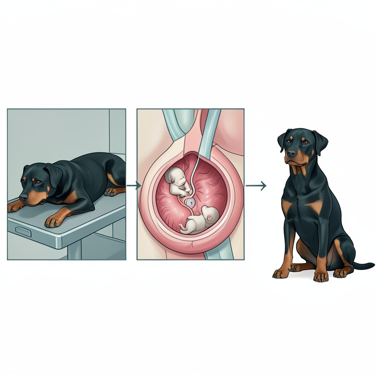
Surgical Intervention Workflow
AI-generated conceptual illustration (Rottweiler dog). It explains the idea and does not show study data.
View source evidence ↗Figures & tables
From the original paper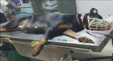 Figure 1A
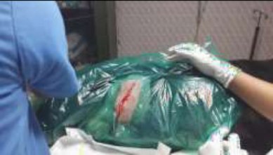 Figure 1B
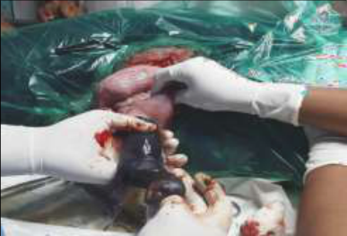 Figure 1C
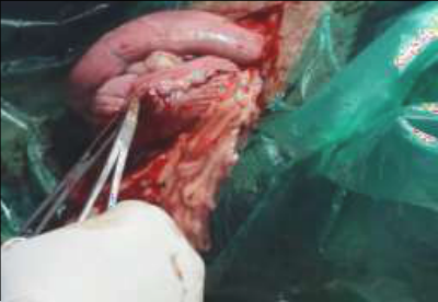 Figure 1D
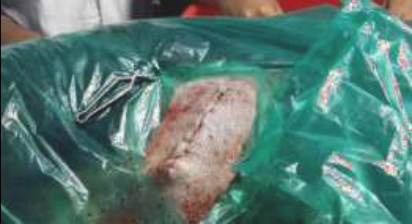 Figure 1E
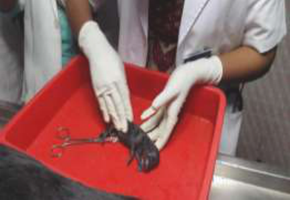 Figure 1F
Introduction
Dystocia refers to difficult birth or inability to expel the fetus through the birth canal without assistance. Dystocia is a common emergency in canine patients. A caesarean section is major surgery performed to remove puppies from uterus (Arthur, 1986) [1] . A 3 year old Rottweiler dog was presented with hanging water bag and greenish discharge from vulva. Animal exhibited severe straining for last 5 hours, and delivered 3 dead puppies. Animal continued to show straining for more than 2 hours without delivery. On clinical examination animal revealed a rectal temperature of 101.2 0 F. Body weight of the animal was 25 kg. On per vaginal examination bony prominence could be detected at the pelvic brim. Administered Oxytocin injection and animal delivered 2 puppies. No further straining observed and on ultrasonography foetal heart beat could be detected and was decided to perform caesarean section.
Surgical management and medicinal care
Animal was dull and dehydrated, stabilized by giving 200ml of D10 i/v. Inj Ceftiofur 1.1 ml s/c, Inj Pantoprazole 6ml i/v. Animal was premedicated with Inj Atropine (.06%) 1.8 ml i/m. and Inj Xylazine 1 ml i/m. Induction was done by using Ketamine (5%) and Inj Diazepam (.5%) as 4 ml i/v. Anesthesia was maintained by using Propofol. Animal was positioned on lateral recumbency (fig1a.) Draped the site and secured it using towel clamp. Made a 5 inch long linear skin incision in middle upper flank (fig1b) using No:10 BP blade loaded on BP handle no:3. Extended the incision using double blunt Mayo scissors. Bluntly separated the subcutaneous tissue using Mayo blunt scissors. incised obliques abdominis externus, obliques abdominis internus, transverse abdominis muscles using BP blade no:10 and entered the abdominal cavity. Identified and exteriorised the gravid uterus and packed the abdomen with sterile mops. An incision was made on the body of uterus and milked out the foetus present in left horn (fig1c.). Removed the foetal membranes and closed the uterine incision using vicryl (1-0) in Cushing followed by Cushing pattern(fig1d). Muscles were apposed by continuous interlock suture using vicryl (1-0). Subcutis was sutured with vicryl (1-0) in subcuticular pattern. Skin was apposed with Intradermal sutures(fig1e) using vicryl (1-0). Dressed the site using tincture benzoin and applied abdominal bandage.
Post-operative care and management
Animal was kept lying with its head and neck extended until recovery from anaesthesia.
Puppies were fed with colostrum .Given Inj Ceftiofur 1.1 ml s/c, Meloxicam 1 ml i/m. Continued wound dressing using Neosporin powder and antibiotic injection for 5 days. Advised bland diet for 3 days.
Discussion
In the present case under study, animal showed restlessness, refusal of food, vomiting and gazing at flank as symptoms of difficulty whelping agreeing with the observations stated by Freak (1975) [2] . As described by Concanon (1986) [3] two days prior to parturition the bitch became restless, it showed reduced food intake, during twelve to twenty four hours prior to parturition there was an increased restlessness, panting, scratching, chewing and nesting behavior as reported by the owner. Linde and Entroth (2000) [4] observed a decline in rectal temperature below 99.7°F indicated that parturition will take place within next twelve to twenty four hours. In the present case, the rectal temperature of animal was 101.2°F. Roberts (1986) [5] pointed out that brachycephalic breed with broad heads are more prone to dystocia. The presented animal was a Rottweiler, which is a brachycephalic breed. Krzyzanowski et al, (1975) [6] opinioned that the interval from day of mating to day of parturition may vary from 58 to 72 days in normal bitches of all breeds. In this case animal showed signs of parturition on 60th day of mating. Cause of dystocia may be maternal or foetal. Walett and Forsberg (1994) [7] in a study found that 75% of the causes of canine dystocia is of maternal origin. In the presented case also the dystocia was of maternal origin.
Figures and tables from the paper
References
7 in the original paper- Arthur GH, England GCW, Noakes DE, Parkinson TJ. Veterinary Reproduction and Obstetrics (8 th ed). Harcourt Health sciences company, Pennysilvania 2001.
- Freak MJ. Practiotioner's -breaders approach to canine parturition. Vet. Rec 1975;96:303-308.
- Concanon. Reproduction in Domestic animals (4 th ed).Academic press Ltd. Oval road, London 1986.
- Linde-Forsberg C, Eneroth A. Abnormalities in pregnancy, parturition and the periparturient period In: Ettinger, S. J. and Feldman, E. C. (ed.), Text book of Veterinary Internal Medicine (5 th Ed.). Sounders, Philadelphia 2000, 1527-1539
- Roberts SJ. Veterinary Obstetrics and Genital Diseases (2 nd ed). CBS Publishers & Distributors, New Delhi, 1986.
- Krzyzanowski J, Malinowski E, Studnicki W. Examinations on the period of pregnancy in dogs of some breeds. Med. Wet 1975;31:373-374.
- Linde-Forseberg C. Abnormalities in pregnancy, Parturition and the periparturient period. In: Ettinger, S.J. and Feldman. E. C. (ed)., Text Book of Veterinary Internal Medicine (6 th Ed.). Saunders, Philadelphia 2005, 1655.