
Research article · 2021 · Full text
International Journal of Veterinary Sciences and Animal HusbandryRising antimicrobial resistance pattern among the bacteria isolated from wound cases of domesticated animal in India
Volume 6(2) · Pages 28–30 · DOI: 10.22271/veterinary.2021.v6.i2a.332 ↗
Research areasMicrobial infections and disease research
Abstract
Author-writtenAntimicrobial resistance gained by the microorganisms is a global threat, which leads to economic loss as there will be treatment failure, loss of life, discovery of newer and better antimicrobials of any infections as there is need for discovery of newer and better antimicrobials. A total of forty five samples were collected from clinical wound cases in and around Parbhani Veterinary Hospitals. The total of 39 Gram negative and 17 Gram positive isolates includes Pseudomonas (24), E. coli (15) and Staphylococcus (17) spp, were confirmed by biochemical characterization. Susceptibility of isolates to seven antibiotics was tested using disc diffusion method according to the guidelines defined by Clinical Laboratory Standard Institute. Pseudomonas Isolates showing resistance to antibiotic for Ceftazidime, Levofloxacin were for E.coli isolates showing resistance to antibiotics Erythromycin, Tetracycline and For Staphylococcus isolates Penicillin and Enrofloxacin were resistance.
Summary
Source-checkedPlain-language reading of the full paper, prepared with AI and checked against the text. Each point links to the authors' own words.
Why it matters
- Antimicrobial resistance is a global threat that causes economic losses, treatment failures, and loss of life. View evidence ↗
- Wounds are common and difficult to manage in both humans and animals, particularly when they are non-healing or infected with multidrug-resistant bacteria. View evidence ↗
- Managing wound infections is increasingly difficult due to high bacterial resistance to antibiotics and the frequent occurrence of polymicrobial infections. View evidence ↗
The question
- The study aimed to investigate the patterns of rising antimicrobial resistance in bacteria isolated from wound cases in domesticated animals in India. View evidence ↗
What they did
- Sample collection took place between March 2018 and April 2019. View evidence ↗
- The 45 samples were obtained from 14 dogs, 11 cattle, 8 buffalo, 7 goats, and 5 horses. View evidence ↗
- The susceptibility of the isolates to seven different antibiotics was tested using the agar disc diffusion method following Clinical Laboratory Standard Institute guidelines. View evidence ↗
What they found
- Mixed infections involving two bacteria were more common (30 cases) than monomicrobial infections (15 cases). View evidence ↗
- Gram-negative bacteria were isolated more frequently than Gram-positive bacteria (52.8%), with Pseudomonas aeruginosa being the most common isolate at 37.3%. View evidence ↗
- For P. aeruginosa, Gentamicin showed 100% susceptibility, while Levofloxacin showed the lowest susceptibility at 28%. View evidence ↗
- E. coli isolates were 100% sensitive to Gentamicin, but showed low sensitivity to Tetracycline (28%) and Norfloxacin (20%). View evidence ↗
- Staphylococcus isolates were 100% sensitive to Ciprofloxacin, but highly resistant to Penicillin (5% sensitivity) and Enrofloxacin (10% sensitivity). View evidence ↗
- Gentamicin was found to be effective (sensitive) against all three major isolated organisms: Pseudomonas, Staphylococcus, and E. coli. View evidence ↗
What it means
- The authors conclude that there is an increase in the level of multidrug resistance to antibiotics commonly used in veterinary hospitals. View evidence ↗
- The study highlights high resistance rates: Pseudomonas to Levofloxacin (90%), Staphylococcus to Penicillin (95%), and E. coli to Erythromycin (88%). View evidence ↗
Visual summary
1 visualsIllustrations explain the paper; they do not show study data.
Bacterial Distribution and Resistance
AI-generated conceptual illustration (Bacteria isolated from domesticated animal wounds (Pseudomonas aeruginosa, Staphylococcus aureus, and Escherichia coli)). It explains the idea and does not show study data.
View source evidence ↗Figures & tables
From the original paper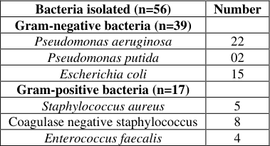 Table 1
 Table 2
Introduction
Wound is defined as the loss or breaking of cell and anatomic or functional continuity of living tissues which might not handiest be prompted by using the external factors, but also as a hardship of a few disorder processes and different internal factors (Ayello, 2005) [2] . Wounds are common and may be challenging to manipulate in each human and animals, specifically non-recuperation wounds and wounds with secondary infection by using multidrug resistant bacteria. (Atiyeh et al., 2009) [1] . Wound therapies have evolved significantly over the past three decades and provided more effective and easily healing methods such as dressings, wound bed preparations, quaterization and ligation. Technology and product proliferation, however, varied by geography, usually as a function of economics and health care policy. A number of research conducted recently discovered that for septic wounds, the most common isolates were Staphylococcus aureus, Pseudomonas aeruginosa, Escherichia coli, Klebsiella spp. and Acinetobacter spp. The management of wound infections has grown to be difficult because of the great bacterial resistance to antibiotics and extra incidence of infections because of poly-microbial plant life (Pallavali et al., 2017) [8] . The use of antibiotics in the treatment of bacterial infections increasingly encounters difficulties caused by the emergence and rapid spread of pathogenic bacteria exhibiting multidrug resistance.
Materials and Method 2.1 Sample collection
Forty five wound swab and pus samples were collected in sterile vials for collection of samples from various injured animals. The samples were marked with position, date of collection and age of the animal. The sampling area includes local dairies, animal farms and state veterinary clinics. The samples were transported on ice to the laboratory and stored at 4 °C until processing. The samples were collected from March 2018 to April 2019.
Bacterial examination and confirmation
The cultural characters of the typical colonies were studied. The smears were crafted from various colonies and stained by means of Gram's staining method. Hanging drop methods were used to determine the motility of organisms. Primarily, organisms were recognized by their morphological, cultural and staining characters and confirmation was made by method explained by Cruickshank et al., (1975) [4] 2.3 Antimicrobial susceptibility test 2.3.1 Agar diffusion method All the bacterial isolates were subjected to antibiotic sensitivity test using standard antibiotic discs by the method described by Bauer et al. (1966) [3] . The zone of inhibition (diameter in mm) of each antibiotic disc was then correlated with sensitivity as per standard chart supplied along with disc and results recorded in terms of either sensitive or resistant bacteria to the respective antibiotics.
Results
In the present study a total of 45 infected wound cases in which 11-cattle,7-goat, 8-Buffalo, 5-Horse, 14-Dog samples were included in this study. Monomicrobial infection was present in 15cases, whereas mixed infection by two bacteria was observed in 30 cases. Gram negative bacteria were isolated frequently [30/45 (52.8%)] than gram positive bacteria. The most common Gram-negative isolates were Pseudomonas aeruginosa (24). Dhawan et al., (2006) [5] reported similar findings though it was 5.63% isolated from wound cases were Escherichia coli (15) and Pseudomonas putida (02). Among Gram-positive bacteria that is Coagulase positive Staphylococcus aureus (5) and Coagulase negative staphylococcus (8) and Enterococcus faecalis (4) were observed (Table 1). Acharya et al., (2008) in their studies conducted in Nepal reported Gram positive cocci constituted 57.06% and Gram negative bacteria constituted 42.94% isolates. In this study the bacterial pathogens of wound were isolated and it was found that, among major isolates Pseudomonas aeruginosa was the most common isolate (37.3%). This finding is in contrast with the findings of Tiwari et al., (2016) [9] who reported Staphylococcus aureus as major bacteria isolated from the wound cases and The bacteria which were identified are presented in Table 2.
| Sr. No. | Pathogen | Media used | Colony characteristics | Gram stain and morphology |
|---|---|---|---|---|
| 1. | Pseudomonas aeruginosa | Cetrimide agar | Yellow green, fluorescent | Negative (Rod) |
| 2. | Escherichia coli | EMB agar | Metallic sheen coloured | Negative (Rod) |
| 3. | Enterococcus faecalis | Nutrient agar | Smooth, cream/white | Positive (Cocci) |
| 4. | Staphylococcus aureus | Mannitol salt agar | Smooth, Golden yellow | Positive (Cocci) |
Table text extracted from the original PDF; superscript letters are shown inline. The original table image is below.
View original table image
| Bacteria isolated (n=56) | Number |
|---|---|
| Gram-negative bacteria (n=39) | |
| Pseudomonas aeruginosa | 22 |
| Pseudomonas putida | 02 |
| Escherichia coli | 15 |
| Gram-positive bacteria (n=17) | |
| Staphylococcus aureus | 5 |
| Coagulase negative staphylococcus | 8 |
| Enterococcus faecalis | 4 |
Table text extracted from the original PDF; superscript letters are shown inline. The original table image is below.
View original table image
Antimicrobial susceptibility of Pseudomonas spp, E. coli and Staphylococcus spp.
Antimicrobial susceptibility test was performed on 45 bacterial spp. by agar disc diffusion method and as many as 7 antibiotics for Pseudomonas, 7 antibiotics for E.coli and 7 antibiotics for Staphylococcus spp. were used for studying the resistance and sensitivity pattern of Bacterial species. Among the P. aeruginosa isolated, all the antibacterial antibiotics were most lively with 100% susceptibility to Gentamicin followed by Ciprofloxacin (80%), Ceftazidime (28.5), Meropenem (54.7%), Imipenem (38%), Levofloxacin (28%) and Amoxicillin (57.7%). For E. coli isolates were 100% sensitive to Gentamicin, followed by Chloramphenicol (95%), Ceftriaxone (85%), Norfloxocin (20%), Erythromycin (37%), Ciprofloxacin (57%), and Tetracycline (28%). and for Staphylococcus isolates were 100% sensitive for ciprofloxacin followed by Chloramphenicol (87%), Gentamicin (67%), Amoxicillin (37%), Penicillin (5%), Enrofloxacin (10%), Ampicillin (28%).
Discussion
In the present study the wound infection is the most dynamic that retards the normal wound healing. Wounds are caused by various reasons such as trauma, injury during surgery etc. A wound is either simple with superficial injury or complicated with involvement of deeper structures e.g., muscle, tendons, vessels, bones etc. Infected wounds illustrate deferred epithelial growth and migration, superior cellular necrosis and lysis of collagen tissue. Bacteria are proficient of producing collagenase enzyme which degrades collagen tissue synthesized by fibroblast to decrease the tensile strength, and ultimately to case wound disruption. Wounds are congregation by a polymicrobial flora comprising synergistic bacteria, such as Pseudomonas, Staphylococci and Escherichia coli spp bacteria. The injudicious uses of antibiotics throw in to the development of super infection and resistant microbial species. When a wound is treated with antimicrobial drug, a selective pressure is applied to all bacteria exposed to the drug. Pallavali et al., (2017) [8] who demonstrated for Pseudomonas spp a relatively higher sensitive to gentamicin, Cefotaxime and the antibiotic resistance pattern demonstrated to ampicillin (80.7%), amoxicillin (88.4%). The sensitivity patterns exhibited by E. coli had been highly decrease than the ones in previous reports, which demonstrated ampicillin (96.6%), tetracycline (79%) and gentamicin (51.7%)) a particularly higher percent of drug resistance than our findings within the current examine as quoted by Mama et al., (2014) [6] The study conducted in Andhra Pradesh verified a relatively greater percentage of multi-drug resistance in comparison to our observations (95.5 to 100%) S. aureus was 100% resistant to benzyl penicillin, ampicillin and had variable resistances to other tested drugs, including kanamycin (13.6%), vancomycin (9%), cefotaxime (9%) and tobramycin (4.7%) as reported by Pallavali et al., (2017) [8] .
Conclusion
It was observed that from clinical wound cases Pseudomonas (60%), Staphylococcus (30%) and E. coli (10%) was present.
The level of multidrug resistance to the commonly used antibiotics in veterinary hospitals has increased. In the present study Pseudomonas spp were resistant to antibiotics such as Ceftazidime (78%), Imipenem (88%) and Levofloxacin (90%). Staphylococcus showed resistance to antibiotics viz. Penicillin (95%), Enrofloxacin (90%), Ampicillin (78%). and E. coli were resistant to antibiotics Tetracycline (80%) and Erythromycin (88%). The antibiotic Gentamicin was observed to express sensitivity against Pseudomonas, Staphylococcus and E. coli organisms isolated from wound
Acknowledgement
This study was supported by Faculty of Veterinary Microbiology, Department of Veterinary Microbiology, Parbhani (MAFSU, Nagpur). The authors would like to thank the staff of COVAS, Parbhani for their guidelines as well as my family for their support during research work.
References
9 in the original paper- Atiyeh BS, Dibo SA, Hayek SN. Wound cleansing, topical antiseptics and wound healing. Journal of Inter Wound 2009;6(6):420-30.
- Ayello EA. What does the wound say? Why determining etiology is essential for appropriate wound care. Journal of Advances Skin Wound Care 2005;18(2):98-109.
- Bauer AW, Kirby WM., Sheris JC, Truck M. Antibiotic susceptibility testing by a standardized single disc method. Journal of Am. J Clin. Path 1966;145:225-230.
- Cruickshank R, Duguid JP, Marmion BP, Swain RHA. Medical Microbiology. 12 th Edition, Vol. II Churchill Livingstone, Edinburgh, London and New York 1975;2;1767-183.
- Dhawan B, Gadepalli R, Sreenivas V, Kapil A, Ammini AC, Chaudhry RA. A clinico-microbilogical study of diabetic foot ulcers in an Indian tertiary care hospital. Journal of Diabetes care 2006;29:1727-32.
- Mama M, Abdissa A, Sewunet T. Antimicrobial susceptibility pattern of bacterial isolates from wound infection and their sensitivity to alternative topical agents at Jimma University Specialized Hospital, South-West Ethiopia. Annals of clinical microbiology and antimicrobials 2014;13:1-7.
- Mokate AA, More SM. Isolation, Identification and Characterization of Pseudomonas species from Lonar lake for Production of Lipase. Journal of Int. J Curr. Microbiol. App. Sci. 2013;2(11):36-42.
- Pallavali RR, Degati VL, Lomada D, Reddy MC. Isolation and in vitro evaluation of bacteriophages against MDR-bacterial isolates from Septic Wound infections. Journal of PLOS ONE 2017;12(7):0179245.
- Tiwari R, Yadav SK, Singh S. Methicillin Resistant Staphylococcus aureus Isolated from Wounds of Livestock and Companion Animals of Uttar Pradesh India: A Preliminary Study. Journal of Int. J Pharmacol 2016;12(8):821-829.