
Research article · 2021 · Full text
International Journal of Veterinary Sciences and Animal HusbandryTraumatic ruminal fistula in cow: Case report
Volume 6(2) · Pages 23–25 · DOI: 10.22271/veterinary.2021.v6.i2a.330 ↗
Research areasAgriculture and Farm Safety
Abstract
Author-writtenAccidental fistulation of forestomachs in cattle is relatively infrequent. Trauma due to sharp objects can result in injuries even to the internal organs. Rumenotomy is a procedure to treat fistulation of forestomachs. This case report summarizes the management of traumatic rumen fistula between the left 11th and 12th intercostal space on cow admitted to VTH by performing rumenotomy. On examination, the rumen wall was found adhered to the flank wall at the wounded area and ruminal discharges from the injured site. The rumen was exteriorized through the left flank incision and from there, the fistula was sutured. The cutaneous wound between 11th and 12th ribs intercostal space was managed as an open wound, postoperative antibiotics were given intramuscularly daily for five days and after surgery amount of water and feed was limited at least for a week. Slight rumen tympany occurred after the first and second days of surgery. Sutures were removed on 12th postoperative day, the ruminal fistula was closed at 20th follow up day and the animal recovered completely. Hence, treating traumatic ruminal fistula by rumenotomy with good postoperative management could be considered as a successful surgical procedure.
Summary
Source-checkedPlain-language reading of the full paper, prepared with AI and checked against the text. Each point links to the authors' own words.
Why it matters
- Sharp object trauma to a cow's flank can cause internal organ injuries, including the formation of fistulas. View evidence ↗
- Accidental fistulation of the forestomach in cattle is considered a relatively rare occurrence. View evidence ↗
- There are very few documented reports specifically regarding traumatic fistulation of the rumen in cattle. View evidence ↗
The question
- The objective of this case report is to document the successful surgical repair of a traumatic rumen fistula in a cow. View evidence ↗
What they did
- The subject was an 8-year-old cow weighing approximately 200 kg with an exudating wound on its left side between the ribs. View evidence ↗
- A rumenotomy was performed in the paralumbar fossa using a standard procedure under local analgesia with 1000 mg of 2% lignocaine hydrochloride. View evidence ↗
- Postoperative care included the intramuscular administration of 10 ml of Penstrep (1ml/20kg) once daily for five days. View evidence ↗
What they found
- Initial examination showed that the rumen wall had adhered to the flank wall at the site of the wound, with ruminal discharges present. View evidence ↗
- The cow experienced slight rumen tympany on the first and second days after surgery, which resolved by the third day due to restricted feed and water. View evidence ↗
- Sutures were removed on the 12th postoperative day, and the rumen fistula was fully closed by the 20th day of follow-up, resulting in complete recovery. View evidence ↗
What it means
- The authors conclude that rumenotomy combined with proper postoperative management is a successful surgical procedure for treating traumatic ruminal fistulas. View evidence ↗
Visual summary
1 visualsIllustrations explain the paper; they do not show study data.
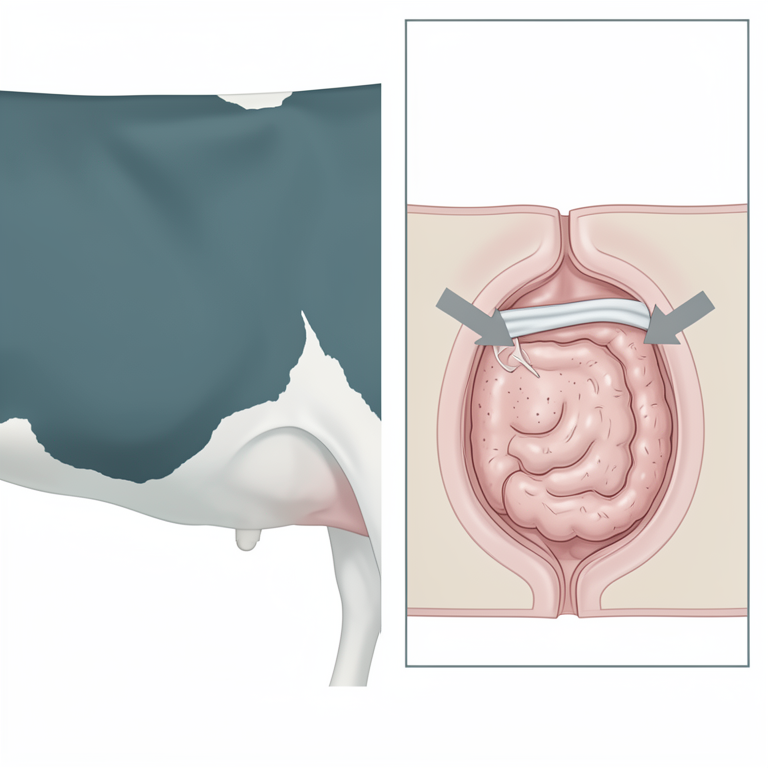
Surgical Approach for Rumen Fistula Repair
AI-generated conceptual illustration (Holstein cow). It explains the idea and does not show study data.
View source evidence ↗Figures & tables
From the original paper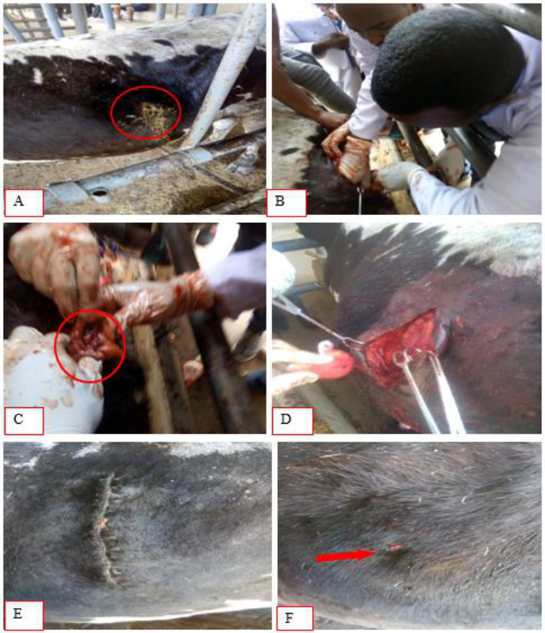 Figure 1
Introduction
Rumenotomy is a routine procedure for many diseases in cattle, such as traumatic reticuloperitonitis, acute and recurrent bloat, ingestion of toxic plants, chemicals, spoiled roughage, or fetal membranes after parturition; placement of a temporary or permanent rumen cannula to relieve bloat; creation of a permanent rumen fistula and impactions. Other reasons include ingestion of foreign bodies, such as nylon ropes or plastic bags that are obstructing the reticulo-omasal orifice, foreign bodies lodged in the distal esophagus and carbohydrate engorgement (Fubini and Ducharme, 2004) [4] . Experimental fistulation is performed in many farm animals as a part of research linked with feeding trials. However, accidental fistulation of forestomach in cattle is relatively infrequent. Trauma due to sharp objects on the flank can result in injuries even to the internal organs. Such injuries causing fistulas have been found to be associated mostly with abomasum (Costa et al., 2002) [3] . To the best of my knowledge, there are very few reports on traumatic fistulation of rumen in cattle. Hence, in this case, successful repair of a traumatic rumen fistula in a cow has been reported.
Case history and clinical examination
A cow aged about 8 years and weighting about 200 kg was presented to the VTH with a history of an exudating wound at its left side between ribs space (Fig 1A ). A penetrating wound was formed due to sharp material by someone resulting from revenge when the cow ate his cereal. The discharge was started two week ago, greenish in color and the animal was apparently normal with usual appetite. The animal was said to have been treated by the local veterinarian. Clinical examination revealed a penetrating wound at the middle of the left side eleventh and twelfth ribs inter costal space. Ruminal discharges were evident from the injured site which were normal in color and foul in odour. The temperature, heart rate and respiratory rates were within the physiological ranges. On examination, the rumen wall was found injured and adhered to the flank wall at the wounded area. Auscultation of the lungs revealed no abnormality. Careful examination of the discharge established the fact that it was rumen cud and hence, the condition was diagnosed as rumen fistula.
Surgical treatment and postoperative care
Prior to surgery, the animal was kept off from feed and water for one complete day to evacuate the rumen and pre operative antibiotic (penstrep 10 ml, IM) was administered before surgery. The fistulation site and paralumbar fossa were prepared aseptically in standard fashion. Rumenotomy was done by the standard procedure as suggested by Fubini and Ducharme (2004) [5] . It was performed on the paralumbar fossa following standard procedure under local analgesia was achieved by direct line infiltration of 1000 mg 2 % lignocaine hydrochloride. After completing incision of the abdominal muscle and peritoneum, the adhesions between the rumen and at the site of fistulation were carefully broken down. Procaine pencillin powder was distributed into abdominal cavity through the paralumbar fossa incision. Then the ruminal wall was pulled caudally to the level of flank incision (Fig 1B ), the fistulous orifice on the rumen was identified (Fig 1C ), derided, incision was extended vertical to remove certain amount of rumen content, presence of foreign material was explored and repaired with a double row of inversion sutures (cushing followed lambert) using number 2 chromic catgut. Peritoneum and muscles were sutured with chromic catgut number 2 by simple interrupted pattern (Fig 1D ) and subcutaneous tissue was closed with chromic catgut number 1 by simple continuous pattern. Skin edges were approximated with silk number 1 by simple interrupted pattern. For postoperative care, the animal owner was advised after surgery to limit amount of water and feed at least for week. Slight rumen tympany was occurred at first day and second of surgery as postoperative complication and it was absent after third day due to limitation amount of feed and water. The cutaneous wound between 11 th and 12 th ribs intercostal space was managed as open wound by dressing with medvlone and povidone iodine for five days. Penstrep, 10 ml (1ml/20kg) was given IM, SID for five days. Sutures were removed on
Discussion
Fistulas of compound stomach have been frequently observed and usually associated with trauma. Among various compartments, abomasum appears more common to become fistulated traumatically followed by rumen (Sharma et al., 2011) [8] . Similar to the present case, Prakash and Ravi (2009) [7] and Devi Prasad et al. (2014) [4] also recorded a successful case of acquired traumatic ruminal fistula in cattle and treated a similar case of rumen fistula at left paralumbar fossa nearer to last intercostal space in cow. Contrary to these findings, Costa et al. (2002) [3] treated abomasal ulceration and abomaso-pleural fistula in an old beef master bull, which died later due to development of pneumo-peritoneum. The success in treatment of the present case could be attributed partly to the fact that the diaphragm was not involved. Peritonitis is a major complication associated with rumen surgery. Any spillage of rumen contents in the abdomen will result in some degree of peritonitis. The degree of peritonitis is dependent on the amount of contamination, blood and tissue levels of antibiotics and the health status of the animal. In addition, animals undergoing rumen surgeries will frequently develop incisional infections, seromas, hemorrhage, fever, intestinal obstruction, physiological bloat, abscesses and death (Fubini and Ducharme, 2004; Andrew, 2008) [5] . Azari and Ali Asghar (2014) [2] reported long-term involvement and existence of fibrotic and necrotic tissues around the perforation site. In the current case, only rumen tympany seen at first and second day after surgery as postoperative complication. Because formation of adhesions between the rumen and abdominal wall might have prevented the theoretical probability of peritonitis. William et al. (1990) [9] and Ismail et al. (2007) [6] also reported this type of complication following rumenotomy. Hence, treating traumatic ruminal fistula by rumenotomy with good postoperative management could be considered as a successful surgical procedure.
Figures and tables from the paper
References
9 in the original paper- Andrew J. Rumenotomy. Vet Clin Food Anim 2008;24:341-347.
- Azari O, Ali Asghar M. Reticulo cutaneous fistula due to the ingestion of a long metallic rod in a cow. Asian Pac J Trop Biomed 2014;4(7):586-588.
- Costa LRR, Gill MS, Williams J, Jhonson AT, Angel KL, Mirza MH. Abomasal ulceration and abomasopleural fistula in an 11 month old beef master bull. Canadian Vet. J 2002;43(3):217-219.
- Devi Prasad V, Devarathnam J, Mahesh R, Kamalaka G, Sumiran N. Surgical management of traumatic ruminal fistula in an Ongole Cow. Intern. J. Liv. Research 2014;4(4):43-46.
- Fubini S, Ducharme N. Surgery of ruminant forestomach compartment. In: Farm Animal Surgery, Saunders, Elsevier 2004, 161-240.
- Ismail ZB, Al-Majali A, Al-Qudah K. Clinical and surgical findings and outcome following rumenotomy in adult dairy cattle affected with recurrent rumen tympany associated with non-metallic foreign bodies. Am. J. Ani. Vet. Sci 2007;2(3): 66-71.
- Prakash BVS, Ravi R. Acquired traumatic fistula in bovine. Indian Vet. J 2009;86(5):87-88.
- Sharma A, Upadhyay SV, Kour K. Congenital abomasal fistula in a buffalo calf and its successful treatment. Buffalo bulletin 2011;30(1):2.
- William BJ, Balchandran S, Kannan C. Postoperative complications of rumenotomy. Indian. Vet. J 1990;67(12):1161-1162.
Cited by 3
From OpenAlex, OpenCitations and reference lists read by Scinovex
- Ruminal Fistulation and Cannulation: A Necessary Procedure for the Advancement of Biotechnological Research in Ruminants ↗
Cristina Castillo, Joaquín Hernández Bermúdez · Animals · 2021
Rumen content is a complex mixture of feed, water, fermentation products, and living organisms such as bacteria, fungi, and protozoa, which vary over time and with different feeds. As it is impossible to reproduce this complex system in the laboratory, surgical fistulation and cannulation of the rumen is a powerful tool for the study (in vivo and in situ) of…
❝ 35PDF - Clinical characteristics and surgical outcomes of full-thickness abdominal wall injuries complicated by evisceration and fistulation in dromedary camels (Camelus dromedarius) ↗
El-Sayed El-Shafaey, Mahmoud Najeb, Mohamed Abdo Rizk, Mahmoud G. El Sebaei · Frontiers in Veterinary Science · 2026
Background: Abdominal wall injuries in dromedary camels often result in life-threatening evisceration or persistent fistulation. While well documented in equines and bovines, standardized surgical management protocols and survival outcomes for camelids remain scarce in veterinary literature. This retrospective study evaluated the clinical patterns, visceral involvement, surgical treatments, and survival outcomes of full-thickness abdominal wall injuries complicated by evisceration or fistulation…
❝ 0PDF - Clinico-epidemiological study of bovine forestomach disorders
S A BIRADAR, RH Bhatt, SH Zalavadiya, MB Bhesaniya, Rama Chandra Reddy K · International Journal of Veterinary Sciences and Animal Husbandry · 2025
The present study was conducted to determine the clinico-epidemiological status of bovine forestomach disorders on 11,047 bovines which were presented during the period January 2021-December 2023 at Veterinary Clinical Complex, Junagadh (Gujarat). Among them, 2279 (20.63%) were suffering from different surgical diseases and out of these animals, 239 (2.16%) were diagnosed with different forestomach disorders. The age wise incidence revealed, highest incidence was recorded in the age group of 6 to 10 years (60.66%), followed by up to 5 years (26.77%), 11 to 15 years (10.87%) and 16 to 20 years