
Research article · 2021 · Full text
International Journal of Veterinary Sciences and Animal HusbandryA mini review on semen sexing in bovine
Volume 6(1) · Pages 04–10 · DOI: 10.22271/veterinary.2021.v6.i1a.315 ↗
Research areasSperm and Testicular FunctionReproductive Biology and FertilityAnimal Genetics and Reproduction
Abstract
Author-writtenSexing of mammalian sperm is a long-standing goal in agricultural industries including cattle breeding. Being able to pre-select the sex of offspring at the time of conception, ranks among the most sought-after reproductive biotechnologies of all time. Sexed semen is commercially offered by most global A.I. companies yielding with 90% accuracy. Fertility of sexed sperm is typically lower compared with conventional sperm. Sexed semen is recommended for breeding only virgin heifers followed by 1st or 2nd lactational cow. The success of sexed semen industry depends upon the sorting speed, accuracy and the fertility of sorted spermatozoa. The use of sexed semen is expected to affect the structure of the dairy industry by creating a greater supply of replacement heifer if reduction in conception rate can be minimized. Today, Flow cytometry is the only successful technique for the sorting of sperm.
Summary
Source-checkedPlain-language reading of the full paper, prepared with AI and checked against the text. Each point links to the authors' own words.
Why it matters
- Predetermining the sex of livestock is a long-standing goal for producers due to the financial advantages it provides. View evidence ↗
- Female calves are prioritized for dairy production, whereas males are preferred for beef production due to superior lean-to-fat ratios and feed conversion efficiency. View evidence ↗
- A critical application of sperm sexing is the reduction of sex-linked genetic diseases within a population. View evidence ↗
The question
- The primary objective of semen sexing is to enable the production of calves of a specific, desired sex. View evidence ↗
What they did
- Sperm separation methods rely on differences between X- and Y-bearing sperm, including DNA content, surface charge, weight, density, size, swimming characteristics, and survival rates in different pH media. View evidence ↗
- The Percoll Density Gradient Method utilizes the fact that X-bearing sperm tend to sediment faster than Y-bearing sperm. View evidence ↗
- Free flow Electrophoresis separates sperm based on surface charge at pH 7.4, where X sperm move faster toward the anode and the slower peak consists of 83-89% Y sperm. View evidence ↗
- Immunological sexing uses monoclonal anti-H-Y antibodies (IgG) to specifically bind to male-determining spermatozoa. View evidence ↗
- Flow cytometry sorts sperm by using the fluorescent dye Hoechst 33342 to stain DNA, then detecting and sorting individual cells based on their fluorescence intensity. View evidence ↗
What they found
- Commercial sexed semen provided by global AI companies typically achieves 90% accuracy. View evidence ↗
- In human sperm, the Percoll method achieved up to 94% purity for X-bearing sperm with a recovery rate of nearly 30%. View evidence ↗
- Using Sephadex column albumin gradient separation, the proportion of X spermatozoa increased from a mean of 60% to 74%. View evidence ↗
- Flow-cytometric sorting has reported purities of 70-90% for X- and Y-bearing sperm when verified by PCR and FISH. View evidence ↗
- Sexed semen exhibits lower fertility (70-80%) compared to standard doses of unsexed semen. View evidence ↗
What it means
- The authors conclude that flow cytometry is currently the only successful technique for sorting sperm. View evidence ↗
- The authors recommend using sexed semen primarily for virgin heifers, followed by cows in their first or second lactation. View evidence ↗
- Flow cytometry is limited by high equipment costs, the need for skilled operators, risks of mutagenic effects, reduced fertility and blastocyst formation, and a lack of 100% accuracy. View evidence ↗
Visual summary
1 visualsIllustrations explain the paper; they do not show study data.
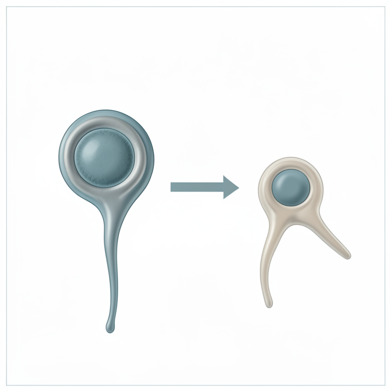
Basis of Sperm Sexing
AI-generated conceptual illustration (bovine sperm cells). It explains the idea and does not show study data.
View source evidence ↗Introduction
The aim of sexed semen is to supply a calf of a specific sex. In the livestock industry, predetermination of sex of animals has been a main goal of producers for generations, because of its financial advantage. Females are essential for dairy products and production of calves, while males are usually required for beef or meat production, because of the better feed conversion efficiency and lean-to-fat ratio. In addition, a male of high genetic merit is still required as sires in AI programs and by sexed semen fastens the genetic progress and allows the farm manger to increase selectively the amount of heifers or steers supported the necessity of the farm. It also reduces calving difficulty in first calvers (Seidel, 2007) [34] and reduces the replacement cost besides maintaining the biosecurity in the farm. There is an urgent need to expand the genetic merit of our cattle and buffaloes with the decreasing land productivity, crop yield and other feed resources; and also, due to a steady increase in the demand and consumption of high quality dairy products.
Importance of semen sexing
The most important application of sexing is to minimize sex-linked genetic disease in the population. The sex pre-selection to increase the chance of having a female child gives the reasonable assurance that they can avoid expression of the disease in their offspring. Some of the sex linked diseases are Haemophilia, Baldness and Eye colour in Drosophila flies. Fertilization of ovum with Y sperm produces male off-spring and X sperm produces female off-spring. So if we could able to separate X and Y sperm then it would be possible to produce the calf of the desired sex. In progeny testing program sires are selected based on the performance of the female progenies. So if ovum could be fertilized with X sperm then we can eliminate the chances of production of male progenies. If only a few individuals of an endangered species are available, chance of production of a few males instead of a few females often would greatly diminish chances of survival of the species. Sex preselection for particular matings also becomes crucial in developing breeding systems that minimize inbreeding with few breeding animals. A commercial dairy farmer mates a heifer or cow for two reasons. The commercial beef cattle farmer produces calves for eventual sale as slaughter animals and for female herd replacements. Depending upon the role that is envisioned for a calf, males and females are of quite different value; so sexed semen potentially is a powerful technology to affect biological and economic efficiency (Hohenboken, 1999) [14] .
Seedstock breeders produce breeding animals, semen and embryos for sale. The primary role of their animals is to produce profitable and efficient offspring. To be competitive in achieving this goal, they must maximize the rate of genetic improvement in their herds, control inbreeding accumulation, and accomplish these conflicting goals as economically as possible. They must seek the optimum balance between male and female selection intensities, selection accuracies and generation intervals (Hohenboken, 1999) [14] . Only live, membrane intact sperms sorted: With flow-cytometry sorting of spermatozoa, only live membrane spermatozoa takes the stain and remaining dead spermatozoa will not take colour of stain. So dead spermatozoa are discarded or eliminated at the time of sperm sorting. The matured oocytes Sex-sorted bovine sperm were used for in vitro fertilization (IVF) to generate embryos from in vitro. Sex-sorted sperm is used in embryo transfer technology which will be useful to produce desired sex from genetically superior animals (Cran et al., 1993) [30] .
Methods or approaches of sperm sexing
More sophisticated methods have been offered over the last few decades. Apart from recommendations concerning sexual behavior, most methods have relied on in vitro spermseparation techniques. Successful separation depends on the existence of fundamental differences between sperm cells containing either X-or Y-chromosomes, such as Variations in relative survival rates in a different pH medium (Shettles, 1960) [33] , Contrasts in sperm-surface charge that allow separation of spermatozoa according to their differences in electrophoretic motility (Engelmann et al, 1988) [4] , Difference in weight, density, size and sperm swimming characteristics, Difference in the DNA content -Flow cytometric sorting of the semen.
Sexing on the basis of the difference in mass and density is
Percoll Density Gradient Method. In a preliminary experiment, it was found that human X-bearing sperm tend to sediment faster than Y-bearing sperm in a density gradient centrifugation using Percoll (Kneko et al., 1984) [20] . Percoll is silica coated material with polyvinylpyrilidone used for density gradient. By more careful examination of the conditions for sedimentation of the two classes of sperm in a discontinuous density gradient with 12 steps of Percoll, selective isolation of human X-bearing sperm (purity of up to 94%) as the sediment has been achieved with the best recovery rate of almost 30% of the total sperm applied. The recovered X-bearing sperm showed active forward movement and were free of other contaminating cells found in the semen. Using the X-bearing sperm fraction for artificial insemination, at least six girls were born. The ratio of Ybearing sperm in the upper layers of Percoll, however, did not increase remarkably under the same conditions. In spite of such a difference in sedimentation velocity between the two classes of sperm, their apparent densities showed little difference when examined by equilibrium sedimentation in a continuous density gradient of Percoll. This means some other factor (s) than apparent density affect the sedimentation velocity of X-and Y-bearing sperm. Density Gradient Centrifugation Density Gradient Centrifugation is a method of separation of X and Y chromosomes bearing sperm. In this procedure, sperm is centrifuged through increasingly dense layers of a solution, on the theory that heavier X-sperm will sediment to the bottom, and lighter Y-sperm will migrate to top. A density medium, a liquid solution available in various thicknesses, is layered in a conical centrifuge tube, with the densest layer at the bottom and the least dense layer at the top. In the standard procedure of preparing sperm for assisted reproduction, 2 layers are used. For gender selection, 3 to 12 layers are used. Diluted semen is layered on top, and centrifuged for 30 minutes. To conceive a woman, only rock bottom layer, or "pellet", is retained, during which the heavier X-sperm have sedimented. To conceive a boy, only the highest layer of the liquid "supernatant" is retained, containing the lighter Y-sperm. The remainder is washed and centrifuged again to remove the density medium. Centrifugal countercurrent distribution is methods of separation of human X-and Y-chromosomes sperm on their centrifugal density. It's specially designed machine on the basis of the invention by Akerlund (1984) [1] . The apparatus contains 60 chambers arranged in a circle, which allows transfer of the upper (mobile) phases relative to the lower (stationary) phases. In this system, CCD is performed during centrifugation by keeping the denser (bottom) phases in the outer half while the lighter (upper) phases are in the inner half of each chamber. Because no elution or pumping of any phase takes place, the overall process consists of a circular multistep transfer of 60 uppers-over 60 bottom-batch phases. Each transfer in this centrifugal-enhanced CCD includes shaking the phases at unit gravity to thoroughly mix them and then separating them by centrifugation. (Ollero, et al., 2000). Seminal plasma was removed from the semen samples by a sucrose washing method. An aliquot of semen was diluted with 1.5 sucrose buffers (222 mm sucrose, 10 mm NaCl, 10 mm glucose, 20 mm HEPES, and 2.5 mm KOH). One milliliter of diluted sample was layered over 7.5 ml of the same medium and centrifuged for 5 minutes at 200 g and for 10 minutes at 1000 g; the supernatant layer was removed to leave a 1-ml volume in which the loose pellet of spermatozoa was suspended. Sperm samples were loaded in chamber 0 to 59 transfers were carried out. Shaking and centrifugation time was 60 seconds and all operations were performed at 20 °C. After the run, the solutions were transformed into a one-phase system by the addition of 1 volume of a dilution buffer (polymer-free medium). The fractions were then collected and the cells were counted under a light microscope. Partition results are expressed as the number of cells recovered in each fraction. Total viable cells in each fraction were assessed by the following: % viability recovered cells in each fraction/100. As a consequence of the separation procedure, sperm cell populations with a marked affinity for the lower dextran-rich phase (mainly due to a low hydrophobicity) partition in the left part of the profile. Sperm cells that partition almost equally in both phases distribute in the central sector. Finally, a sperm population with a high affinity for the upper PEG-rich phase (mainly due to a high hydrophobicity) and partition in the right sector of the profile was separated (Harrison et al., 1982) [13] .
Sexing on the basis of difference in motility is Albumin
Gradient Method, Successful separation of X-and Ychromosome-human spermatozoa using an albumin gradient was first reported by Ericsson et al. (1973) [6] . The method is based on size that Y-chromosome spermatozoa are smaller in size and exhibit a greater downward swimming velocity than X-chromosome-bearing spermatozoa within vertical columns of high density human serum albumin (Ericsson et al., 1973) [6] . A fraction enriched with Y-chromosome bearing spermatozoa can be obtained by obtaining the first 22% of spermatozoa to swim to the bottom of the gradient, and discarding the remainder. The latest version of this technique improved the separation accuracy. (Ericsson and Ericsson, 1999) [5] . This technique has never been shown to sex spermatozoa accurately from mammals aside from humans (Beal et al., 1984; White et al., 1984) [2,42] . Sephadex column separation: Sephadex is the methods of the separation of human X-and Y-chromosomes sperm. Sephadex column separation By means of the Sephadex gel filtration method we were ready to separate a fraction rich in X human spermatozoa, in contrast with the tactic of Ericsson et al. (1973) [6] , which allows to obtain a fraction rich in Y sperm. The method, which is simple and not time-consuming, may allow control of sex in cases of expressing sex desire in off-spring. The separation of X-and Y sperm in human spermatozoa by means of Sephadex gel filtration was performed with Locke's solution at room temperature. Smears of semen and smears of the filtrate, and washed spermatozoa were stained with fluorochrome Quinacrine, in which fraction rich of X-bearing spermatozoa was obtained. The average number of F-bodies detected in semen before gel filtration was 38.8%, whereas the average number of F-bodies in filtrate samples was 4.7%, and 64.7% in washed spermatozoa. The concentration of spermatozoa in the collected fraction was about 25% of the sample concentrations prior to gel filtration. It might be due to that, immobile sperm cells are retained by the lack of an active passage, while Y-bearing spermatozoa are most likely attached to the gel particles by adsorption (Steeno et al., 1975) [35] . Sephadex albumin gradient separation: Sephadex column albumin gradient is a method of separation of human X-and Ybearing sperm. In which the semen was passed through the columns of Sephadex G50 in order to increase the proportion of X spermatozoa. The X spermatozoa increased from a mean of 60% to 74%. The degree of separation in individual samples showed considerable variation in this method, but in some samples only a 6% change in the proportion of X and Y spermatozoa could be obtained, whereas in others the increase was 37%. The proportion of motile spermatozoa showed a marked increase with the Sephadex method (Quinlivan, et al, 1982) [27] .
Sexing on the basis of differences in surface charge is free flow Electrophoresis Method: Electrophoretic separation of X-and Y-bearing sperm has been tried by many investigators, on the basis of the difference in electric charge on their surface. Sperm moved towards the anode at pH 7.4 and were separated into two main peaks, the faster peak containing only X sperm and the slower peak consisting mainly (83-89%) of Y sperm. In other words, X sperm have a higher net negative charge on the cell surface than do Y-bearing sperm. Contamination of immature sperm and/or agglutinated sperm appeared to reduce the yield of Y sperm in the slower peak. The results were reproducible provided that normal semen samples were properly processed. Sperm motility was reduced after free-flow electrophoresis. Improvement of composition of buffers and other media would result in separation of the two classes of sperm without loss of motility. In fact, it was confirmed that bull sperm were fertile after the electrophoresis. The negative charge of the cell surface is mainly due to projection into the surrounding medium of glycoproteins with neuraminic acid and sulfate group. When human sperm were treated with neuraminidase prior to freeflow electrophoresis, the electrophoretic mobilities of the two classes of sperm were much reduced, and the two separated peaks finally joined into a single peak in which the yield of Y-bearing sperm was 43-46%. The observed difference in net negative charge appeared to be due at least in part to a larger content of glycoproteins possessing neuraminic acid on the surface of X-bearing sperm. Although X-bearing sperm have a larger surface area than Y-bearing sperm, it is possible that the surface difference between the two classes of sperm reflects the haploid gene expression. There was no remarkable difference in morphology of the separated human X-and Ychromosome bearing sperm. Examination of separated X-and Y-sperm fractions by means other than quinacrine mustard staining is now in progress. Countercurrent galvanic separation: Convection Countercurrent galvanization is a method and apparatus for controlling the sex of mammalian offspring through separation of X-chromosome and Ychromosome bearing sperm. The separation is accomplished by producing a thermal convection counter stream within a sedimentation column containing a universal medium with sperm suspended therein, and subsequently allowing the two sperm populations to sediment into different fractions according to different densities. A method of separating sperm cells of differing densities and electrical potentials from semen comprising to the mixing fresh semen with a liquid suspending medium and separating the mixture by thermal convection counter streaming sedimentation according to different sperm densities into three fractions, a first fraction containing predominately X-sperm of a normal genotype, a second fraction containing predominately Ysperm of a normal genotype, and a third fraction containing both X and Y-sperm carrying defective genes; and subjecting a desired fraction of said medium to convection galvanization thus further separating and concentrating all X-sperm of a normal genotype and all Y-sperm of normal genotype (Gledhil, 1988) [7] .
Sexing on the basis of immunological difference in sex chromosomes
Separation on the Basis of Presence of H-Y Antigen: Expose live, native spermatozoa to an excess concentration of an antibody that binds substantially exclusively to male determining spermatozoa. Specifically, freshly ejaculated spermatozoa are diluted. The diluent used varies depending on the species of the spermatozoa being separated. Specific examples of diluents include sodium citrate solution for bovine spermatozoa, creme gel buffer for equine spermatozoa and test buffer for human spermatozoa. The diluent used, however, should be free of protein as any protein in the diluent tends to coat the spermatozoa and block the H-Y antigen binding sites on the surface of the male determining spermatozoa. Thus, by substantially eliminating this blocking protein provides conditions for optimal cross reactivity between the antibody and antigen for the best possible immuno separation results. An excess concentration of the male specific antibody is contained in or added to the diluent and spermatozoa. The male specific antibody is a monoclonal anti-H-Y antibody (immunoglobulin G) prepared by outgrowth of primary hybridomas and recovered from culture medium by stepwise precipitation and dialysis. So only male determining spermatozoa express the H-Y antigen on their surface. The female determining spermatozoa do not express the H-Y antigen, believed associated with the Y or male determining chromosome. Since the monoclonal antibody is absolutely specific for the H-Y antigen, it binds only to the male determining spermatozoa and not any of the female determining spermatozoa. Following a short incubation period for immunoreaction of 45 minutes, the spermatozoa/monoclonal H-Y antibody dilution is centrifuged and collect the exposed spermatozoa from the diluent. The exposed spermatozoa are then washed using fresh, pure protein free diluent in order to remove unbound monoclonal antibody. After washing, the exposed spermatozoa are suspended in an equal volume of protein free diluent (as used during the exposing step) substantially without the presence of unbound H-Y-antibody. Immediately thereafter, antibody/Immuno absorbent conjugates are added to the dilution so as to form a conjugate/spermatozoa suspension preparation. The antibody is coupled to the Immuno absorbent material or agarose beads binds immunoglobulin G and, therefore, binds the monoclonal anti-H-Y antibody already bound to the male determining spermatozoa. Again, just as with the first antibody the female determining spermatozoa do not immunoreaction with the immunoglobulin G antibody bound to the beads. In order for the second, immunoglobulin G antibody, to properly react with the monoclonal H-Y antibody, the immunoglobulin G antibody must be species specific for the monoclonal antibody. Thus, if spermatozoa are exposed to monoclonal H-Y antibody produced by mice lymphocyte cells as described above, the second antibody bound to the conjugate must be anti-mouse immunoglobulin G. The anti-immunoglobulin G/agarose bead conjugate is added to the diluted spermatozoa at an approximate concentration of 0.4 milliliters of conjugate beads per milliliter of diluted spermatozoa. The conjugate/spermatozoa preparation is incubated at approximately 37 °C and agitated to prevent the beads from settling and thereby maintain the beads and spermatozoa in physical contact for complete immunoreaction. During this time, the anti-immunoglobulin G antibody of the conjugate binds the monoclonal H-Y antibody bound to the male determining spermatozoa. Thus, the antibody/bead conjugate agglutinates the monoclonal antibody/male determining spermatozoa complexes. Following immunoreaction and agglutination, the female determining spermatozoa are recovered. This step may be completed by draining the conjugate/spermatozoa preparation solution from the antibody/agarose bead conjugates now bound to the male determining spermatozoa. Since the female determining spermatozoa do not bind to the beads, the female determining spermatozoa are contained in this drained solution. The solution is then gently centrifuged to collect the female determining spermatozoa in a pellet at the bottom of the centrifuge tube without significantly adversely affecting spermatozoa viability. The pelleted female determining spermatozoa are then resuspended in a diluent and added into the filtering column. Viable spermatozoa pass through or are eluted from the column at a substantially faster rate than dead or weak spermatozoa and, therefore, excellent filtering results are obtainable. The spermatozoa eluted from the column may be collected in a protein containing medium for maintaining the spermatozoa for freezing and/or subsequent use in artificial insemination. An example of such a protein containing medium is the protein free diluent used above during the exposing and suspending steps mixed with egg yolk, 20% by volume. The male determining spermatozoa are also recovered from the agarose beads. The methods include competitive inhibition using a solution of male specific antibody, enzymatic digestion of the beads, and alteration of the pH or salt concentration of the diluent. Following recovery, the male determining spermatozoa is filtered through a sephadex column in the same manner as the female determining spermatozoa so as to increase the efficacy of the male determining spermatozoa fraction or sample. Sex Specific Proteins (SSPs): A viable immunological sperm sexing procedure which is non-invasive can be evolve using a more efficient method to isolate sex-specific proteins (SSPs) (Blecher et al., 1999) [3] . A large proportion of genes on the mammalian X chromosome are highly evolutionarily conserved; an X-linked genes in any one species predicts the existence of a homologue in all others. This proposition is known as Ohno"s law (Ohno and Watchtel, 1979) [26] . The hypothesis is that 1) the sex chromosomes encode or control the expression of X-and Y-sex chromosome-specific proteins (X-or Y-SCSPs) that are expressed on the surface of somatic and sperm cells, and 2) as an extrapolation of Ohno"s law, that these proteins are more highly conserved between species than non-specific molecules. As a consequence of the latter, when a same-sex recipient is immunized with tissue from a special species, the recipient's system would mainly perceive SSPs as "self" and would not raise antibodies to these molecules. The letter would reflect species differences in the non SSPs; these antibodies could be used to immune precipitate non-SSPs and leave SSPs in a partially purified form. Based on this hypothesis the non-SSPs were removed immunologically before the attempted isolation of SSPs because they are likely to be more highly conserved than non-SSPs. Antibodies to SSPs were grown and used to identify SSPs by affinity chromatography (Blecher et al., 1999) [3] . In these methods Inject sperm and Freund"s incomplete adjuvant s/c in female and male rabbits respectively and semen of anti "X" or anti "Y" antisera and Incubated for 60 min at 38.5̊ C. Advantages of these methods are free swimming semen separated, only takes 20-30 minute and in vitro fertilization -92% male embryos can produce (Moore and Thatcher, 1988). DNA Based Methods for Sperm Sexing Sorting based on volumetric differences: Spermatozoa containing an X chromosome are theoretically bigger than those containing a Y chromosome. Van Munster et al., (1999) [38] recently used interference microscopy and subsequent image analysis to demonstrate a difference in sperm head volume that matched differences in DNA content between X-and Y-chromosome carrying bovine spermatozoa. A method based on this principle has been evolved for sorting live spermatozoa by using interference microscopy optics with a flow cytometer (Van Munster, 2002) [37] .
Flow-cytometric sorting of semen: It was first reported by Johnson, (1989) [16] in rabbits. Flow cytometry for selection of gender is based on variability in chromatin staining by the DNA-binding fluorescent dye Hoechst 33342, detection of fluorescence from individual cells, and sorting of individual cells based on fluorescence. Selected spermatozoa are then used for IUI, IVF or ICSI. However, purities of X-and Ybearing sperm of 70-90%, as judged by PCR and FISH methods have been reported (Johnson, 1995, Vidal et al., 1998) [17,39] . The difference of their DNA-content can be used by flow-cytometry to produce populations of either sex at high accuracy. As the method is predicated on single cell identification, the output of cells is restricted, although major improvements were made by high-speed flow-cytometry and improved cell orientation ahead of the UV-Laser. Flow cytometer Principles and cell sorter use for semen sexing: The spermatozoon passes two fluorescence detectors at 90°angles to each other, each detector measures the intensity of fluorescence resulting from the excitation of the DNA-bound dye molecules by light, which is generated by a laser. The wavelengths of light used depend on the source of light. The strength of the fluorescence signals clearly depends on the number of fluorescing molecules bound to DNA. This is the basis for sexing spermatozoa. In addition, the signal depends on a number of other parameters, including the laser intensity (Guthrie et al., 2002) [8] , whether and how the laser is pulsed, optical properties of the entire system, the sensitivity of detectors and electronic noise. The main obstacle to accurate quantification of spermatozoa DNA with this approach is the geometry of the sperm head, which is paddleshaped in most species of interest. The intensity of fluorescence is lowest if the flat face of the paddle is oriented toward a detector, and highest when the edge is so oriented. The most accurate discrimination of X and Y chromosome spermatozoa are results when its orientation is flat. The second detector at 90° to the laser is used to diagnose orientation (Senger, 2003) [32] . As the fluorescence signal is highest for spermatozoa oriented with their paddle edge toward this 90° detector, only the population of spermatozoa that emit peak fluorescence to the 90° detector are considered oriented appropriately for sexing by the contemporaneous signal to the 0°detector. A considerable effort, therefore, has been made to increase this percentage by modifying the cylindrical geometry of the fluid stream (Johnson and Welch, 1999; Rens et al., 2001) [18,29] . DNA-binding dyes: Hoechst 33342 (H33342) may be a live cell stain that permeates the cell wall and binds selectively to A -T base pairs along the minor groove of dsDNA. H33342 usually is happy with the 351 or 364 nm lines of an argon-ion laser Most DNA stains intercalate between the base pairs of the DNA, thereby presumably increasing their mutagenicity. H33342 is carefully used as it might be toxic to workers at high doses. Dead or moribund cells in the population of spermatozoa stained with H33342 can be identified by adding propidium iodide (Johnson et al., 1994). More recently, this classical dead-cell stain has been replaced with red food dye (FD&C40) to avoid potential mutagenic effects of propidium iodide (Johnson and Welch, 2001; Schenk et al., 1999) [29,30] . Other food dyes also are effective. Two basic sperm populations: Living (Stained with Hoechst 33342) Dead (Stained with Red food Dye) These methods are independent of the type of cell sorter, sample preparation begins with the staining of spermatozoa using Hoechst 33342 stain at 34-38 °C for 60-90 minutes. Labelled spermatozoa are introduced into the flow cytometer across the sample line under high pressure. Two streams of the sheath fluid with pressure just above the sample pressure surround the core stream in the injection tube. Oriented spermatozoa exit through the vibrating nozzle tip, which produces small droplets, theoretically containing one labelled spermatozoa only. Immediately after their exit, the laser light excites the fluorescent dye and the emitted light is collected through detectors in position 0° and 90°. The electric signals of the photocells are processed in a computer. Cells failing to fulfill preset criteria are excluded from further analysis. The proper positioned cells are selected as X-or Y-chromosome bearing spermatozoa, according to the amount of emitted light from the flat sperm surface (0° detector). Based on this signal, the computer sends the signal to the wire loop to electrically charge the droplets accordingly. Charged droplets pass an electro-static field and are separately collected into tubes prefilled with a collection extender. Sorted spermatozoa are then washed from the sheath fluid by centrifugation and the remaining sperm pellet is extended in a suitable medium. Composition of media differ in among species and also according to the following preservation process (Hollinshead et [31,15,23,28,30] . Flow-cytometry based sperm sorting is the only available method that has been shown to produce high purity of sex chromosome selection. Several major improvements have been made in the past years, especially by invention of high speed cell sorting and improved orientation of cells in front of the Laser. Nevertheless, the limitations of the technology due to the principle of single sperm cell analysis have to be recognized and even with further technical improvements it is unlikely that the output will reach the amount of a normal insemination dosage, especially in the porcine. In combination with other biotechnologies, bovine sexed spermatozoa are already commercially available. In other species intensive research is required to provide sufficient sexed spermatozoa.
Limitations of Flow-Cytometry: It require specialized, expensive equipments and highly skilled operators, Risk of cyto-toxic mutagenic effect, Reduction in fertility, Reduction in rate of blastocyst formation (Merton et al., 1997) [25] , Cost of semen dose is higher, Present day methods do not result in 100% accuracy (Johnson, 1991) [19] .
Evaluation of Sperm Separation Efficacy
Quinacrine Staining: Y-bearing spermatozoa exhibit a fluorescent spot or Y body. X-bearing spermatozoa remain unstained. However, quinacrine produces false positive (two Y bodies) and false negative results. This technique can produce misleading and inaccurate results with human spermatozoa. (Van kooij and Vanoost, 1992) [36] .
Sort reanalysis: Sort reanalysis for DNA has an advantage over FISH and PCR since both techniques may take 3 to 4 h, whereas reanalysis in the flow cytometer requires less than 40 minutes. For reanalysis in the sorter 100,000 spermatozoa are taken preferable from the originally sorted material or can be taken from an extra sort for reanalysis. Cells are sonicated to remove tails and Hoechst 33342 is added at a 10 th of the original concentration in order to maintain staining uniformity. Spermatozoa are then analyzed, but not sorted at very low speed in order to maximize orientation. With the proper set up of the sorter and uniformly stained sperm population, more than 90% of the sperm sorted will be the desired sex. (Johnson and Welch, 1999) [18] Polymerase Chain Reaction (PCR): It is based on using primers from putative testis -determining SRY on Ychromosome to discriminate male DNA. It controls primers from sperm receptor gene on chromosome 7. It has been used to determine the ratio of X-to Y-bearing spermatozoa in the semen sample. It is very specific and highly sensitive. Its application to population of cells is of limited use in the evaluation of sex selection procedures (Lobel et al., 1993, Welch et al., 1993) [24] .
Fluorescence In-situ Hybridization (FISH): FISH is a technique in which chromosome-specific DNA probes are hybridized to chromosomes in cells and are then visualized using reagents which produce a colored, fluorescent or radioactive signal. Fluorescence in situ hybridization (FISH) allows the detection of specific nucleic acid sequences on morphologically preserved spermatozoa, is an ideal method for quantitatively and qualitatively assessing the purity of sorted sperm samples (Piumi et al. 2001, Rens et al. 2001) [29] . The most important reagents on a FISH procedure are the fluorescent DNA probes. After or during DNA amplification steps by PCR, specific DNA must be labelled by incorporating fluorescent nucleotides. It is important to use a labelling method that allows the production of probes with good specificity that is easy to visualize producing optimal hybridization results. The type of probe and labelling method should be chosen according to the sensitivity requirements of the situation. FISH can be used on cells at either interphase or metaphase, so it is well suited to the detection of chromosomes in interphase sperm nuclei. Early studies used auto radiographic detection systems (Joseph et al., 1984), but more recently, fluorescence in-situ hybridization (FISH) has become the method of choice for studying chromosomes in spermatozoa (Kawarasaki et al., 1998) [22] . FISH using a single probe to either the X or Y chromosome has been used to confirm that the ratio of X-to Y-bearing spermatozoa in human semen is 1:1 (Guttenbach and Schmid, 1990; Wyrobek et al., 1990; Han et al., 1992) [9,43,11] . Single label FISH has also been used to evaluate human sperm fractions recovered from albumin gradients and sephadex columns (Vidal et al., 1993) [40] . However, when only a single probe is used, only some of the spermatozoa exhibit a hybridization signal and an assumption has to be made that the unlabelled spermatozoa carry the other sex chromosome. This is an unacceptable assumption because spermatozoa can remain unlabelled for several reasons: (i) they carry the other chromosome; (ii) they are aneuploid and do not carry either sex chromosome; or (iii) hybridization failed to occur. Furthermore, the overall hybridization efficiency cannot be determined using single label FISH, so the proportion of spermatozoa that were unlabelled due to hybridization failure cannot be determined. To obtain unequivocal identification of X-and Y-bearing sperm, it is necessary to use a double label FISH procedure in which probes to both chromosomes are simultaneously hybridized to sperm nuclei (Han et al., 1993) [10] . This enables accurate and unequivocal identification of the gender of each spermatozoon. Ideally, a third autosomal probe should be included to identify spermatozoa which are nullisomic for the sex chromosomes. Advantages of FISH: Chromosome specific DNA probes are used, Individual spermatozoa are analyzed rather than population spermatozoa and large number of spermatozoa can be scored quickly and accurately. Advantage of Semen Sexing over Embryo Sexing: ETT in remote areas -difficult as compared to AI, Less technical expertise needed in AI, Wastage of unwanted embryos and Time required for production of embryo animal has to cycle, the rest to be given.
Limitations of semen sexing
Low sorting rate: Full day's sort of intact spermatozoa will only yield 20 X 10 6 spermatozoa. It would take 1-2 hour to sort the sperm in a typical artificial insemination dose. Expense: Flow cytometer is very expensive to purchase and maintain. Sex specific sperm will cost more and may be packaged differently for AI than conventional semen. Required skill: as with many new technologies, effective commercial use of sex specific sperm will require greater management and labour skills, including well trained inseminator and properly design livestock handling facility (Kansara et al, 2009) [21] . Mutagenic effects of DNA binding dyes such as Hoechst 33342 bind loosely to the DNA in the genome but May still cause chromosomal aberrations. Fluorochrome dyes are a potential cause of low embryo viability and hence low pregnancy rates.
Low sperm viability/ fertility: To facilitate access of DNA stain to the genome, the integrity of the cell membrane has to be compromised either by digestion with papain or by light sonication, sonication increases cell survival rate but at the expense of sperm motility as sonication causes loss sperm flagellum, necessitating sperm microinjection. Fertilization and embryo development rates in IVF programs with sex semen are lower than the unsexed semen. Low Conception: low conception rate achieved with sex semen in high producing lactating cow could be unsatisfactorily. Sexing semen having low fertility (70-80%) than the normal doses of unsexed semen. (Kansara et al, 2009) [21] . Sexing semen comes with two handicaps: limited number and stresses due to the number of steps and manipulation, they undergo during the sexing process. It can conclude that sexed semen is commercially offered by most global A.I. companies yielding with 90% accuracy. Fertility of sexed sperm is usually lower compared with conventional sperm. Sexed semen is recommended for breeding only virgin heifers followed by 1st or 2nd lactational cow. The success of sexed semen industry depends upon the sorting speed, accuracy and the fertility of sorted spermatozoa. The use of sexed semen is expected to affect the structure of the dairy industry by creating a greater supply of replacement heifer if the reduction in conception rate can be minimized. Today, Flow cytometry is the only successful technique for the sorting of sperm.
References
43 in the original paper- Akerlund HE. An apparatus for counter-current distribution in a centrifugal acceleration field J Biochem Biophys Methods 1984;9:133-141.
- Beal WE, White LM, Garner DL. Sex ratio after insemination of bovine spermatozoa isolated using a bovine serum albumin gradient J Anim Sci 1984;58:1432-1436.
- Blecher SR, Howie R, Li S, Detmar J, Blahut L. A new approach immunological sexing of sperm Theriogenology 1999;52:1309-1321.
- Engelmann U, Krassing F, Schatz H, Schill WB. Separation of human X-and Y-spermatozoa by Free Flow electrophoresis Gamete Res 1988;19:151-159.
- Ericsson RJ, Ericsson SA. Sex ratios In: Encyclopedia of Reproduction 4 th Ed Knobil E and Neill JD Academic Press 1999,431-437.
- Ericsson RJ, Langevin CN, Nishino M. Isolation of fractions rich in human Y sperm Nature 1973;246:421424.
- Gledhill BL. Selection and separation of X-and Ychromosome-bearing mammalian sperm Gamete Research 1988;20(3):377-395.
- Guthrie HD, Johnson LA, Garrett WM, Welch GR, Dobrinski JR. Flow cytometric sperm sorting: effects of varying laser power on embryo development in swine Mol Reprod Dev 2002;61:87-92.
- Guttenbach M, Schmid M. Determination of Y chromosome aneuploidy in human sperm nuclei by nonradioactive in situ hybridization Am J Hum Genet 1990;46:553-558.
- Han TL, Flaherty SP, Ford JH, Matthews CD. Detection of X-and Y-bearing human spermatozoa after motile sperm isolation by swim-up Fertil Steril 1993;60:10461051.
- Han TL, Webb GC, Flaherty SP. Detection of chromosome 17-and X-bearing human spermatozoa using fluorescence in situ hybridization Mol Reprod Dev 1992;33:189-194.
- Harley CB, Vaziri H, Counter CM, Allsopp RC. Mutational analysis of SRY in Y-Chromosomes Exp 1992;27:375-382.
- Harrison RA, P Dott HM, Foster GC. Bovine serum albumin sperm motility and the ''dilution effect'' J Exp Zoo 1982;222:81-88.
- Hohenboken WD. Applications of sexed semen in cattle production Theriogenology 1999;52:1421-1433.
- Hollinshead FK, O'Brien JK, Maxwell WMC, Evans G. Assessment of in vitro sperm characteristics after flow cytometric sorting of frozen-thawed bull spermatozoa Theriogenology 2004;62:958-968.
- Johnson LA. Sex Preselection in rabbit: Live births from X and Y bearing sperm separated by DNA and cell sorting Biol Reprod 1989;41:199-203.
- Johnson LA. Sex Preselection by flow cytometric separation of X-and Y-chromosome bearing sperm based on DNA difference: A review Reprod Fertil Dev 1995;7:893-903.
- Johnson LA, Welch GR. Sex Preselection: High-Speed Flow Cytometric Sorting of X and Y Sperm for Maximum Efficiency Theriogenology 1999;52:13231341.
- Johnson LA. Sex selection in swine Altering sex ratios in offspring following surgical insemination with flow sorted X-and Y-bearing sperm Reprod Domest Anim 1991;26:195-204.
- Kaneko S, Oshiro S, Kobayashi T, Mohri H, Iizuka R. Selective isolation of human X-bearing sperm by differential velocity sedimentation in Percoll density gradients Biomed Res 1984;5:187-194.
- Kansara JD, Patel SB, Patel AV. Adevent of semen sexing Technology-Future impact on dairy industry of India Guj Vet 2009;1(2):1-7.
- Kawarasaki T, Welch GR, Long CR, Yoshida M, Johnson LA. Verification of flow cytometorically-sorted X-and Y-bearing porcine spermatozoa and reanalysis of spermatozoa for DNA content using the fluorescence in situ hybridization (FISH) technique Theriogenolgy 1998;50:625-635.
- Lindsey AC, Schenk JL, Graham JK, Breummer JE, Squires EL. Hysteroscopy insemination of low numbers of flow-sorted fresh and frozen/thawed stallion spermatozoa Equine Vet J 2002;34:121-127.
- Lobel SM, Pomponio RJ, Mutter GL. The sex ratio of normal and manipulated human sperm quantitated by the polymerase chain reaction Fertil Steril 1993;59:387-392.
- Merton JS, Haring RM, Stap J, Hoebe RA, Aten JA. Effect of flow cytometrically sorted frozen ⁄ thawed semen on success rate of in vitro bovine embryo production Theriogenolgy (Abstract) 1997;47:295.
- Ohno S, Wachtel S. The selective elimination of Ybearing sperm Immunogenetics 1979;7:13-16.
- Quinlivan WLG, Preciado K, Long TL, Sullivan H. Separation of human X and Y spermatozoa by albumin gradients and Sephadex chromatography Fertil Steril 1982;37:104-107.
- Rath D, Sieg B, Leigh J, Klinc P, Besseling M, Krüger C et al. Current perspectives of sperm sorting in domestic farm animals In: Proceedings of the 19th Meeting Association Europeenne de Transfert Embryonnaire Rostock Germany 2003,125-128.
- Rens W, Yang F, Welch S, Revell S, O'Brien PCM, Solanky N et al. An X-Y paint set and sperm fish protocol that can be used for validation of cattle sperm separation procedures Reproduction 2001;121:541-546.
- Schenk JL, Suh TK, Cran DG, Seidel GE Jr. Cryopreservation of flow-sorted bovine sperm Theriogenology 1999;52:1375-1391.
- Seidel GE, Jr Allen CH, Brink Z, Holland MD, Cattell MB. Insemination of heifers with very low numbers of frozen spermatozoa J Anim Sci 1996;74(1):235.
- Senger PL. Pathways to Pregnancy and Parturition: 2 nd Edition Ephrata PA: Cadmus Professional Communications 2003.
- Shettles LB. Nuclear morphology of human spermatozoa Nature 1960;186:648-649.
- Seidel Jr GE. Overview of sexing sperm Theriogenology 2007;68:443-446.
- Steeno O, Adimoelja A, Steeno J. Separation of X-and Y-bearing human spermatozoa with the Sephadex column separation method Andrologia 1975). 7(2): 95-7.
- Van Kooij RJ, Van Oost BA. Determination of sex ratio of spermatozoa with a deoxyribonucleic acid-probe and quinacrine staining: a comparison Fertil Steril 1992;58(2):384-386.
- Van Munster EB. Interferometry in flow to sort unstained X-and Y-chromosome-bearing bull spermatozoa Cytometry 2002;47:192-199.
- Van Munster EB, Stap J, Hoebe R, Te Meerman GJ, Aten JA. Differences in volume of X-and Y-chromosome bearing bovine sperm heads matches differences in DNA content Cytometry 1999;35:125-128.
- Vidal F, Fugger EF, Blanco J, Keyvanfar K, Catala V, Norton M et al. Efficiency of Microsoft flow cytometry for producing sperm populations enriched in X-or Ychromosome haplotypes: a blind trial assessed by double and triple colour fluorescent in-situ hybridization Hum Reprod 1998;13:308-312.
- Vidal F, Moragas M, Cataliá V, Torelló MJ, Santaló J, Caderón G et al. Sephadex filtration and human serum albumin gradient do not select spermatozoa by sex chromosome: a fluorescence in situ hybridization study Hum Reprod 1993;8:1740-1743.
- Welch GR, Johnson LA. Sex Preselection: Laboratory validation of the sperm sex ratio of flow sorted X-and Ysperm by sort reanalysis for DNA Theriogenolgy 2004;61:103-115.
- White IG, Mendoza G, Maxwell WHC. Preselection of sex of lambs by layering spermatozoa on protein columns In Reproduction in Sheep Ed DR Lindsay and DT Pearce Cambridge University Press Cambridge 1984,299-300.
- Wyrobek AJ, Alhborn T, Balhorn R. Fluorescence in situ hybridization to Y chromosomes in decondensed human sperm nuclei Mol Reprod Dev 1990;27:200-208.
Cited by 1
From OpenAlex, OpenCitations and reference lists read by Scinovex
- Microfluidic innovations driving the future of sperm motility and sex sorting techniques ↗
Vidhi Das, Abinaya Rajendran, Lakshmi Krishnan, Fan‐Gang Tseng, Tuhin Subhra Santra · Biomaterials Advances · 2026
❝ 2