
Research article · 2021 · Full text
International Journal of Veterinary Sciences and Animal HusbandryMorphological studies on the axial skeleton of the African Lion (Panthera leo)
Volume 6(2) · Pages 09–14 · DOI: 10.22271/veterinary.2021.v6.i2a.328 ↗
Research areasMorphological variations and asymmetry
Abstract
Author-writtenThe African lion (Panthera leo) is the largest carnivore. Despite being the best studied mammalian species in African ecosystem, there still exist a dearth of information on the morphology of its skeletal components, which is vital for diagnoses of bone diseases and archeological study. To this end, this study was aimed at documenting the morphological features and number of the bones making up its axial skeleton. It was conducted using two (2) lions that were obtained at different times after post mortem examinations of their carcasses at the Department of Veterinary Pathology, University of Ilorin, Nigeria. Bone preparation was achieved via cold water maceration after proper defleshing. The Skull presented an elongated structure consisting of flat paired and unpaired cranial and facial bones accompanied with processes and foramina. The orbital margin was incomplete as the supraorbital foramen was absent with prominent orbital processes of the frontal and malar bone. The paired nasal bones formed the straight dorsum of the nasal cavity entirely such that it articulated ventro-cranially and ventro-caudally with the premaxilla and maxilla respectively. The Vertebral segments with a formula C7 T13 L7 S3 C20-21, presented a butterfly shaped wing of Atlas with a small foramen associated with the transverse foramen; cranially directed transverse process of the lumbar with the 7th making a 90-degree curve; semi fused transverse process and unfused spinous processes of the sacrum. The average total number of bones making up the axial skeleton was accounted to be 85. In conclusion, this study has provided a baseline datafor further biological, archaeological and comparative anatomical studies.
Summary
Source-checkedPlain-language reading of the full paper, prepared with AI and checked against the text. Each point links to the authors' own words.
Why it matters
- Detailed information on the skeletal morphology of the African lion is limited, yet it is essential for archaeological research and diagnosing bone diseases. View evidence ↗
- Studying the anatomy of the African lion helps scientists understand the adaptive features of the species. View evidence ↗
The question
- The study aimed to document the number and morphological characteristics of the bones in the African lion's axial skeleton. View evidence ↗
What they did
- The research was conducted using two lion carcasses obtained from the Department of Veterinary Pathology at the University of Ilorin, Nigeria. View evidence ↗
- Bones were prepared by defleshing the carcasses followed by cold water maceration. View evidence ↗
- The two adult lions used in the study weighed 173kg and 112kg. View evidence ↗
What they found
- The axial skeleton of the African lion consists of an average of 85 bones. View evidence ↗
- The skull's orbital margin is incomplete, characterized by the absence of a supraorbital foramen. View evidence ↗
- In the lumbar region, the transverse process of the 7th vertebra forms a 90-degree curve, whereas the 1st does not curve. View evidence ↗
- While the sacrum consists of fused bones, the spinous processes remain distinct and unfused. View evidence ↗
- The sternum is composed of 7 sternebrae. View evidence ↗
- The lion's occipital bone has a prominent median crest that ends approximately 3cm above the foramen magnum, differing from cats and dogs. View evidence ↗
- The reduction in height of the thoracic spinous processes from the 2nd to the 13th vertebra creates the lion's characteristic saddle-like back. View evidence ↗
What it means
- The authors conclude that this study establishes baseline data to support future comparative anatomy, biological, and archaeological research. View evidence ↗
Visual summary
1 visualsIllustrations explain the paper; they do not show study data.
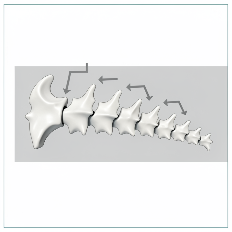
Thoracic Vertebrae Gradient
AI-generated conceptual illustration (African Lion (Panthera leo) thoracic vertebrae). It explains the idea and does not show study data.
View source evidence ↗Figures & tables
From the original paper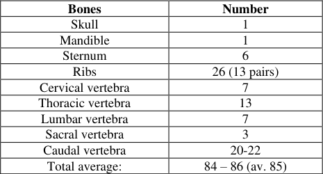 Table 1
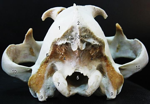 Figure 1
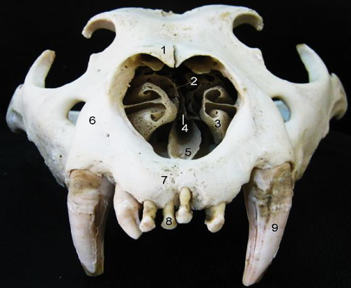 Figure 2
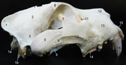 Figure 3
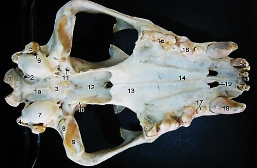 Figure 4
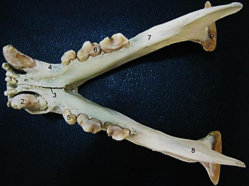 Figure 5
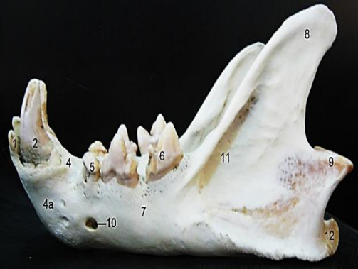 Figure 5
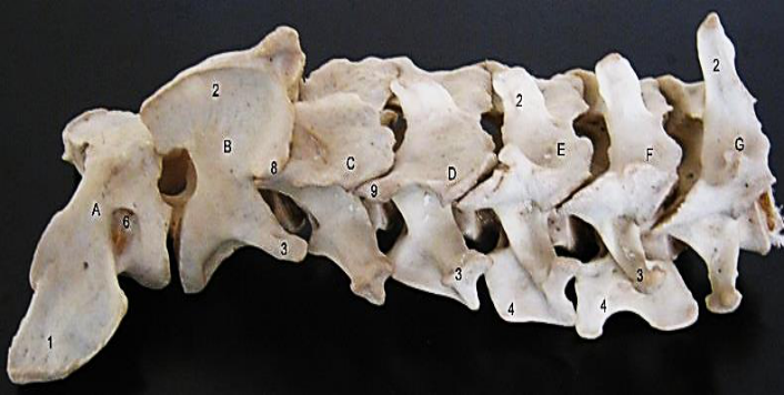 Figure 6
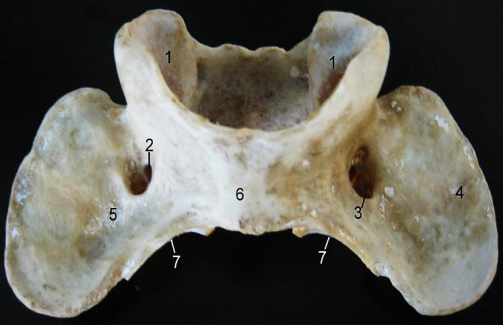 Figure 7
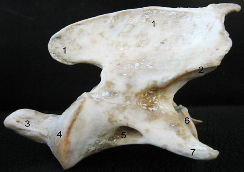 Figure 8
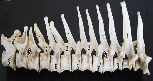 Figure 9
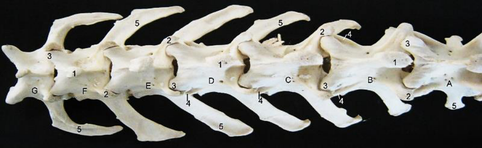 Figure 10
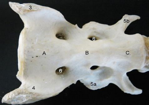 Figure 11
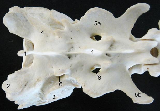 Figure 11
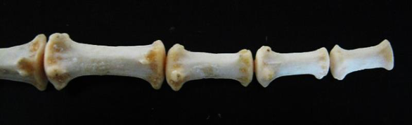 Figure 2
Introduction
The African lion (Panthera leo) is a representative of the family Felidae of the order Carnivora and suborder Feliformia [1] . It is closely related to the leopard, tiger and jaguar which are members of the same genus Panthera [2] . It is the largest carnivore and best studied mammalian species in African ecosystem [3,4,5] having been in existence for almost 3 million years in Laetoli, Tanzania [6] . The African Lion (Panthera leo) has cultural significance in entertainment; national flag symbols and literatures because of its recognition in Human culture [7,8] . Lions have been kept in exhibition since the time of the Roman Empire and have been kept in zoos worldwide since the late 18th century [9] . Its characteristics, significance and behaviour further increases its interests in scientific investigations with anatomy not excluded; which exposes adaptive features of this species. To this end, this study on the axial skeleton was conducted to investigate other specific gross features and number of bones making up this region of the animal skeleton. It also serves to enhance better understanding of its features when comparing with other species of felidae.
Materials and Methods
Two adult African lions (Panthera leo) weighing 173kg and 112kg were obtained as postmortem carcasses at different periods from the Department of Veterinary Pathology, University of Ilorin and transported to the Department of Veterinary Anatomy gross laboratory, University of Ilorin, Nigeria for bone preparation as museum specimens. They were carefully de-fleshed using sharp knife and scalpel blade to remove the skin and tease out muscle leaving the bones with minimal soft tissue attachment before being transferred to a large container of cold water at room temperature enough to submerge the bones. The container was then covered airtight and placed under shade throughout the period of maceration with a change of water regularly. After completion, the water was drained, the bones recovered and sun dried. Photographs of bones recovered were taken individually. They were also articulated using glue while noting the bones that constituted each segment of the vertebral column. The procurement of specimen carcass and experiments conformed to the European Convention for the protection of Vertebrate animals used for scientific purposes (Council of Europe No. 123, Strasbourg 1985). The experimental protocols described were approved by the Ethics review committee for Animal experimentation of University of Ilorin, Ilorin, Nigeria.
Results
The axial skeleton comprised the skull (made up of small flat paired and unpaired bones joined together) and the vertebral column (Cervical vertebrae, Thoracic vertebrae, lumbar vertebrae, sacral vertebrae and caudal vertebrae) which gave a formula of C7 T13 L7 S3 C20-21. They exhibited general and peculiar features similar to and different from other domestic animals studied. The average number of bones of the axial skeleton was established to be an average of 85.
The Skull
The Skull comprised of flat bones joined together by sutures.
It presented an elongated structure consisting of cranial and facial bones (fig. 3). Dorsocaudally, it presented an occipital bone that continued cranially with the parietal bones. It formed a crest medially that eventually ends as a spine (fig. 1). The crest of the parietals extended caudally to join that of the occipital forming the external occipital protuberance (fig. 1). There was no obvious suture separating the occipital from parietal bone. The occipital bone extended on the caudal surface to present squamous and lateral parts. The squamous part bounded the large foramen magnum dorsally presenting a medial crest (that terminated 3c.m above the foramen magnum) and a lateral crest that continued with the temporal bone. Each lateral side presented a condyle and a paramastoid process (attaching cranioventrally with tympanic bulla). These two components were separated by a wide, shallow condyloid fossa and placed lateral to the foramen magnum.
The parietal bone articulated rostrally with the frontal bone (via the parietofrontal suture) which presented a somewhat butterfly shape. The frontal bone presented dorsolaterally the zygomatic processs of the frontal bone. It articulated rostrally with the nasal, maxilla and lacrimal bones on either side. The nasal bones joined by the internasal suture formed the roof of the nasal cavity and articulated laterally with the maxilla and rostroventrally with the premaxilla which extended rostrally to house the incisors in its ventral alveoli. The maxilla occupied the rostro-lateral aspect of the skull. Its dorsal border met with the nasal bones, the caudal border met with the lacrimal and malar bones while its ventral border presented alveoli for housing the canine, premolar and molar teeth of the upper jaw. Close to its caudal border, the maxilla presented a large infraorbital foramen. The lacrimal bone presented a small triangular process and a lacrimal foramen on its orbital surface. Cranially, the nasal cavity presented perforated bony structure known as turbinate bone or nasal concha laterally with the perpendicular plate of ethmoid seen dividing the cavity into two equal halves. The floor of the cavity was occupied by the vomer medially and palatine bone laterally.
On the lateral view, the malar bone articulated with the maxilla rostrally. It extended caudo dorsally and caudo ventrally to form the frontal and temporal processes respectively. The frontal process forms an incomplete orbital margin while the temporal process meets with the zygomatic process of the temporal bone to form the Zygomatic arch. The squamous part of the temporal bone articulated with the ventral border of the parietal, continued the occipital crest to form the mastoid process before detaching laterally its zygomatic process. On close examination of the orbital wall cranial to the squamous temporal, the following foramina were observed on the orbital wing of sphenoid namely: ethmoid foramen (dorsal), optic foramen (middle), and orbital foramen (ventral). On the perpendicular part of the palatine were the large and caudal sphenopalatine foramen, the caudal palatine foramen and more rostral maxillary foramen. Ventrally, the skull presented at its cranial part the basilar part of the occipital bone which continued medially with the basisphenoid and presphenoid. The petrous part of the temporal bone lodged laterally having a large tympanic bulla, small muscular process with no visible styloid process.
Between the bulla and the basisphenoid cranially were two foramina the small hypoglossal foramen and large jugular foramen. The external acoustic meatus and foramen lacerum were located lateral and ventral to the tympanic bulla respectively. Lateral to the basisphenoid presented the alar foramen. The pterygoid bone anchored ventrally from the wing of sphenoid to end in a process and a hamulus. Rostral to the presphenoid, the vomer projected into the nasal cavity while the palatine (horizontal part) having the palatine foramen greatly reduced the size of the posterior choarnae.
The palatine process of the maxilla extended rostrally from the palatine to the incisors while creating an opening, the palatine fissure.
The Mandible (Fig. 5) presented a mandibular symphysis rostro-medially that joins each mandibular half forming the body of the mandible that anchored the lower canine and incisor teeth. It also presented the horizontal ramus which anchored the premolar and molar dorsally with the mental foramen visible laterally. The vertical ramus formed a deep masseteric fossa laterally and continued caudally to form the coronoid process dorsally, the mandibular condyle at the middle and the angular process ventrally. Rostral to the middle of the mandibular condyle and angular process on the medial aspect presented the mandibular foramen.
The Vetebral Column
The Vertebral column consisted of 7 cervical vertebrae, 13 thoracic vertebrae, 7 lumbar vertebrae, 3 sacral vertebrae and 20-21 caudal vertebrae. The Typical cervical vertebrae (3 rd -7 th ) presented a body and an arch which enclosed a vertebral foramen. Emerging from the arch were the spinous process dorsally, the transverse processes laterally, the articular processes with the facet cranially and caudally. On the transverse process, close to the body presented transverse foramen. The 4 th to 6 th cervical vertebrae had in addition to the above features a ventral process while the 7 th cervical had no transverse foramen. The Atlas presented a modified transverse process, the butterfly shaped wings which formed a depression (the Atlanta fossa) and housed the transverse foramen on its ventral view. A small foramen was also seen associated with the transverse foramen which led to a canal that opened on the caudal border of the wings. The alar notch and lateral vertebral foramen were seen on the dorsal surface.
The Axis presented an odontoid process that projected cranially from the body medial to the cranial articular facets. Its prominent feature was the large spinous process with a crest that hangs (but not completely) over the odontoid process. The Thoracic vertebrae presented a prominent long spinous process that reduced in length caudally and inclined obliquely from the 7 th . It also presented short transverse processes that lacked transverse foramina. The bodies of each vertebra presented coastal facets cranially and caudally for articulation with the head of rib. The transverse processes of the 1 st to 11 th presented coastal facets on their lateral borders for articulation with the tubercle of the ribs. The 13 th thoracic vertebrae presented a mammillary process on the cranial articular process, the shortest spinous process and lacked a caudal coastal facet. The lumbar vertebrae presented prominently, long transverse processes that increased and curved cranially from the 2 nd to the 7 th . The 7 th lumber's transverse process specifically made a 90° curve while that of the 1 st didn't curve at all. Accessory process was seen lateral to the caudal articular processes of the 1 st to 4 th but was rudimentary on the 5 th to 7 th lumber vertebrae. The sacrum presented 3 fused bones. The first one presented cranial articular processes (with facets for articulation with the 7 th lumbar vertebra) and wings laterally (with an auricular surface for articulation with the pelvis). The transverse process of the second and third were fused but distinguishable. While the second was broad with a convex lateral border, that of the third was slender and curved backwards. Only 2 dorsal and ventral sacral foramina were visible on each side. Despite the fusion, the spinous processes were easily identified and not fused. The caudal vertebrae presented three different parts of varying shapes and sizes craniocaudally. The spinous process was absent in all the caudal vertebrae. Transverse, cranial and caudal articular were present in the cranial parts, rudimentary in the middle (forming haemal arches) and absent in the caudal parts of the vertebrae.
The Thorax
The ribs formed the skeleton of the lateral thoracic wall; which presented a dorsal bony and ventral cartilaginous part; the costal cartilage. The proximal extremity of each rib presented a head, neck, and a tubercle which articulated with the corresponding thoracic vertebrae. The body curved downwards forming an angle with a small depression along the curved surface called costal groove. On its medial surface the costal cartilage articulated with the sternum. The distal end of the body articulated with the costal cartilage forming the costochondral junction.
The sternum presented a somewhat fused structure with bones separated by cartilage. The bones known as sternebrae were 7 in number. The 1st sternebra presented a triangular shaped pointed cranial end with a straight caudal part looking like the 2 nd to 6 th . The 7 th was rod shaped ending with a xyphoid process.
Discussion
The elongated structure of the skull with flat paired and unpaired cranial and facial bones adopts similar presentation to most animals especially the cat and dog. However, salient differences were observed. The paired nasal bones formed the straight dorsum of the nasal cavity entirely such that it articulated ventro-cranially and ventro-caudally with the premaxilla and maxilla respectively. This is similar to the cat [10] with its nasal bone curving dorso-craniad. The dog's nasal bone articulates laterally with the premaxilla and maxilla [11] .
The prominent median crest of the occipital bone and its termination about 3cm above the foramen magnum presents another significant difference when compared to the dog and cat [12] The incomplete margin of the orbit and absence of supraorbital foramen were similar to that of the cat, dog and pig though the supraorbital groove is present in the pig [13] .
The Vertebral formula C7 T13 L7 S3 C20-21 was typical of the feline and canine family only with the exception of the caudal vertebrae which is variable in number [14] . The Atlas with its butterfly shaped wings presented similar structure to that seen in the dog and cat with the exception of small foramen associated with the transverse foramen. The spinous process of its axis does not project completely over the odontoid process as seen in the dog [15] . The arrangement of the thoracic vertebrae such that the spinous processes reduce in height from the 2 nd to 13 th gives the saddle-like structure of the lion's dorsum. The cranially directed orientation of the lumbar vertebra's transverse processes was seen to be similar to the cat and dog while in other species like the Ox, horse and pig, they were placed horizontally [13] . The 90-degree angle curve of the 7 th was only similar to the cat [10] . Though the number of fused bones of the sacrum are similar to the cat and dog, its semi fused but distinguishable transverse processes and non-fused distinct spinous processes were perculiar features different from that of the dog which presents completely fused processes [14] . The presentations of the caudal vertebra, thorax and sternum was similar to that of the cat and dog [15] .
Conclusion
This study presented the numerical and morphological information on bones of the Lion's axial skeleton highlighting specific features, similarities and differences from other feline and canine species.
Acknowledgement
The authors wish to acknowledge the technologists and academic staff of Department of Veterinary Pathology, Faculty of Veterinary Anatomy, University of Ilorin, Nigeria, for their assistance in acquiring the carcass and in the bone preparation process.
Figures and tables from the paper
| Bones | Number |
|---|---|
| Skull | 1 |
| Mandible | 1 |
| Sternum | 6 |
| Ribs | 26 (13 pairs) |
| Cervical vertebra | 7 |
| Thoracic vertebra | 13 |
| Lumbar vertebra | 7 |
| Sacral vertebra | 3 |
| Caudal vertebra | 20-22 |
| Total average: | 84 – 86 (av. 85) |
Table text extracted from the original PDF; superscript letters are shown inline. The original table image is below.
View original table image
References
15 in the original paper- Wozencraft WC. "Panthera leo" In Wilson, D.E., Reeder, D.M (eds.). Mammal Species of the World: A Taxonomic and Geographic Reference 3rd edition Johns Hopkins University Press 2005, 546.
- Tseng ZJ, Wang X, Slater GJ, Takeuchi GT, Li Q, Liu J et al. "Himalayan fossils of oldest known pantherine establish ancient origin of big cats". Proceedings of the Royal Society B: Biological Sciences 2014;281:(1774).
- Bygott JD, Bertram BCR, Handy JP. Male lions in large coalitions gain reproductive advantages. Nature 1979;282(20-27):839-841.
- Packer C, Hilborn R, Mosser A, Kissui B, Borner M, Hopcraft G. Ecological change, group territoriality and non-linear population dynamics in Serengeti lions. Science 2005;307:390-393.
- Schaller GB. The Serengeti Lion; study of predator-prey relations. University of Chicago press 2009, 28-30.
- Werdelin L, Harrison T, Dehghani R. Paleontology and Geology of Laetoli: Human Evolution in Context Vertebrate Paleobiology and Paleoanthropology. lars werdelin publikationer ny naturhistorika riksmuseet. Springer: Netherlands 2011, 189-232.
- Hogarth C, Butler N. "Animal symbolism (Africa)" In Walter, M.N. (ed.) Shamanism: An encyclopedia of World beliefs, practices and culture 2004;1:3-6.
- Pinch G. "Lion" Egyptian mythology A to Z. Infobase publishing 2004, 63.
- Guggisberg CAW. Wild Cats of the World. New York: Tap linger Publishing 1975, 138-179.
- Sebastiani AM, Fishbeck DW. Mammalian Anatomy: The Cat. Morton Publishing Company. Colorado, U.S.A. 2006, 6-27.
- Adams DR. Canine anatomy: A systemic study. Iowa state Press. U.S.A 2004, 113-124.
- Dyce KM, Sack WO, Wensing CJG. Text book of Veterinary Anatomy. Saunders Elseviers, St. Lois Missouri. U.S.A. 4 th edition 2010, 407-414.
- Adams B, Crabtree P. Comparative Osteology: A laboratory and Field Guide of common North American Animals. Academic press. Oxford, U.K. 2012, 156-214.
- Konig HE, Liebich H. Veterinary Anatomy of Domestic mammals: Textbook and colour Atlas. Schattauer, Stuttgart, Germany 2004, 27-90.
- Evans HE, De-lahunta A. Miller's Anatomy of the Dog. Elsevier Saunders. U.S.A 2013, 85-126.
Cited by 2
From OpenAlex, OpenCitations and reference lists read by Scinovex
- Allometry and Atlas Shape Analysis Between Tekir and Mix-breed Cats ↗
Ermiş Özkan, Burak Ünal, Yusuf Altundağ, Zihni MUTLU, Kozet Avanus · Kafkas Universitesi Veteriner Fakultesi Dergisi · 2023
The first cervical vertebra, the atlas, connects the skull to the body and plays a crucial role in supporting the skull and holds a distinct position within the atlanto-axial complex.In this study was aimed to examine the shape variation of the atlas in cats and to reveal shape changes related to variation in size via geometric morphometry.A total of 61…
❝ 4PDF - Use of epidural anaesthesia as analgesic management in an asiatic lion (Panthera leo persica) ↗
Cristina Sánchez, Eva Martínez‐Nevado, Mario Arenillas · Veterinary Research Communications · 2025
❝ 1PDF