
Research article · 2020 · Full text
International Journal of Veterinary Sciences and Animal HusbandryUltrastructural studies on the infundibulum of Oviduct in emu (Dromaius novaehollandiae)
Volume 5(2) · Pages 19–21
Research areasPhotosynthetic Processes and MechanismsPlant Reproductive BiologyProtist diversity and phylogeny
Abstract
Author-writtenThe Ultrastructure of infundibulum was studied in eight adult apparently healthy emu birds. The mucosal surface contained spirally oriented unbranched longitudinal ridges in the funnel part of infundibulum with shallow furrows. The epithelial cells contained cylindrical cilia of uniform length and microvilli, in which clumps of cilia were observed. Four types of cells were observed in the lining epithelium, Viz., ciliated, non-ciliated, basal and mucous secretary cells (Goblet cells). The goblet cell was distended with secretion and contained elongated mitochondria throughout the cytoplasm. The roots of cilia were found to be concentrated with prominent mitochondria. The columnar ciliated cells showed little evidence of secretary activity by membrane bound electron dense secretary granules.
Summary
Source-checkedPlain-language reading of the full paper, prepared with AI and checked against the text. Each point links to the authors' own words.
Why it matters
- Emus are the second largest birds and are commercially raised globally for high-value products including meat, oil, skin, and feathers. View evidence ↗
- While ultrastructural studies of the infundibulum have been performed on ostriches, there was a need to examine this structure in emus. View evidence ↗
The question
- The objective of this study was to examine the ultrastructure of the infundibulum in the emu (Dromaius novaehollandiae). View evidence ↗
What they did
- The study examined the infundibulum of eight adult, apparently healthy emu birds. View evidence ↗
- Scanning Electron Microscopy (SEM) was used to analyze the surface morphology using a JOEL-JSM 5600 model. View evidence ↗
- Transmission Electron Microscopy (TEM) was used to analyze internal cellular structures using a Hitachi H-7500 model. View evidence ↗
- TEM preparation involved semi-thin sections (200-300 nm) stained with Toludine blue and ultra-thin sections (50-70 nm) mounted on copper grids. View evidence ↗
What they found
- SEM revealed that the funnel part of the infundibulum's mucosal surface has shallow furrows and unbranched longitudinal ridges oriented spirally. View evidence ↗
- The epithelial cells were characterized by microvilli and cylindrical cilia of uniform length, sometimes appearing in clumps. View evidence ↗
- The lining epithelium of the infundibulum consists of four cell types: ciliated, non-ciliated, basal, and mucous secretory (Goblet) cells. View evidence ↗
- Goblet cells were found to be distended with secretions and contained elongated mitochondria throughout their cytoplasm. View evidence ↗
- Prominent mitochondria were found concentrated around the roots of the cilia. View evidence ↗
- Columnar ciliated cells exhibited minimal secretory activity, indicated by the presence of membrane-bound electron-dense secretory granules. View evidence ↗
- Tubular glands were observed to open onto the surface epithelium in the spaces between mucosal folds. View evidence ↗
Visual summary
1 visualsIllustrations explain the paper; they do not show study data.
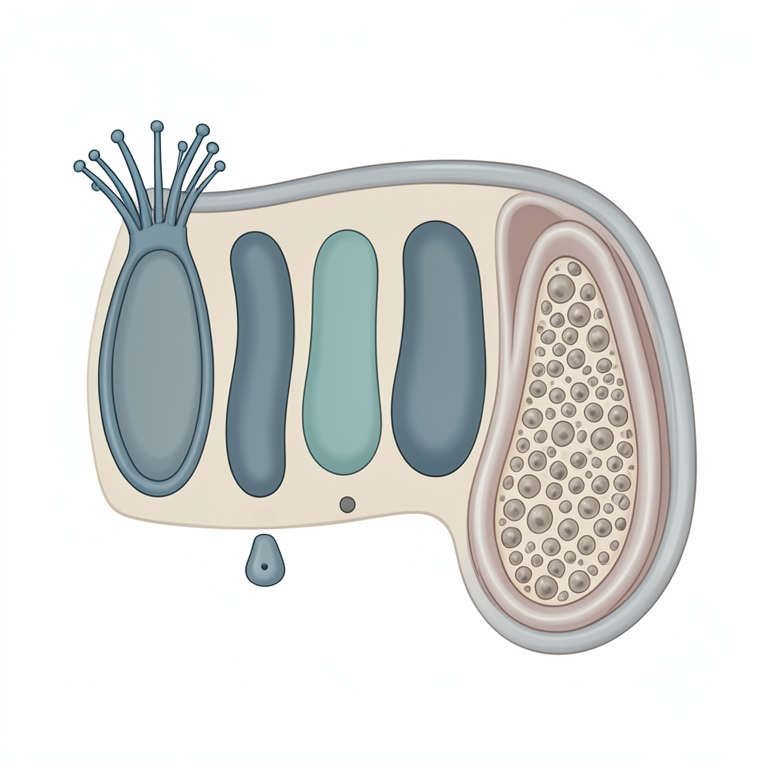
Epithelial Cell Diversity
AI-generated conceptual illustration (emu (Dromaius novaehollandiae) oviduct infundibulum epithelium). It explains the idea and does not show study data.
View source evidence ↗Figures & tables
From the original paper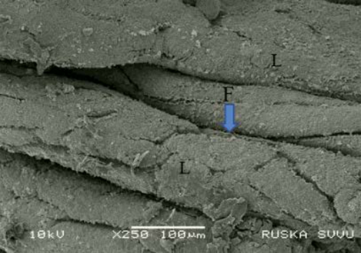 Figure 1
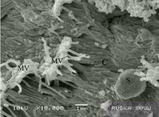 Figure 2
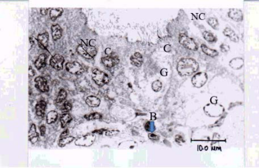 Figure 3
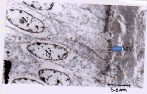 Figure 4
Introduction
The emu is the second largest bird and belonged to order Ratite. These birds are reared commercially in many parts of the world for their meat, oil, skin and feathers, which are of high economic value (Sibley and Ahlquist, 1990; Patodkar et al., 2009; Sreedevi et al, 2012; Supriya Shukla et al, 2013). The ultra structural studies on the infundibulum have been carried out in Ostrich (Sharaf et al. 2012).So the present study was initiated to examine the ultrastructure of the infundibulum in emu (Dromaius novaehollandiae).
Materials And Methods
The present work on “Micro anatomical and Histochemical studies on the Oviduct of Emu bird (Dromaius novaehollandiae)” was conducted at the Department of Veterinary Anatomy, College of Veterinary Science, Rajendranagar, Hyderabad. Fresh tissue samples were collected immediately from different regions of the oviduct like infundibulum. For SEM, fixed samples collected from different regions of oviduct were dehydrated in series of graded alcohol and were dried with CPD unit. The dried samples were mounted over the stubs with double-sided conductivity tape and were coated by a thin layer of gold metal over the samples using an automated sputter coater for about 3min (Bozzola et al., 1999).
The samples were scanned under Scanning Electron Microscope (model: JOEL-JSM 5600, Japan). For TEM, the tissues from different regions of the oviduct were dehydrated in series of graded alcohol from 50% to 100% for 40 minutes each, infiltrated in 1:1 alcohol and resin, pure resin and later embedded in pure Spurr resin. Both semi thin and ultra thin sections were cut with a glass knife on a Leica Ultra cut UCT-GA-D/E-1/00 ultramicrotome. Semi thin sections of 200- 300 nm were stained with Toludine blue whereas, ultra thin sections (50-70 nm) were mounted on copper grids.
Then the sections were stained with saturated aqueous Uranyl acetate for 30 minutes and counter stained with 4% Lead citrate for 20minutes (Bozzola et al., 1999) [9] and were later observed under Transmission Electron Microscope (Model:Hitachi,H-7500, Japan).
Results And Discussion
Scanning electron microscopy The mucosal surface contained spirally oriented unbranched longitudinal ridges in the funnel part of infundibulum. The tubal part of infundibulum contained small secondary folds on the http://www.veterinarypaper.com Fig 3: Transmission Electron micrograph (TEM) of infundibulum showing lining epithelium containing ciliated (C), non ciliated (NC), basal (B) and mucous secretary cells (Goblet cells) (G), X1930 larger primary ridges. Parto et al. (2011) [11] also reported that the infundibulum of the turkey showed spirally oriented longitudinal ridges and contained small secondary folds on them. The surface of infundibulum was lined by ciliated columnar epithelium with deep furrows in between.
Similar finding was reported in ostrich by Saber et al. (2009) [12]. In the present study, the epithelial cells contained cylindrical cilia of uniform length and microvilli. Transmission electron microscopy In the present study, the tubular glands were opened onto the surface epithelium between the mucosal folds. However Wyburn et al. (1970) [13] reported that the tubular glands in sub-epithelial layer were lined with cells containing large granular endoplasmic reticular (GER) space filled with homogeneous material of low electron density.
Similar observations were reported in fowl. In the present study, four types of cells were observed in the lining epithelium of the infundibulum, Viz., ciliated, non ciliated, basal and mucous secretary cells (Goblet cells). However, Wyburn et al. (1970) [13] reported that the surface ridges of infundibulum in fowl was lined with columnar epithelium and contained only ciliated cells, with little secretary activity and granular cells with electron dense granules. Fig 4: Transmission Electron micrograph (TEM) of the lining epithelium of infundibulum showing ciliated cells with achromatic oval nuclei (N) in central or apical portions and long, complex and microvilli (MV) on apical surface and prominent mitochondria (M) around the roots of the cilia, X3860 Fig 1: Scanning Electron micrograph (SEM) of infundibula funnel surface showing spirally oriented longitudinal ridges (L) with furrows (F) in between, X250
Figures and tables from the paper
References
13 in the original paper- Aitken R.N.C. The oviduct. In Bell J.D and Freeman B.M. (eds) Physiology and Biochemistry of the Domestic Fowl London, Academicals Press 1971; 3:1237-1289.
- Aitken R.N.C, Johnston H.S. Observations on the fine structure of the infundibulum of the avian oviduct, Journal of Anatomy. 1963; 97:87-99.
- Arta M.E, Daghoglu S. Tavuk, Keklik ve bildircinda yumurta Yolunum mikroskopik yapis uzerinde karsilastirmali bir calisma Istanbul University Fak Derg. 1984; 10:17-28.
- American Emu Association. Unpublished data P.O Box 8174, DaUas, TX 75205, 1994.
- Bradley O. Notes on the histology of the oviduct of the domestichen J Anatomy. 1928; 62:339-345.
- Bradley O.C, Grahame T. The Structure of the Fowl 4th EDN, Edinburgh, Oliver and Boyd, London, 1960
- Banks W.J. Applied Veterinary Histology 1st EDN, Williams and Wilkins, Baltimore, London, 1981.
- Bacha W.J, Bacha L.M. Colour Atlas of Veterinary Histology 2nd EDN, Lippincott Williams and Wilkins, Philadelphia Opp, 1990, 240-243.
- Bozzola J.J Lonnie D, Russel. Electron Microscopy: Pinciples and Techniques for Biologists 2nd, Jones and Bartlett Publishers, Canada, 1999. Fig 2: Scanning Electron micrograph (SEM) of the infundibulum showing cylindrical cilia (C) of uniform length and microvilli (s- secretory cell), X10.000 http://www.veterinarypaper.com
- Bacha W.J, Bacha L.M. Colour Atlas of veterinary histology. Lippincott Williams and Wilkins, London 2nd EDN, 2000, 223-243.
- Parto P, Khaksar Z, Akramifard A, Moghii B. The microstructure of oviduct in laying turkey hen as observed by light and scanning electron microscopies World J Zoo. 2011; 2:120-125.
- Saber A.S, Emara S.A.M, Abosaeda O.M.M. Light scanning and transmission electron microscopically study on the oviduct of the ostrich (Struthio camelus) J Vet A nat. 2009; 2(2):79-89.
- Wyburn G.M, Johnston H.S, Draper M.H. Davidson The fine Structure of the infundibulum and magnum of the oviduct of Gallus domestic us O.J Exp physiology, 1970, 213-232.