
Research article · 2020 · Full text
International Journal of Veterinary Sciences and Animal HusbandryEpidemiological surveillance of Dengue fever: An overview
Volume 5(6) · Pages 01–10
Research areasMosquito-borne diseases and controlDengue and Mosquito Control ResearchViral Infections and Vectors
Abstract
Author-writtenDengue fever is the most common of all arthropod-borne viral diseases and has emerged as a major public health problem in recent years. Though the disease has been referred in ancient Chinese medical encyclopedia, the first case was reported in 1789 during the epidemics in Asia, Africa and North America. It was then when Benjamin Rush coined the term ‘break bone fever’ because of the symptoms of myalgia and arthralgia. However, the term dengue fever came into general use only after 1828 (Anonymous, 2010) [6]. Dengue viruses (DENVs) belong to family Flaviviridae, which are transmitted through mosquito; Aedes aegypti and also by Ae. albopictus. There are four sero types of the virus referred to as DENV-1, DENV-2, DENV-3 and DENV-4, actually originated in monkeys and independently jumped to humans in Africa or Southeast Asia between 800 and 100 years ago (Brett et al. 2007) [19]. Each dengue virus is an encapsulated RNA virus and is composed of three structural protein genes, which encode the nucleocapsid or core (C) protein, a membrane-associated protein, an enveloped (E) glycoprotein and seven non-structural (NS) proteins (Chaturvedi et al. 2005) [23, 24, 25, 26, 27, 28, 45, 94]. All four strains are capable of causing three spectra of disease – Dengue fever (DF), Dengue Haemorrhagic Fever (DHF) and Dengue Shock Syndrome (DSS) (David et al. 2010; Gupta et al.2012) [8, 34, 39, 44, 45].Today, about 2.5 billion people live in areas where, there is a risk of dengue transmission. It is endemic in over 100 countries in Asia, the Pacific, the Americas, Africa, and the Caribbean. The World Health Organization (WHO) estimates that 50 to 100 million infections occur each year, including 500,000 DHF cases and 22,000 deaths, mostly affecting children (Brett et al. 2007) [19].In India, the first confirmed case reports of dengue dates back in the year 1940. Since then many cases have been reported from various states (Gupta et al.2012) [8, 34, 44, 45]. The first major wide spread epidemics of DHF/DSS occurred in India in 1996 involving areas around Delhi and Lucknow and then it spreaded to all over the country (Anonymous, 2010) [6].
Summary
Source-checkedPlain-language reading of the full paper, prepared with AI and checked against the text. Each point links to the authors' own words.
Why it matters
- Dengue fever is the most frequent viral disease transmitted by arthropods and is currently a significant global public health issue. View evidence ↗
- The WHO estimates that between 50 and 100 million infections happen annually, resulting in 500,000 cases of Dengue Hemorrhagic Fever (DHF) and 22,000 deaths, primarily among children. View evidence ↗
- The global resurgence of dengue is attributed to factors such as rapid population growth, uncontrolled urbanization, increased air travel, poor public health infrastructure, and ineffective mosquito control. View evidence ↗
The question
- The paper aims to provide a comprehensive overview of the epidemiological surveillance of dengue fever. View evidence ↗
What they found
- Dengue viruses belong to the Flaviviridae family and are transmitted to humans primarily by Aedes aegypti and Aedes albopictus mosquitoes. View evidence ↗
- The dengue virus exists in four distinct serotypes: DENV-1, DENV-2, DENV-3, and DENV-4. View evidence ↗
- All four serotypes can cause a range of illness, from standard dengue fever (DF) to more severe forms like Dengue Hemorrhagic Fever (DHF) and Dengue Shock Syndrome (DSS). View evidence ↗
- In northern India, DV-2 genotype V was replaced by genotype IV over the last decade. View evidence ↗
- During a 2004 epidemic in northern India, DV serotype-3 (subtype III) became dominant, replacing the previously circulating serotype-2 (subtype IV). View evidence ↗
- Mosquitoes become infected when biting a person during their 2- to 10-day febrile period; the virus then reaches the mosquito's salivary glands approximately 8–10 days later. View evidence ↗
- The risk of severe disease increases during a secondary infection, particularly when a person previously infected with DENV-1 acquires DENV-2 or DENV-3, or when someone previously infected with DENV-3 acquires DENV-2. View evidence ↗
- Among the tested substances for controlling Aedes aegypti, Temephos was found to be the most effective, followed by fenthion, malathion, and DDT. View evidence ↗
What it means
- The authors conclude that health education programs are necessary to increase community knowledge and encourage participation in integrated vector control. View evidence ↗
- The authors note that many clinical studies in India are limited because they rely on diagnostic kits with questionable sensitivity and specificity. View evidence ↗
Visual summary
1 visualsIllustrations explain the paper; they do not show study data.
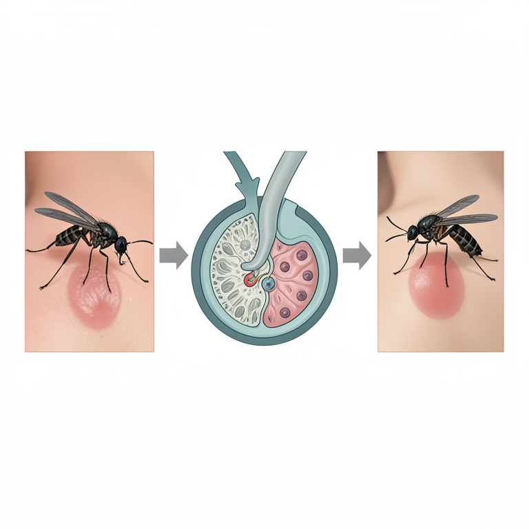
Dengue Transmission Cycle
AI-generated conceptual illustration (Aedes aegypti mosquito and human skin). It explains the idea and does not show study data.
View source evidence ↗Figures & tables
From the original paper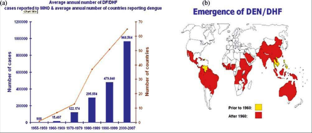 Figure 1
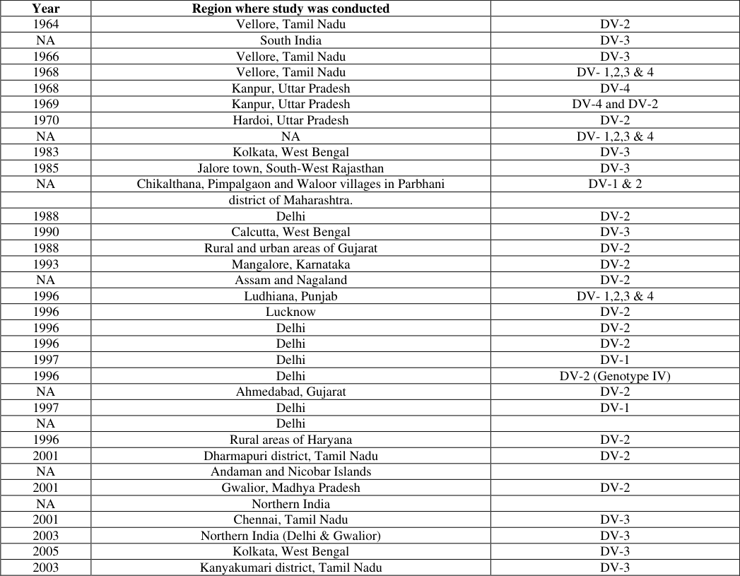 Table 1
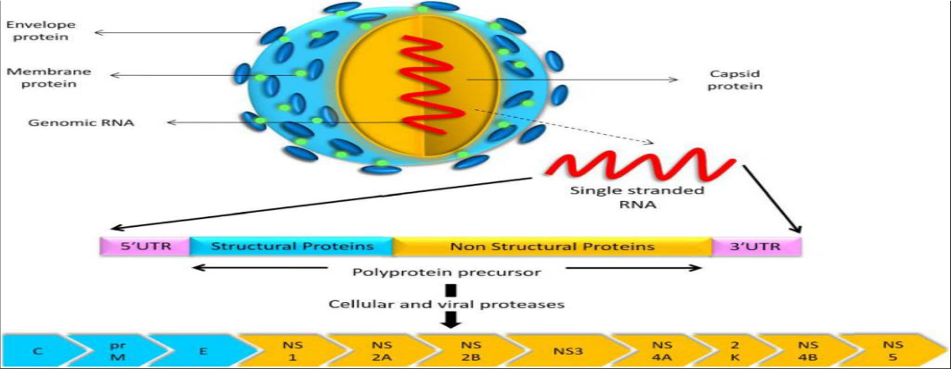 Figure 2
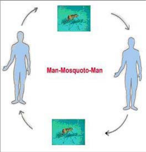 Fig. 3
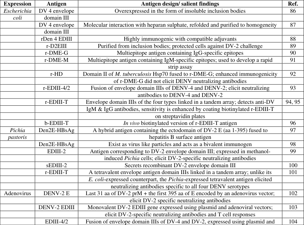 Table 2
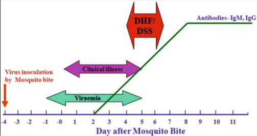 Fig. 4
Introduction
Dengue viruses (DV) belong to family Flaviviridae and have four serotypes (1 to 4). They are transmitted mainly by the Aedes aegypti mosquito and also by Aedes albopictus. Biologically, DV are highly adapted to the mosquito and are maintained by vertical transmission. DV produces from a subclinical infection to a mild self-limiting disease, the dengue fever (DF) and a severe disease that may be fatal, the dengue hemorrhagic fever/ dengue shock syndrome (DHF/DSS). The mosquito vectors are present in tropical and subtropical regions of the earth that determines the prevalence of DV in a region. Prior to 1970, only 9 countries had experienced cases of DHF; since then the number has increased more than 4-fold and continues to rise (figure 1a). The WHO published a global map of the distribution of the dengue epidemic activity during the year 2006 that shows whole India in red colour (figure 1b). Similar maps prepared in earlier years shows that the activity of both the vector and the virus has spread to newer areas, acquiring global public health importance. An estimated 2.5 billion people in more than 100 countries are at risk of acquiring dengue viral infection with more than 50 million new infections being projected annually, 500000 cases of DHF that must be hospitalized and 20000-25000 deaths, mainly in children (Kumar et al. 2010; Halstead 2002; Halstead 2007) [21,35,67,68] . Dengue has been an urban disease but now has spread to rural areas of India as well (Chaturvedi et al. 2004; Mehendle et al. 1991; Kumar et al. 2001; Arunachalam et al. 2004) [7,21,23,24,25,26,27,28,35,45,67,68,94] . The factors considered responsible for Global resurgence of DF/DHF are unprecedented population growth, unplanned and uncontrolled urbanization, increased Air travel, absence of an effective mosquito control programme and deterioration of Public Health infrastructure. The risk factors for infection with DV are the increased density of the mosquito vector, reinfestation with Ae. aegypti of a new geographical area, warm and humid climate, increased population density, water storage pattern in houses, storage of junk in open spaces, including tyres, coconut shells etc that trap rain water and introduction of new serotype of the virus, etc. Vaccines or antiviral drugs are not available for dengue viruses; the only effective way to prevent epidemic DF/DHF is to control the mosquito vector, Ae. Aegypti and prevent its bite.
History
Dengue virus was isolated in Japan in 1943 by inoculation of serum of patients in suckling mice (Tewari et al. 2004) [52,81,97] and at Calcutta (now Kolkata) in 1944 from serum samples of US soldiers (Whitehorn et al. 2010) [102] . [62,90] . The first major epidemic of the DHF occurred in 1953-1954 in Philippines followed by a quick global spread of epidemics of DF/DHF (Sarkar et al.1964) [22,91,92] . DHF was occurring in the adjoining countries but it was absent in India for unknown reasons as all the risk factors were present. [16,20,53,54] . The first major wide spread epidemics of DHF/DSS occurred in India in 1996 involving areas around Delhi (Dal et al. 1999) and Lucknow (Agraval et al. 1999) and then it spread to all over the country (Shah et al. 2004) [93] .
Epidemiology and geographical distribution
The epidemiology of dengue fevers in the Indian subcontinent has been very complex and has substantially changed over almost past six decades in terms of prevalent strains, affected geographical locations and severity of disease. The very first report of existence of dengue fevers in India was way back in 1946 (Karamchandani, 1946) [55] . Thereafter, for the next 18 years, there was no significant dengue activity reported anywhere in the country. [9,22,65,85] , it spread northwards and reached Delhi in 1967 (Balaya et al. 1969) [9] and Kanpur in 1968 (Chaturvedi et al.1968; Chaturvedi et al.1970) [23,24,25,26,27,28,45,94] . Simultaneously it also involved the southern part of the country (Myers et al. 1968; Ghosh et al. 1968) [42,43,74,75,76,77,78] and gradually the whole country was involved with wide spread epidemics followed by endemic/hyperendemic prevalence of all the four serotypes of DV. The epidemiology of dengue virus and its prevalent serotypes has been ever changing. The epidemic at Kanpur during 1968 was due to DV-4 (Chaturvedi et al. 1968) [23,24,25,26,27,28,45,94] and during 1969 epidemic, both DV-2 and DV-4 were isolated (Chaturvedi et al. 1972) [23,24,25,26,27,28,45,94] . It was completely replaced by DV-2 during 1970 epidemic in the adjoining city of Hardoi (Chaturvedi et al. 1974) [23,24,25,26,27,28,45,94] . Myers et al (Myers et al. 1968; Myers et al. 1970) [74,75,76,77,78] had reported the presence of DV-3 in patients and Ae. aegypti at Vellore during the epidemic of 1966 while during the epidemic of 1968, all the four types of DV were isolated from patients and mosquitoes (Myers et al. 1970) [74,75,76,77,78] . In another study Myers & Varkey (Myers et al. 1971) [74,75,76,77,78] reported an instance of a third attack of DV in one individual. DV-2 was isolated during the epidemics of dengue in urban and rural areas of Gujarat State during 1988 and 1989 (Mahadev et al. 1993) [43,70] . Outbreaks of dengue occurred in Rajasthan by DV-1 and DV-3 (Ghosh et al. 1974) [42,43] , DV-3 (Chouhan et al. 1990) [32] , Madhya Pradesh by DV-3 (Rodrigues et al. 1973) [32,89], Gujarat by DV-2 (Mahadev et al. 1993) [43,70] and in Haryana by DV-2 (Kumar et al. 2001) [21,35,67,68] . DV-2 was the predominant serotype circulating in northern India, including Delhi, Lucknow and Gwalior ( [15,20,33,34,35,38,44,82,98,99,100] while DV-1 was isolated during the 1997 epidemic at Delhi (Kurukumbi et al. 2001) [69] . The phylogenetic analysis by the Molecular Evolutionary Genetics Analysis programme suggests that the 1996 Delhi isolates of DV-2 were genotype IV. The 1967 isolate was similar to a 1957 isolate of DV-2, from India, and was classified as genotype V. This study indicates that earlier DV-2 strains of genotype V have been replaced by genotype IV (Singh et al. 1999) [43,95,96] . The Gwalior DV-2 viruses were classified into genotype-IV, and were most closely related to Delhi 1996 DV-2 viruses and FJ 10/11 strains prevalent in the Fujian State of China. However, two earlier Indian isolates of DV-2 were classified into genotype-V. Genotype V of DV-2 has been replaced by genotype IV during the past decade, which continues to circulate silently in north India, and has the potential to re-emerge and cause major epidemics of DF and DHF (Dash et al. 2004) [35,36,37,38,82] . DV-2 has also been reported from southern India -in Kerala alongwith DV-3 (Anoop et al. 2010) [1] . DV-3 has been isolated during the epidemics at Vellore in 1966 (Myers et al. 1968; Myers et al. 1969) [74,75,76,77,78] , at Calcutta in 1983 (Mukherjee et al. 1987) [16,73] and in 1990 (Bhattacharjee et al. 1993) [16] , at Jalore city, Rajasthan in 1985 (Chouhan et al. 1990) [32] at Gwalior in 2003 and 2004 (Dash et al. 2005; Paramasivan et al. 2010) [35,36,37,38,81,82] and at Tirupur, Tamil Nadu in 2010 (Paramasivan et al. 2010). [81] Phylogenetic analysis showed that the Madurai isolates were closely related to Gwalior and Delhi isolates. The emergence of DV-4 has been reported in Andhra Pradesh (Dash et al. 2011) [35,36,37,38,82] and Pune, Maharashtra (Dayaraj et al. 2011) [40] , which was also implicated in increased severity of disease. At Delhi, till 2003, the predominant serotype was DV-2 (genotype IV) but in 2003 for the first time all four dengue virus subtypes were found to co-circulate in Delhi thus changing it to a hyperendemic state (Dar et al. 2006) [15,20,33,34,37,38,44,100] followed by complete predominance of DV serotype 3 in 2005 (Gupta et al. 2006) [8,34,44,45] . During the 2004 epidemic of DHF/DSS in northern India a sudden shift and dominance of the DV serotype-3 (subtype III) occurred replacing the earlier circulating serotype-2 (subtype IV) (Dash et al. 2005) [35,36,37,38,82] . Cocirculation of DV serotypes in Delhi in 2003-2004 has also been reported (Dash et al. 2005) [35,36,37,38,82] , which may have implications for increased DHF/DSS. Emergence of a distinct lineage of DV-1, having similarity with the Comoros/Singapore 1993 and Delhi 1982 strains, but quite different from the Delhi 2005 lineage and microevolution of the pre-circulating DV-3 has been reported (Kukreti et al. 2008). Co-circulation of several serotypes of dengue viruses has resulted in concurrent infection in some patients with multiple serotypes of DV (Bharaj et al. 2008) [15,20] . Further, replacement of DV-2 and 3 with DV-1 as the predominant serotype in Delhi over a period of three years (2007-2009) has been reported (Ryes-Aldasoro, 2017). Concurrent infection by Chikungunya and DV-2 was reported from Vellore (Chakravarti et al. 2008) [16,20,22,73] and Delhi (Myers et al. 1967) [74,75,76,77,78] (Table 1).
| Year | Region where study was conducted | — |
|---|---|---|
| 1964 | Vellore, Tamil Nadu | DV-2 |
| NA | South India | DV-3 |
| 1966 | Vellore, Tamil Nadu | DV-3 |
| 1968 | Vellore, Tamil Nadu | DV- 1,2,3 & 4 |
| 1968 | Kanpur, Uttar Pradesh | DV-4 |
| 1969 | Kanpur, Uttar Pradesh | DV-4 and DV-2 |
| 1970 | Hardoi, Uttar Pradesh | DV-2 |
| NA | NA | DV- 1,2,3 & 4 |
| 1983 | Kolkata, West Bengal | DV-3 |
| 1985 | Jalore town, South-West Rajasthan | DV-3 |
| NA | Chikalthana, Pimpalgaon and Waloor villages in Parbhani | DV-1 & 2 |
| district of Maharashtra. | ||
| 1988 | Delhi | DV-2 |
| 1990 | Calcutta, West Bengal | DV-3 |
| 1988 | Rural and urban areas of Gujarat | DV-2 |
| 1993 | Mangalore, Karnataka | DV-2 |
| NA | Assam and Nagaland | DV-2 |
| 1996 | Ludhiana, Punjab | DV- 1,2,3 & 4 |
| 1996 | Lucknow | DV-2 |
| 1996 | Delhi | DV-2 |
| 1996 | Delhi | DV-2 |
| 1997 | Delhi | DV-1 |
| 1996 | Delhi | DV-2 (Genotype IV) |
| NA | Ahmedabad, Gujarat | DV-2 |
| 1997 | Delhi | DV-1 |
| NA | Delhi | |
| 1996 | Rural areas of Haryana | DV-2 |
| 2001 | Dharmapuri district, Tamil Nadu | DV-2 |
| NA | Andaman and Nicobar Islands | |
| 2001 | Gwalior, Madhya Pradesh | DV-2 |
| NA | Northern India | |
| 2001 | Chennai, Tamil Nadu | DV-3 |
| 2003 | Northern India (Delhi & Gwalior) | DV-3 |
| 2005 | Kolkata, West Bengal | DV-3 |
| 2003 | Kanyakumari district, Tamil Nadu | DV-3 |
Table text extracted from the original PDF; superscript letters are shown inline. The original table image is below.
View original table image
Causative agent
Dengue virus (DENV) belongs to the family Flaviviridae, genus Flavivirus, and is transmitted to humans by Aedes mosquitoes, mainly Aedes aegypti. Flaviviruses have a (+) sense RNA genome and replicate in the cytoplasm of the host cells. The genome mimics the cellular mRNA molecule in all aspects except for the absence of the poly-adenylated (poly-A) tail. This feature allows the virus to exploit cellular apparatus to synthesise both structural and non-structural proteins, during replication. The cellular ribosome is crucial to the replication of the flavivirus, as it translates the RNA, in a similar fashion to cellular mRNA, resulting in the synthesis of a single polyprotein. In general, the genome encodes 3 structural proteins (Capsid, prM, and Envelope) and 7 non-structural proteins (NS1, NS2A, NS2B, NS3, NS4A, NS4B, NS5) (Chahar et al. 2009) [15, 20] . The genomic RNA is modified at the 5′ end of positivestrand genomic RNA with a cap-1 structure (me 7 -GpppAme 2 ).
Dengue virus and its serotypes
DV-1 was isolated in 1956 at Vellore. All the Indian DV-1 isolates belong to the American African (AMAF) genotype. The Indian DV-1 isolates are distributed into four lineages, India I, II, III and the Africa lineage. Of these, India III is the oldest and extinct lineage; the Afro-India is a transient lineage while India I is imported from Singapore and India II, evolving in situ, are the circulating lineages (Mehendale et al. 1991) [71,72] . The American genotype of DV-2 which circulated predominantly in India during the pre-1971 period, was subsequently replaced by the Cosmopolitan genotype. Post-1971 Indian isolates formed a separate subclade within the Cosmopolitan genotype. DV-2 strains were isolated in India over a time span of more than 50 years (1956-2011). The re-emergence of an epidemic strain of DV type-3 in Delhi in 2003 and its persistence in subsequent years marked a changing trend in DV circulation in this part of India (Dash et al. 2004) [35,36,37,38,82] . Occasional reports of circulation of DV-4 are also seen, though it is not the predominant type in India (Padbidri et al. 1995; Barua et al. 1996) [10,68,79,80] . Dengue virus is primarily transmitted by Aedes mosquitoes, particularly A. aegypti. These mosquitoes usually live between the latitudes of 35° North and 35° South below an elevation of 1,000 metres (3,300 ft). They typically bite during the early morning and in the evening, but they may bite and thus spread infection at any time of day. Other Aedes species that transmit the disease include A. albopictus, A. polynesiensis and A. scutellaris (Kaur et al. 1997) [56] . Humans are the primary host of the virus, but it also circulates in nonhuman primates. An infection can be acquired via a single bite. A female mosquito that takes a blood meal from a person infected with dengue fever, during the initial 2-to 10-day febrile period, becomes itself infected with the virus in the cells lining its gut. About 8-10 days later, the virus spreads to other tissues including the mosquito's salivary glandsand is subsequently released into its saliva (Agrawal et al. 1998). The virus seems to have no detrimental effect on the mosquito, which remains infected for life. Aedes aegypti is particularly involved, as it prefers to lay its eggs in artificial water containers, to live in close proximity to humans, and to feed on people rather than other vertebrates (Kabra et al. 1999) [20,53,54] . Dengue can also be transmitted via infected blood products and through organ donation. In countries such as Singapore, where dengue is endemic, the risk is estimated to be between 1.6 and 6 per 10,000 transfusions. Vertical transmission (from mother to child) during pregnancy or at birth has been reported. Other person-to-person modes transmission have also been reported, but are very unusual. The genetic variation in dengue viruses is region specific, suggestive that establishment into new territories is relatively infrequent, despite dengue emerging in new regions in recent decades (Chahar et al. 2009; Vajpayee et al. 1999) [15, 20, 100] .
Transmission of dengue virus
Transmission of dengue virus
Predisposition
Severe disease is more common in babies and young children, and in contrast to many other infections, it is more common in children who are relatively well nourished. Other risk factors for severe disease include female sex, high body mass index, and viral load. While each serotype can cause the full spectrum of disease, virus strain is a risk factor (Joshi et al.
2000) [51,79,80] . Infection with one serotype is thought to produce lifelong immunity to that type, but only short-term protection against the other three. The risk of severe disease from secondary infection increases if someone previously exposed to serotype DENV-1 contracts serotype DENV-2 or DENV-3, or if someone previously exposed to DENV-3 acquires DENV-2. Dengue can be life-threatening in people with chronic diseases such as diabetes and asthma (Singh et al. 2001) [43,95,96] .
Clinical features and pathogenesis
The patients initially develop an abrupt onset of high fever (39-40°C) with headache, retro-orbital pain, malaise, nausea, vomiting, and myalgia. The acute febrile stage lasts 2-7 days and may be followed by recovery but patients feel weakness. During defervescence some patients develop hemorrhagic manifestation that may be mild petechial haemorrhage, and bleeding at the nose, gastrointestinal tract and gums, which may be severe. Menorrhagia has been more prevalent due to the increasing number of affected adolescents, but haematuria is rare. Hepatomegaly is common with soft and tender liver. Thrombocytopoenia and rising haematocrit due to plasma leakage are usually detectable before the onset of the subsequent stage of shock (DSS) with an abrupt fall to normal or subnormal levels of temperature, varying degrees of circulatory disturbances lasting for 24-48 h. In recent publications a number of atypical manifestations of dengue, such as encephalitis/encephalopathy (Victor et al. 2002) [101] and myocarditis, hepatitis and cholecystitis etc. have been reported (Padbidri et al. 2002) [68,79,80] . The majority of patients have rapid uneventful recovery without sequelae in the convalescent stage. The sequence of events in a patient after bite of infected mosquito is summarized in figure 2. The clinical diagnosis of DHF is based on four main characteristic manifestations (Sarkar et al. 1964) [22,91,92] : (i) continuous high fever lasting 2-7 days; (ii) hemorrhagic tendency as shown by a positive tourniquet test, petechiae or epistaxis; (iii) thrombocytopoenia (platelet count less than 100 × 10 9 /l); and (iv) evidence of plasma leakage manifested by hemoconcentration (an increase in haematocrit 20% above average for age, sex and population), pleural effusion and ascites etc. But some patients with bleeding or anemia may not have a rising haematocrit. Therefore, close observation, serial haematocrit and daily platelet count monitoring are suggested to fulfill the clinical diagnostic criteria. The severity of DHF is categorized into four grades (Sarkar et al. 1964) [22,91,92] : grade I, being the mildest and grade IV being most severe, with circulatory failure manifested by a rapid and weak pulse with narrowing of pulse pressure (20 mmHg) or hypotension, with the presence of cold clammy skin and restlessness. There may be profound shock in which pulse and blood pressure are not detectable (DSS). In such patients the mortality rate is high.
Diagnosis
Diagnosis of dengue virus infection is confirmed in the laboratory. During the stage of fever there is viraemia (figure 2) with presence of NS1 antigens in blood. The presence of virus in blood is detected either by isolation of the virus using infant mice or in tissue culture or by RT-PCR and the NS1 is detected by ELISA. During the post-febrile stage lasting a few weeks, IgM and IgG antibodies are present and are detected by Capture-ELISA (Figure 4). During primary infection, viraemia and fever coincides, but during a secondary infection (second time infection with DV), the viraemia is present for 2 to 3 days, and NS1 antigens in blood lasts little longer. With a newer approach, artificial NS1 receptors have been implanted on a reuseable microchip that can capture and identify NS1 instantly and may be used for bedside diagnosis of dengue virus infection (Kabilan et al. 2004) [7,52] .
Treatment
The management of dengue virus infection is essentially supportive and symptomatic. No specific treatment is available. However, there are Indian studies which have contributed in terms of better management of DHF/DSS. A rapid response to platelet and fresh frozen plasma (FFP) transfusion is reported in a study. Anti-D has been used in children with DHF and severe refractory thrombocytopenia. In experimental study pre-feeding mice with trivalent chromium picolinate (CrP) in drinking water could abolish the adverse effects of DV infection on most of the haematological parameters. Hippophae rhamnoides (Seabuckthorn, SBT) leaf extract has been shown to have a significant anti-dengue activity.
Vaccine for dengue virus
Dengue vaccines have been under development since the 1940s, but a tetravalent vaccine which simultaneously provides long-term protection against all DV serotypes is round the corner 205. A tetravalent antigen was designed by splicing the EDIIIs of DV-1, DV-2, DV-3 and DV-4 using flexible pentaglycyl linkers. A synthetic gene encoding this tetravalent antigen was expressed in Pichia pastoris and purified to near homogeneity. This tetravalent antigen when injected into inbred BALB/c mice, elicited neutralizing antibodies specific to each of the four DVs in plaque reduction neutralization tests206. Efforts are underway to present the tetravalent antigen on a chimeric VLP platform. Some promising dengue antigens have been developed using different systems (Table 2). The EDIII-based tetravalent antigen expressed using plasmid and adenoviral vectors 105 elicit neutralizing antibodies and T cell responses specific to four DV serotypes
| Expression | Antigen | Antigen design/ salient findings | Ref. |
|---|---|---|---|
| Escherichia | DV 4 envelope | Overexpressed in the form of insoluble inclusion bodies | 86 |
| coli | domain III | ||
| DV 4 envelope | Molecular interaction with heparan sulphate, refolded and purified to homogeneity | 87 | |
| domain III | |||
| rDen 4 EDIII | Highly immunogenic with compatible adjuvants | 88 | |
| r-D2EIII | Purified from inclusion bodies; protected cells against DV-2 challenge | 89 | |
| r-DME-G | Multiepitope antigen containing IgG-specific epitopes | 90 | |
| r-DME-M | Multiepitope antigen containing IgM-specific epitopes; used to develop a rapid | 91 | |
| strip assay | |||
| r-HD | Domain II of M. tuberculosis Hsp70 fused to r-DME-G; enhanced immunogenicity | 92 | |
| of r-DME-G did not elicit DENV neutralizing antibodies | |||
| r-EDIII-4/2 | Fusion of envelope domain IIIs of DENV-4 and DENV-2; elicit neutralizing | 93 | |
| antibodies to DENV-4 and DENV-2 | |||
| r-EDIII-T | Envelope domain IIIs of the four types linked in a tandem array; detects anti-DV | 94, 95 | |
| IgM & IgG antibodies, sensitivity is enhanced by coating biotinylated r-EDIII-T | |||
| on streptavidin plates | |||
| b-EDIII-T | In vivo biotinylated version of r-EDIII-T antigen | 96 | |
| Pichia | Den2E-HBsAg | A hybrid antigen containing the ectodomain of DV-2 E (aa 1-395) fused to | 97 |
| pastoris | hepatitis B surface antigen | ||
| Den2E-HBsAg | Exist as virus like particles and acts as a bivalent immunogen | 98 | |
| EDIII-2 | Antigen corresponding to DV-2 envelope domain III; expressed in methanol- | 99 | |
| induced Pichia cells; elicit DV-2-specific neutralizing antibodies | |||
| sEDIII-2 | Secrets recombinant DV-2 envelope domain III | 100 | |
| r-EDIII-T | A tetravalent envelope antigen domain IIIs linked in a tandem array; unlike its | 101 | |
| E. coli-expressed counterpart, the Pichia-expressed tetravalent antigen elicited | |||
| neutralizing antibodies specific to all four DENV serotypes | |||
| Adenovirus | DENV-2 E | Last 31 aa of DV-2 prM + the first 395 aa of E encoded by an adenovirus vector; | 102 |
| elicit DV-2 specific neutralizing antibodies | |||
| DENV-2 EDIII | Monovalent DV-2 EDIII gene expressed using plasmid and adenoviral vectors; | 103 | |
| elicit DV-2-specific neutralizing antibodies and T cell responses | |||
| EDIII-4/2 | Fusion of envelope domain IIIs of DV-4 and DV-2, expressed using plasmid and | 104 |
Table text extracted from the original PDF; superscript letters are shown inline. The original table image is below.
View original table image
Recombinant dengue virus antigens
Several studies have contributed in terms of developing new reagents or technology for diagnostic purposes (Table 2). A recombinant DV3 envelope domain III (rDen 3 EDIII) protein has been produced in Escherichia coli for potential use in diagnosis. A biotinylated chimeric dengue antigen to exploit the high affinity of biotin-streptavidin interaction to detect anti-dengue antibodies has been developed which incorporates the envelope domain III of all four DV serotypes. Immunosensor has been established for label free and real time assay for the serological diagnosis of DV infection. Scope for development of biosensors for diagnosis was demonstrated. The recombinant dengue multiepitope (rDME-M) protein specific to IgM in E. coli was produced in a 5-L fermentor for use in diagnostic purpose.
Prevention and control
Aedes aegypti is the commonest vector of DV in India, followed by Ae. albopictus. Larval indices indicate that Ae. aegypti is well established in peri-urban areas and is beginning to displace Ae. albopictus. Water-holding containers, viz. plastic, metal drums and cement tanks facilitate breeding of Ae. aegypti. Expansion in the risk area of diseases borne by it in the context of urbanization, transport development and changing habitats is a major concern.
Vector control
Vector control is known to be a good method for prevention of vector borne diseases. There are several reports from India which have demonstrated resistance of mosquito vector with anti larval substances like DDT and dieldrin but susceptibility to malathione is reported. Temephos is relatively more effective in controlling Ae. aegypti, followed by fenthion, malathion and DDT. Peridomestic thermal fogging reduced the resting and biting for the 3 days after treatment, whereas indoor fogging suppressed adult populations for 5 days. Plant based repellent against mosquito borne diseases have also been described. Flavonoid compounds derived from Poncirus trifoliata compounds have various activities against different life stages of Ae. aegypti. Larvicidal and ovicidal activities of benzene, hexane, ethyl acetate, methanol and chloroform leaf extract of Eclipta alba have shown potential for controlling Ae. aegypti mosquito. Hydrophobic nanosilica at 112.5 ppm is effective against mosquito species.
Public health importance
Dengue is one of the major public health problems which can be controlled with active participation of the community. Need is to organize health education programmes about dengue disease to increase community knowledge and sensitize the community to participate in integrated vector control programmes.
Conclusions
Dengue disease continues to involve newer areas, newer populations and is increasing in magnitude, epidemic after epidemic. Every aspect of dengue viral infection continues to be a challenge; the pathogenesis of severe dengue disease is not known, no vaccine is yet available for protection and the vector control measures are inadequate. Dengue virus was isolated in India in 1944, but the scientific studies addressing various problems of dengue disease have been carried out at limited number of centres. Though clinical studies have reported on dengue disease in India, but these are largely based on diagnosis made by kits of doubtful specificity and sensitivity. A lot more remains to be achieved for creating an impact.
Figures and tables from the paper
References
92 in the original paper- for the detection of anti-dengue antibodies in human sera. BMC Infect. Dis. 2011; 11(3):64.
- Batra G, Raut R, Dahiya S, Kamran N, Swaminathan S, Khanna N. Pichia pastoris-expressed dengue virus type 2 envelope domain III elicits virus-neutralizing antibodies. J Virol. Methods. 2010; 167(1):10-6.
- Batra G, Talha SM, Nemanim SK, Dhar N, Swaminathan S, Khanna N. Expression, purification and characterization of in vivo biotinylated dengue virus envelope domain III based tetravalent antigen. Protein Exp. Purif. 2010; 74(1):99-105.
- Bharaj P, Chahar HS, Pandey A, Diddi K, Dar L, Guleria R. Concurrent infections by all four dengue virus serotypes during an outbreak of dengue in 2006 in Delhi, India. Virol. J. 2008; 5(9):1.
- Bhattacharjee N, Mukherjee KK, Chakravarti SK, Mukherjee MK, De PN, Sengupta M. Dengue hemorrhagic fever (DHF) outbreak in Calcutta -1990. J Commun. Dis. 1993; 25(1):10-4
- Bisht H, Chugh DA, Raje M, Swaminathan S, Khanna N. Recombinant dengue virus type 2 envelope/hepatitis B surface antigen hybrid protein expressed in Pichia pastoris can function as a bivalent immunogen. J Biotechnol. 2002; 99(2):97-110.
- Bisht H, Chugh DA, Swaminathan S, Khanna N. Expression and purification of dengue virus type 2 envelope protein as a fusion with hepatitis B surface antigen in Pichia pastoris. Protein Exp. Purif. 2001; 23(1):84-96.
- Brett D, Lindenbach, Heinz-Jurgen Thiel, Charles M. Rice. Flaviviridae: The Viruses and their Replication. In::Knipe, David M.; Howley, Peter M, editors. Fields Virology. United States: Lippincott Williams & Wilkins; P. 2007,1102-32.
- Chahar HS, Bharaj P, Dar L, Guleria R, Kabra SK, Broor S. Co-infections with chikungunya virus and dengue virus in Delhi, India. Emerg. Infect. Dis. 2009; 15(7):1077-80.
- Chakravarti A, Kumar A, Matlani M. Displacement of dengue virus type 3 and type 2 by dengue virus type 1 in Delhi during 2008. Indian J Med. Microbiol. 2010; 28(4):412-3.
- Chatterjee SN, Chakravarti SK, Mitra AC, Sarkar JK. Virological investigation of cases with neurological complications during the outbreak of hemorrhagic fever in Calcutta. J Indian Med Assoc. 1965; 45(3):314-6.
- Indian J Med. Microbiol. 2004; 22(1):5-6.
- Chaturvedi UC, Shrivastava R, Nagar R. Dengue vaccines: problems and prospects. Indian J Med. Res. 2005; 121(5):639-652.
- Chaturvedi UC, Kapoor AK, Mathur A, Chandra D, Khan AM, Mehrotra RML. A clinical and epidemiological study of an epidemic of febrile illness with hemorrhagic manifestations which occurred at Kanpur, India in 1968. Bull World Health Organ. 1970; 43(2):281-7.
- Chaturvedi UC, Mathur A, Kapoor AK, Mehrotra NK, Mehrotra RML. Virological study of an epidemic of febrile illness with hemorrhagic manifestations at Kanpur, India, during 1968. Bull. World Health Organ. 1970; 43(2):289-93.
- Chaturvedi UC, Mathur A, Kapoor AK, Tandon HO, Mehrotra, RML. Clinicovirological study of the recurrence of dengue epidemic with hemorrhagic manifestation at Kanpur, during 1969. Indian J Med. Res. 1972; 60(3):329-33.
- Chaturvedi UC, Mathur A, Mehrotra RM. Experimentally produced cardiac injury following dengue virus infection. Indian J Pathol. Bacteriol. 1974; 17(4):218-20.
- Chaudhary R, Khetan D, Sinha S, Sinha P, Sonker A, Pandey, P. Transfusion support to Dengue patients in a hospital based blood transfusion service in north India. Transfus. Apher. Sci. 2006; 35(3):239-44.
- Chaudhuri RN, Saha TK, Chaudhuri AD. Dengue-like fever in Calcutta: further preliminary observations. Bull. Calcutta Sch. Trop. Med. 1965; 13(1):2-3.
- Cherian T, Ponnuraj E, Kuruvilla T, Kirubakaran C, John TJ, Raghupathy P. An epidemic of dengue hemorrhagic fever & dengue shock syndrome in & around Vellore. Indian J Med. Res. 1994; 100:51-6.
- Chouhan GS, Rodrigues FM, Shaikhm BH, Ilkal MA, Khangaro SS, Mathur KN. Clinical & virological study of dengue fever outbreak in Jalore city, Rajasthan 1985. Indian J Med. Res. 1985; 91(11):414-8.
- Dar L, Broor S, Sengupta S, Xess I, Seth P. The first major outbreak of dengue hemorrhagic fever in Delhi, India. Emerg. Infect. Dis. 1999; 5(4):589-90.
- Dar L, Gupta E, Narang P, Broor S. Cocirculation of dengue serotypes, Delhi, India. Emerg. Infect. Dis. 2006. 2003; 12(2):352-3.
- Dash PK, Parida MM, Saxena P, Kumar M, Rai A, Pasha ST. Emergence and continued circulation of dengue-2 (genotype IV) virus strains in northern India. J Med. Virol. 2004; 74(2):314-22.
- Dash PK, Saxena P, Abhyankar A, Bhargava R, Jana AM. Emergence of dengue virus type-3 in northern India. Southeast Asian J Trop. Med. Public Health. 2005; 36(2):370-7.
- in India: implications for increased incidence of DHF & DSS. Virol. J. 2006; 3(7):55-65.
- Dash PK, Sharma S, Srivastava A, Santhosh SR, Parida MM, Neeraja M. Emergence of dengue virus type 4 (genotype I) in India. Epidemiol. Infect. 2011; 139(6):857-61.
- Hemorrhagic Fever, Japanese Encephalitis, West Nile Encephalitis, St. Louis Encephalitis, Tick-Borne Encephalitis). In: Mandell, Douglas, And Bennett's editors. Principles and practice of infectious diseases. 7 ed. Philadelphia. 2010, 2133 -56.
- Dayaraj C, Kakade MB, Bhagat AB, Vallentyne Singh J A, Patil JA. Detection of dengue-4 virus in Pune, western India after an absence of 30 years -its association with two severe cases. Virol. J. 2011; 8(2):46-9.
- Etemad B, Batra G, Raut R, Dahiya S, Khanam S, Swaminathan S. An envelope domain III-based chimeric antigen produced in Pichia pastoris elicits neutralizing antibodies against all four dengue virus serotypes. Am. J Trop. Med. Hyg. 2008; 79(3):353-63.
- Ghosh BN. A study on the epidemic of dengue-like fever in Pondicherry (1964-65 and 1965-66). J Indian Med. Assoc. 1968; 51(6):261-4.
- Ghosh SN, Pavri KM, Singh KR, Sheikh BH, D'lima LV, Mahadev PV. Investigations on the outbreak of dengue fever in Ajmer City, Rajasthan State in 1969. Part I. Epidemiological, clinical and virological study of the epidemic. Indian J Med. Res. 1974; 62(4):511-22.
- Gupta E, Dar L, Kapoor G, Broor S. The changing epidemiology of dengue in Delhi, India. Virol. J. 2006; 3(5):92-6.
- Gupta N, Srivastava S, Jain A, Chaturvedi U. Dengue in India. Indian J Med Res. 2012; 136(3):373-90.
- Guy B, Barrere B, Malinowski C, Saville M, Teyssou R, Lang J. From research to phase III: preclinical, industrial and clinical development of the Sanofi Pasteur tetravalent dengue vaccine. Vaccine. 2011; 29(42):7229-41.
- Halstead SB. Dengue. Current Opinion in Infectious Diseases. Halstead SB. Dengue; Lancet. 2007; 370(10):1644-1652
- Hapugoda MD, Batra G, Abeyewickreme W, Swaminathan S, Khanna N. Single antigen detects both immunoglobulin M (IgM) and IgG antibodies elicited by all four dengue virus serotypes. Clin. Vaccine Immunol. 2007; 14 (11):1505-14.
- Jain M, Ganju L, Katiyal A, Padwad Y, Mishra KP, Chanda S. Effect of Hippophae rhamnoides leaf against dengue virus infection in human blood-derived macrophages. Phytomedicine. 2008; 15(10):793-9.
- Jaiswal S, Khanna N, Swaminathan S. A replicationdefective adenoviral vaccine vector for the induction of immune responses to dengue virus type 2. J Virol. 2003; 77(23):12907-13.
- Jaiswal S, Khanna N, Swaminathan S. High-level expression and one-step purification of recombinant dengue virus type 2 envelope domain III protein in Escherichia coli. Protein Expr. Purif. 2004; 33(1):80-91.
- Joshi PT, Pandya AP, Anjan JK. Epidemiological and entomological investigation in dengue outbreak area of Ahmedabad district. J Commun. Dis. 2000; 32(1):22-7.
- Kabilan L, Velayutham T, Sundaram B, Tewari SC, Natarajan A, Rathnasamy R. Field-and laboratory-based active dengue surveillance in Chennai, Tamil Nadu, India: observations before and during the 2001 dengue epidemic. Am. J Infect. Control. 2004; 32(7):391-6.
- Kabra SK, Jainm Y, Pandey RM, Madhulika Singhal T, Tripathi P, Broor S, Seth P, Seth V. Dengue hemorrhagic fever in children in the 1996 Delhi epidemic. Trans. R. Soc. Trop. Med. Hyg. 1999; 93(3): 294-8.
- Kabra SK, Verma IC, Arora NK, Jain Y, Kalra V. Dengue hemorrhagic fever in children in Delhi. Bull. World Health Organ. 1992; 70(2):105-8.
- Karamchandani PV. Dengue group of fevers in India. Lancet. 1946; 1(6386):92.
- Kaur H, Prabhakar H, Mathew P, Marshalla R, Arya M. Dengue hemorrhagic fever outbreak in OctoberNovember 1996 in Ludhiana, Punjab, India. Indian J Med. Res. 1997; 106(7):1-3.
- Khanam S, Pilankatta R, Khanna N, Swaminathan S. An adenovirus type 5 (AdV5) vector encoding an envelope domain III-based tetravalent antigen elicits immune responses against all four dengue viruses in the presence of prior AdV5 immunity. Vaccine 2009. 2009; 27(43):6011-21.
- Khanam S, Etemad B, Khanna N, Swaminathan S. A bivalent antigen composed of linked envelope domains III of two dengue virus serotypes elicits neutralizing antibodies specific to both constituent serotypes. Am. J Trop. Med. Hyg. 2006; 74(2):266-77.
- Khanam S, Khanna N, Swaminathan S. Induction of antibodies and T cell responses by dengue virus type 2 envelope domain III encoded by plasmid and adenoviral vectors. Vaccine. 2006; 24(42-43):6513-25.
- Khanam S, Rajendra P, Khanna N, Swaminathan S. An adenovirus prime/plasmid boost strategy for induction of equipotent immune responses to two dengue virus serotypes. BMC Biotechnol. 2007; 7(2):10.
- Kharya G, Yadav SP, Katewa S, Sachdeva A. Management of severe refractory thrombocytopenia in dengue hemorrhagic fever with intravenous anti-D immune globulin. Pediatr. Hematol. Oncol. 2011; 28(8):727-32.
- Kimura R, Hotta S. Studies on dengue fever (IV) on inoculation of dengue virus into mice. Nippon. Igaku. 1944; 3379:629-33.
- Kolli R, Khanam S, Jain M, Ganju L, Sai Ram M, Khanna NV. A synthetic dengue virus antigen elicits enhanced antibody titers when linked to, but not mixed with, Mycobacterium tuberculosis HSP70 domain II. Vaccine. 2006; 24(22):4716-26.
- Kolli R, Khanam S, Jain M, Ganju L, Sai Ram M, Khanna NV. A synthetic dengue virus antigen elicits enhanced antibody titers when linked to, but not mixed with, Mycobacterium tuberculosis HSP70 domain II. Vaccine. 2006; 24(22):4716-26.
- Krishnamurthy K, Kasturi TE, Chittipantulu G. Clinical and pathological studies of an outbreak of dengue-like illness in Visakhapatnam. Indian J Med. Res. 1965; 53(8):800-12.
- Kukreti H, Chaudhary A, Rautela RS, Anand R, Mittal V, Chhabra M. Emergence of an independent lineage of dengue virus type 1 (DENV-1) and its co-circulation with predominant DENV-3 during the 2006 dengue fever outbreak in Delhi. Int. J Infect. Dis. 2008; 12(5):542-9.
- Kumar A, Rao Pandit CR, Shetty V, Bammigatti C, Samarasinghe CM. Clinical manifestations and trend of dengue cases admitted in a tertiary care hospital, Udipi district, Karnataka. Indian J of Community Medicine. 2010; 35(3):386-90.
- Kumar A, Sharma SK, Padbidri VS, Thakare JP, Jain DC, Datta KK. An outbreak of dengue fever in rural areas of northern India. J Commun. Dis. 2001; 33(4):27481.
- Kurukumbi M, Wali JP, Broor S, Aggarwal P, Seth P, Handa R. Seroepidemiology and active surveillance of dengue fever/dengue hemorrhagic fever in Delhi. Indian J Med. Sci. 2001; 55(3):149-56.
- Mahadev PV, Kollali VV, Rawal ML, Pujara PK, Shaikh BH, Ilkal MA. Dengue in Gujarat state, India during 1988 & 1989. Indian J Med. Res. 1993; 97(7):135-44.
- Mehendale SM, Risbud AR, Rao JA, Banerjee K. Outbreak of dengue fever in rural areas of Parbhani district of Maharashtra (India). Indian J Med. Res. 1991; 93(3):6-11
- Mehendale SM, Risbud AR, Rao JA, Banerjee K. Outbreak of dengue fever in rural areas of Parbhani district of Maharashtra (India). Indian J Med. Res. 1991; 93(1):6-11.
- Mukherjee KK, Chakravarti SK, Dey PN, Dey S, Chakraborty MS. Outbreak of febrile illness due to dengue virus type 3 in Calcutta during 1983. Trans. R. Soc. Trop. Med. Hyg. 1987; 81(6):1008-10.
- Myers RM, Carey DE. Concurrent isolation from patient of two arboviruses, Chikungunya and dengue type 2. Science. 1967; 157(3794):1307-8.
- Myers RM, Carey DE, Banerjee K, Reuben R, Ramamurti DV. Recovery of dengue type 3 virus from human serum and Aedes aegypti in South India. Indian J Med. Res. 1968; 56(6):781-7.
- Myers RM, Carey DE, De Ranitz CM, Reuben R, Bennet B. Virological investigations of the 1966 outbreak of Dengue type 3 in Vellore, Southern India. Indian J Med. Res. 1969; 57(8):1392-401.
- Myers RM, Varkey MJ, Reuben R, Jesudass ES..Dengue outbreak in Vellore, southern India, in 1968, with isolation of four dengue types from man and mosquitoes. Indian J Med. Res. 1970; 58(1):24-30.
- Myers RM, Varkey MJ. A note on sequential dengue infection, presumptive and proved, with report of an instance of a third proved attack in one individual. Indian J Med. Res. 1971; 59(8):1231-6.
- Padbidri VS, Adhikari P, Thakare JP, Ilkal MA, Joshi GD, Pereira P. The 1993 epidemic of dengue fever in Mangalore, Karnataka state, India. Southeast Asian J Trop. Med. Public Health. 1995; 26(4):699-704.
- Padbidri VS, Wairagkar NS, Joshi GD, Umarani UB, Risbud AR, Gaikwad DL. A serological survey of arboviral diseases among the human population of the Andaman and Nicobar Islands, India. Southeast Asian J Trop. Med. Public Health. 2002; 33(4):794-800.
- Paramasivan R, Thenmozhi V, Thangaratham PS, Rajendran R, Tewari SC, Dhananjeyan KJ. An outbreak of dengue fever in Tirupur, Coimbatore district, Tamil Nadu. Indian J Med. Res. 2010; 132(7):105-7.
- Parida MM, Dash PK, Upadhyay C, Saxena P, Jana AM. Serological & virological investigation of an outbreak of dengue fever in Gwalior, India. Indian J Med. Res. 2002; 116(12):248-54.
- Pattnaik P, Babu JP, Verma SK, Tak V, Rao PV. Bacterially expressed and refolded envelope protein (domain III) of dengue virus type-4 binds heparan sulfate. J Chromatogr. B. Analyt. Technol. Biomed. Life. Sci. 2007; 846(1-2):184-94.
- Pattnaik P, Babu JP, Verma SK, Tak V, Rao PV. Bacterially expressed and refolded envelope protein (domain III) of dengue virus type-4 binds heparan sulfate. J Chromatogr. B. Analyt. Technol. Biomed. Life. Sci. 2007; 846(1-2):184-94.
- Paul SD, Dandawate CN, Banerjee K, Krishnamurthy K. Virological and serological studies on an outbreak of dengue-like illness in Visakhapatnam, Andhra Pradesh. Indian J Med.Res. 1965; 53(8):777-89.
- Ramakrishanan SP, Gelfand HM, Bose PN, Sehgal PN, Mukharjee RN. The epidemic of acute hemorrhagic fever, Calcutta, 163: epidemiological inquiry. Indian J Med Res. 1964; 52:633-50.
- Reyes-Aldasoro C. "The proportion of cancer-related entries in PubMed has increased considerably; is cancer truly "The Emperor of All Maladies?" PLOS ONE. 2017; 12(3):e017367. https://doi.org/10.1371/journal.pone.0173671
- Rigau-Perez JG, Clark GG, Gubler DJ, Reiter P Sanders EJ, Vorndam AV. Dengue and dengue hemorrhagic fever. Lancet. 1998; 352(4): 971-7.
- Rodrigues FM, Pavri KM, Dandawate CN, Banerjee K, Bhatt PN. An investigation of the aetiology of the 1966 outbreak of febrile illness in Jabalpur, Madhya Pradesh. Indian J Med. Res. 1973, 61(10):1462-70.
- Sabin AB, Schlesinger MC. Production of immunity to dengue with virus modified by propagation in mice. Science. 1945; 101: 640-2.
- Sarkar JK, Chatterjee SN, Chakravarty SK. Hemorrhagic fever in Calcutta: some epidemiological observations. Indian J Med. Res. 2011; 52(7):651-9.
- Sarkar JK, Pavri KM, Chatterjee SN, Chakravarty SK, Aanderson CR. Virological and serological studies of cases of hemorrhagic fever in Calcutta. Indian J Med. Res. 1964; 52(7):684-91.
- Shah I, Deshpande GC, Tardeja PN. Outbreak of dengue in Mumbai and predictive markers for dengue shock syndrome. J Trop. Pediatr. 2004; 50(5):301-5.
- Shrivastava R, Nagar R, Ravishankar GA, Upreti RK, Chaturvedi UC. Effect of pretreatment with chromium picolinate on haematological parameters during dengue virus infection in mice. Indian J Med. Res. 2007; 126(5):440-6.
- Singh UB, Maitra A, Broor S, Rai A, Pasha ST, Seth P. Partial nucleotide sequencing and molecular evolution of epidemic causing dengue 2 strains. J Infect. Dis. 1999; 180(4):959-65.
- Singh UB, Seth P. Use of nucleotide sequencing of the genomic cDNA fragments of the capsid/premembrane junction region for molecular epidemiology of dengue type 2 viruses. Southeast Asian J Trop. Med. Public Health. 2001; 32(2):326-35.
- Tewari SC, Thenmozhi V, Katholi CR, Manavalan R, Munirathinam A, Gajanana. A Dengue vector prevalence virus infection in a rural area in south India; Trop. Med. Int. Health. 2004; 9(4):499-507
- Tripathi NK, Babu JP, Shrivastva A, Parida M, Jana AM, Rao PV. Production and characterization of recombinant dengue virus type 4 envelope domain III protein. J Biotechnol. 2008; 134(3-4):278-86.
- Tripathi NK, Babu JP, Shrivastva A, Parida M, Jana AM, Rao PV. Production and characterization of recombinant dengue virus type 4 envelope domain III protein. J Biotechnol. 2008; 134(3-4):278-86.
- Vajpayee M, Mohankumar K, Wali JP, Dar L, Seth P, Broor S. Dengue virus infection during post-epidemic period in Delhi, India. Southeast Asian J Trop. Med. Public Health. 1999; 30(3):507-10.
- Victor TJ, Malathi M, Gurusamy D, Desai A, Ravi V, Narayanasamy G. Dengue fever outbreaks in two villages of Dharmapuri district in Tamil Nadu. Indian J Med. Res. 2002; 116(10):133-9.
- Whitehorn J, Farrar J. Dengue Br. Med. Bull. 2010; 95(7):161-73.
Cited by 14
From OpenAlex, OpenCitations and reference lists read by Scinovex
- Dengue virus: Etiology, epidemiology, pathobiology, and developments in diagnosis and control – A comprehensive review ↗
Masoud Pourzangiabadi, Hamideh Najafi, Arezoo Fallah, Aida Goudarzi, Iman Pouladi · Infection Genetics and Evolution · 2024
Dengue flavivirus (DENV) is the virus that causes dengue, one of the most dangerous and common viral diseases in humans that are carried by mosquitoes and can lead to fatalities. Every year, there are over 400 million cases of dengue fever worldwide, and 22,000 fatalities. It has been documented in tropical and subtropical climates in over 100 nations. Unfortunately, there…
❝ 83PDF - Laboratory Evaluation and Field Testing of Dengue NS1 and IgM/IgG Rapid Diagnostic Tests in an Epidemic Context in Senegal ↗
Oumar Ndiaye, Kevin Woolston, Aboubacry Gaye, Cheikh Loucoubar, Michael Cocozza, Cheikh Fall et al. · Viruses · 2023
In Senegal, the burden of dengue is increasing and expanding. As case management and traditional diagnostic techniques can be difficult to implement, rapid diagnostic tests (RDTs) deployed at point of care are ideal for investigating active outbreaks. The aim of this study was to evaluate the diagnostic performance of the Dengue NS1 and Dengue IgM/IgG RDTs on the serum/plasma samples…
❝ 14PDF - Antiviral Effect of Microalgae Phaeodactylum tricornutum Protein Hydrolysates against Dengue Virus Serotype 2 ↗
Bianca Vianey Rivera-Serrano, Sandy Lucero Cabanillas-Salcido, Carlos Daniel Cordero‐Rivera, Ricardo Jiménez-Camacho, Claudia Desireé Norzagaray-Valenzuela, Loranda Calderón-Zamora et al. · Marine Drugs · 2024
Dengue, caused by the dengue virus (DENV), is a global health threat transmitted by Aedes mosquitoes, resulting in 400 million cases annually. The disease ranges from mild to severe, with potential progression to hemorrhagic dengue. Current research is focused on natural antivirals due to challenges in vector control. This study evaluates the antiviral potential of peptides derived from the microalgae…
❝ 12PDF - Mosquito-Borne Dengue Fever-An Update ↗
International Journal of Agriculture and Biosciences · 2023
❝ 3PDF - Review on dengue with homoeopathic management ↗
Girish Khachane, Kavita Khandare · International Journal of Homoeopathic Sciences · 2021
Dengue being a worldwide Health hazard having its impact on human from all parts of world is existing with its killer outbreaks are increasing day by day in India. It is actually a vector- borne disease which is transmitted by the bite of an infected mosquito usually occurs in tropical and subtropical areas of the world., Dengue fever, commonly known…
❝ 1PDF - Exploring the Various Radiological Observations and Appearances in Patients with Dengue Fever; A Retrospective Observational Approach ↗
Naushaba Malik, Shah Bakht Aftab, Noor Aftab, Minnal Ahmed Malik, Hammad Ahmad · Annals of PIMS-Shaheed Zulfiqar Ali Bhutto Medical University · 2024
Objective: To assess the range of radiological observations in patients testing positive for dengue serology. Methodology: This retrospective, observational study was conducted at Islamabad Diagnostic Center, Islamabad from September 2022 to March 2023. The Ethical Committee approved this study. All the individuals who arrived at the IDC, Islamabad, complaining of fever and tested positive for dengue serology, from September 2022…
❝ 0PDF - Neurological and clinical profiles in dengue patients: A cross-sectional observational study at a tertiary care hospital ↗
Dr. N Krishna Geetha, I Mariraj, Jagadeswar Kakumani, Dr. KISN Vaishnavi, Fahad Mohammedamin Dadu, Gowri Shankar A · International Journal of Mosquito Research · 2024
Introduction: Dengue fever is a significant public health issue in tropical and subtropical regions, caused by the dengue virus. The disease presents in various clinical forms, ranging from mild to severe, with potential complications affecting multiple organ systems. This study aims to evaluate the clinical, neurological, hematological, radiological, and ECG findings in patients diagnosed with dengue at a tertiary care…
❝ 0PDF - GAMBARAN TINGKAT PENGETAHUAN MAHASISWA TENTANG PROGRAM WINGKO (WOLBACHIA ING KOTA SEMARANG) ↗
Aerrosa Murenda Mayadilanuari, Silvia Nurvita, Dody Indra Sumantiawan, Dhieo Kurniawan · JURNAL KEPERAWATAN TROPIS PAPUA · 2024
Wolbachia adalah jenis bakteri yang secara alami ditemukan pada serangga dan memiliki kemampuan untuk menekan replikasi virus dalam tubuh nyamuk seperti virus dengue. Kota Semarang menetapkan program Wolbachia Ing Kota Semarang (WINGKO) untuk menekan angka demam berdarah di Kota Semarang. Keberhasilan program WINGKO bergantung pada penerimaan dan kerjasama antara pemerintah dan masyarakat. Penelitian ini bertujuan untuk mengetahui tingkat pengetahuan mahasiswa…
❝ 0PDF - Role of gall bladder wall thickening in predicting dengue haemorrhagic fever before it’s onset ↗
Sachin Khanduri, Riya Jindal, Ashok Kumar Gupta, Dr. Saif Malik, Aayush Varshney · International Journal of Radiology and Diagnostic Imaging · 2022
Aim: The aim of this study is to evaluate role of gall bladder wall thickening in predicting dengue haemorrhagic fever before it’s onset. Material and Methods: Abdominal ultrasound was done for all study participants on admission and during treatment via ultrasound machine (GE LOGIQ P9). Ultrasound was done on an empty stomach (fasting for eight hours or more). This investigation…
❝ 0PDF - Reimagining paediatric health care with m-health technology ↗
S Karthigaieswari, Suja Santosh PS · International Journal of Research in Paediatric Nursing · 2022
m-Health otherwise known as mobile health, a term used for the practice of medicine and public health supported by mobile devices. m-health care services are more popular in Community Pediatrics, which has increased in the last five years especially during the COVID-19 pandemic. This trend runs counter to reluctance to adaptation of the new mode of healthcare that existed prior…
❝ 0PDF - Zika virus: A review ↗
Manish Nathaniel Tadke · International Journal of Advance Research in Medical Surgical Nursing · 2021
The virus was first isolated in April 1947 from a rhesus macaque monkey placed in a cage in the Ziika Forest of Uganda, near Lake Victoria, by the scientists of the Yellow Fever Research Institute. A second isolation from the mosquito A. africanus followed at the same site in January 1948. When the monkey developed a fever, researchers isolated from…
❝ 0PDF - Assessment of cases of Pediatric dengue fever in tertiary care hospital ↗
Bopanna KM · International Journal of Paediatrics and Geriatrics · 2021
Background: Dengue is the most rapidly spreading mosquito-borne viral disease in the world. The present study was conducted to assess cases of dengue fever in Pediatric population. Materials & Methods: 52 confirmed cases of dengue of both genders were studied. Haemoglobin, haematocrit and platelet count was recorded. Results: Out of 52 patients, boys were 32 and girls were 20. Common…
❝ 0PDF - A Study to Assess the Knowledge, Awareness and Preventive Practices Regarding Dengue Fever among Patients Visiting Medical OPD at Civil Hospital, Nadiad City ↗
Dhara Vyas, Pooja Kavaiya, Reeva Parmar, Roshni Patel · Journal of Pharmaceutical Research International · 2021
Background: Dengue, the mosquito borne disease has become global public health problem. The present study was conducted to assess the knowledge, awareness and preventive practices regarding dengue fever among patients visiting medical OPD at civil hospital, Nadiad City. Methods: A cross-sectional study was conducted with 100 randomly selected patients from Civil Hospital, Nadiad during September to November 2021. A structured…
❝ 0PDF - Assess the level of knowledge on mosquito control measures among house wives in selected village, Nellore, Andhra Pradesh, India ↗
Usha Rani Kandula, Sharone Rose · International Journal of Advance Research in Community Health Nursing · 2021
Mosquito control is a vital public health practice throughout the world and especially in the tropics because mosquitoes spread many disease like, malaria, dengue, filarial, chikungunya, continuing as major health problem despite the persistent efforts being taken to control them. Objectives: (1). To assess the level of knowledge regarding mosquito control measures among house wives. (2). To find out the
❝ 0Found by Scinovex