
Research article · 2020 · Full text
International Journal of Advanced Biochemistry ResearchEvaluation of the extracts of Xylopia villosa Chipp (Annonaceae) on the lipid profile of rats rendered diabetic by streptozocin
Volume 4(2) · Pages 28–32 · DOI: 10.33545/26174693.2020.v4.i2a.53 ↗
Research areasNatural Antidiabetic Agents StudiesTraditional and Medicinal Uses of AnnonaceaeDiabetes, Cardiovascular Risks, and Lipoproteins
Abstract
Author-writtenThe objective of this study was to assess the anti-lipid-lowering effect of extracts from the bark of Xylopia villosa stems in rats made diabetic by streptozocin. Daonil® 20 mg/kg body weight reduced the total cholesterol concentration in the same way (P ˃ 0.05) as the hydroethanolic extract 200 mg/kg body weight. The triglycerides concentration (1.68 ± 0.10 g/L) of diabetic rats treated with hydroethanolic extract 200 mg/kg body weight became similar (P ˃ 0.05) to that (1.34 ± 0.01 g/L) of non-diabetic control rats. Aqueous extract at a dose of 200 mg/kg body weight normalized (P ˃ 0.05) HDL cholesterol concentration while hydroethanolic extract at a dose of 200 mg/kg body weight, it strictly increased (P
Summary
Source-checkedPlain-language reading of the full paper, prepared with AI and checked against the text. Each point links to the authors' own words.
Why it matters
- Diabetes is linked to a high risk of vascular disease, with cardiovascular disease being the primary cause of death for individuals with type 1 or type 2 diabetes. View evidence ↗
- Managing cardiovascular risk factors, specifically dyslipidemia in diabetic patients, is an important health objective. View evidence ↗
The question
- The study aimed to evaluate the anti-lipid-lowering effects of Xylopia villosa stem bark extracts in streptozocin-induced diabetic rats. View evidence ↗
- The researchers specifically sought to measure the effects on total cholesterol, triglycerides, and HDL and LDL cholesterol levels. View evidence ↗
What they did
- The experiment used 60 male Wistar albino rats, aged 11 weeks, with an average weight of 149.02 ± 0.10 grams. View evidence ↗
- Diabetes was induced in the rats via an intravenous injection of streptozocin at a dose of 60 mg/kg body weight. View evidence ↗
- Seven days after induction, 42 diabetic rats were divided into seven groups of six and treated via gavage for six days. View evidence ↗
- Treatment groups included a non-diabetic control, an untreated diabetic group, and diabetic groups receiving aqueous extract (100 or 200 mg/kg), hydroethanolic extract (100 or 200 mg/kg), or Daonil® (10 or 20 mg/kg). View evidence ↗
What they found
- Treatment with 200 mg/kg of aqueous extract, 200 mg/kg of hydroethanolic extract, or 20 mg/kg of Daonil® normalized total cholesterol levels to be equivalent to non-diabetic controls. View evidence ↗
- The hydroethanolic extract at 200 mg/kg reduced triglyceride levels (1.68 ± 0.10 g/L) to a level similar to non-diabetic controls (1.34 ± 0.01 g/L). View evidence ↗
- Aqueous extract at 200 mg/kg normalized HDL cholesterol levels (1.32 ± 0.03 g/L) compared to non-diabetic controls (1.37 ± 0.05 g/L). View evidence ↗
- The hydroethanolic extract at 200 mg/kg significantly increased HDL cholesterol (1.68 ± 0.04 g/L) beyond the levels of non-diabetic controls (1.37 ± 0.05 g/L). View evidence ↗
- Aqueous extract at 200 mg/kg reduced LDL cholesterol (1.20 ± 0.02 g/L) to a level slightly lower than that of non-diabetic controls. View evidence ↗
- The hydroethanolic extract at 200 mg/kg was slightly more effective at lowering LDL cholesterol than Daonil® at 20 mg/kg. View evidence ↗
What it means
- The authors concluded that the hydroethanolic extract at 200 mg/kg acts as a lipid-lowering agent by correcting dyslipidemia and raising HDL cholesterol. View evidence ↗
Visual summary
1 visualsIllustrations explain the paper; they do not show study data.
Experimental Grouping
AI-generated conceptual illustration (Wistar rats). It explains the idea and does not show study data.
View source evidence ↗Figures & tables
From the original paper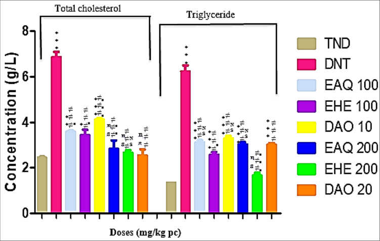 Figure 1
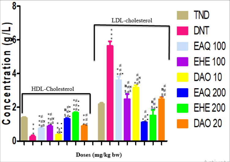 Figure 2
Introduction
Diabetes is associated with a high risk of vascular disease. Cardiovascular disease is the leading cause of death in people with type 1 or type 2 diabetes [1] . Energetic management of all cardiovascular risk factors including dyslipidemia in diabetics [2] . The aim of this study is to assess the anti-lipid-lowering effect of extracts from the bark of Xylopia villosa stems in rats made diabetic by streptozocin. The specific objectives are to measure total cholesterol, triglycerides and HDL and LDL cholesterols.
Materials and Methods
Materials and Methods
Plant Material
Xylopia villosa stems bark were harvested in June 2014 at the National Floristic Center of Felix Houphouët Boigny University where can be found a sample recorded at the number 14712.
Preparation of Extracts
The stems bark of Xylopia villosa were dried for four weeks in the shade of the sun then made powder using an electric grinder IKAMAG RCT®. Hundred (100) grams of powder of Xylopia villosa were macerated for 24 hours in 1 liter of ethylic alcohol (ethylic alcohol and distilled water mixture: 70/30). The macerated obtained was then filtered twice on white cotton and once on Whatman filter paper N°4. The filtrate obtained in 70% ethanol was evaporated to dryness at reduced pressure at temperature of 40°C using a rotary evaporator type Buchi 161 Water Bath. About aqueous extract preparation, 100 grams of Xylopia villosa stem barks powder were added to 100 milliliters of boiling distilled water. Just like ethanolic preparation, the resulting mixture was filtered twice on white cotton and once on Whatman filter paper N°4. The filtrate obtained is preserved at temperature of 40°C in an oven for drying.
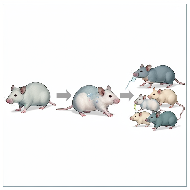
non-diabetic rats received streptozocin intravenously at a dose of 60 mg / kg body weight. The evolution of glycaemia of rats was followed from day D0 to D7 using strip glucose meter. At the 7th day after streptozocin injection, 42 diabetic rats were selected and divided into 7 groups of 6 rats according to their glycaemia. Subsequently, a treatment by gavage took place during 6 days with the different drugs according to the following distribution: Non-diabetic control group → 1 mL of distilled water Untreated diabetic group → 1 mL of distilled water Treated diabetic group → 1 mL of aqueous extract 100 mg / kg body weight (bw) Treated diabetic group → 1 mL of aqueous extract 200 mg / kg bw Treated diabetic group → 1 mL of ethylic alcohol extract 100 mg / kg bw Treated diabetic group → 1 mL of ethylic alcohol extract 200 mg / kg bw Treated diabetic group → 1 mL Daonil ® 10 mg / kg bw Treated diabetic group → 1 mL Daonil ® 20 mg / kg bw
Blood collection
After 6 days of treatment, blood was taken. For this study, the total volume of blood which can be taken with an animal (example of the rat or the mouse) is not enough. Thus, the number of taking away was distributed on several animals. We did not take with an animal more than 20% of its total blood volume. The volume of taken blood took into account the weight and the total volume of blood available to the animal. About 3 mL of blood was collected from puncturing the retro orbital sinus from anesthetized rats. The blood was centrifuged at 2000×g for 10 min to separate serum. This serum was kept at -20°C until the analysis [3] .
Determination of lipids in diabetic rats 2.3.1. Determination of total cholesterol
In an EDTA tube containing 1000 μL of reagent preincubated at 37 ° C for 2 to 3 min, 10 μL of the sample to be assayed are added. After stirring for 1 min and automatic incubation at room temperature (16 to 25 ° C) for 10 min, the optical density is read with a spectrophotometer at 500 nm in order to determine the concentration of cholesterol according to the following expression:
Determination of triglycerides
The determination of triglycerides was carried out according to the enzymatic method of triglycerides (Glycerol phosphate oxidase) using the glycerides reagent kit REF 7D74 (USA) [4] . The triglycerides, following several coupled reactions, give a color complex whose intensity, determined with a spectrophotometer at the wavelength λ equal to 500 nm, is proportional to the amount of triglycerides present in the serum.
Determination of HDL and LDL cholesterols
The determination of HDL (HDL-C) and LDL (LDL-C) cholesterols was performed using the phosphotungustic reagent associated with magnesium chloride after precipitation of LDL cholesterol [4] . In a hemolysis tube containing the phosphotungustic reagent associated with magnesium chloride, 1 μl of the serum to be assayed was added. The tube was centrifuged at 1500 rpm for 5 min. LDL-C precipitates and HDL cholesterol is assayed in the supernatant resulting from centrifugation. The LDL-C is then assayed in the supernatant of the centrifugation of the precipitate obtained previously. The absorbance of the solutions read at the wavelength λ equal to 500 nm is directly proportional to the quantity of the parameters assayed in the serum.
Results
Results
Determination of the lipid profile of diabetic rats 3.1.1. Case of total cholesterol and triglycerides in diabetic rats
Fig. 1 represents the total cholesterol and triglyceride concentrations of diabetic rats treated with the extracts of Xylopia villosa and Daonil ® . The total cholesterol concentration in non-diabetic control rats increased from 2.45 ± 0.04 g/L to 6.87 ± 0.23 and 4.15 ± 0.03 g/L respectively in untreated diabetic rats and those treated with Daonil ® at a dose of 10 mg/kg body weight (bw); these total cholesterol concentrations are extremely increased (P <0.001) compared to the total cholesterol concentration in non-diabetic control rats. The total cholesterol concentration (3.59 ± 0.04 g/L) of the diabetic rats treated with the aqueous extract (100 mg/kg bw) remained significantly high (P <0.01) compared to that of the rats non-diabetic controls. Also, the total cholesterol concentration (3.46 ± 0.22 g/L) in diabetic rats treated with the hydroethanolic extract (100 mg/kg bw) remained slightly elevated (P <0.05) compared to that non-diabetic control rats. However, the total cholesterol concentrations of the diabetic rats treated with the aqueous extract (200 mg/kg bw), the hydroethanolic extract (200 mg/kg bw) and Daonil ® (20 mg/kg bw) respectively gave 2, 86 ± 0.35; 2.68 ± 0.10 and 2.56 ± 0.25 g/L and are equivalent (P ˃ 0.05) to that of non-diabetic control rats. In addition, the total cholesterol concentrations of the rats rendered diabetic and treated with the aqueous extract (100 and 200 mg/kg bw), the hydroethanolic extract (100 and 200 mg/kg bw) and Daonil ® (10 and 20 mg/kg bw) were significantly lower (P <0.001) compared to the total cholesterol concentration of the untreated diabetic rats. The effect of Daonil ® (20 mg/kg bw) on the total cholesterol concentration of diabetic rats is the same (P ˃ 0.05) as that of the hydroethanolic extract (200 mg/kg bw) while it is significantly higher (P <0.01) than that of the aqueous extract (200 mg/kg bw). Regarding the triglyceride concentration, it went from 1.34 ± 0.01 g/L in non-diabetic control rats to 6.24 ± 0.26; 3.14 ± 0.08; 2.59 ± 0.10; 3.31 ± 0.09; 3.04 ± 0.09 and 3.03 ± 0.05 g/L respectively in untreated diabetic rats and those treated with aqueous extract (100 mg/kg bw), hydroethanolic extract (100 mg/kg bw), Daonil® (10 mg/kg bw), aqueous extract (200 mg/kg bw) and Daonil ® (20 mg / kg bw) and are strictly up (P <0.001) compared to the triglyceride concentration of non-diabetic control rats.
On the other hand, the triglyceride concentration (1.68 ± 0.10 g/L) of the diabetic rats treated with the hydroethanolic extract (200 mg/kg bw) became similar (P ˃ 0.05) to that of the control rats. non-diabetic (1.34 ± 0.01 g/L).
In addition, the triglyceride concentrations of the rats rendered diabetic and treated with the aqueous extract (100 and 200 mg/kg bw), the hydroethanolic extract (100 and 200 mg/kg bw) and Daonil ® (10 and 20 mg/kg bw) were strictly reduced (P <0.001) compared to the triglyceride concentration of untreated diabetic rats. The impact of the hydroethanolic extract (200 mg/kg bw) on the triglyceride concentration of diabetic rats is extremely higher (P <0.001) than that of Daonil ® (20 mg/kg bw) and of the aqueous extract (200 mg/kg bw).
Case of HDL and LDL cholesterols in diabetic rats
Fig. 2 represents the HDL and LDL cholesterols concentrations of diabetic rats after six days of treatment with Daonil ® and the extracts (aqueous and hydroethanolic) of Xylopia villosa. The HDL cholesterol concentration increased from 1.37 ± 0.05 g/L in non-diabetic control rats to 0.26 ± 0.07; 0.76 ± 0.03; 0.87 ± 0.06; 0.37 ± 0.03 and 0.89 g/L respectively in untreated diabetic rats and those treated with aqueous extract (100 mg/kg body weight (bw)), hydroethanolic extract (100 mg/kg bw); Daonil® (10 mg/kg bw) and Daonil ® (20 mg/kg bw) and these concentrations are extremely low (P <0.001) compared to the HDL cholesterol concentration of non-diabetic control rats. Furthermore, treatment of diabetic rats with the aqueous extract (200 mg/kg bw) normalized (P ˃ 0.05) the HDL cholesterol concentration (1.32 ± 0.03 g/L) in comparison with that non-diabetic control rats (1.37 ± 0.05 g/L). As for the treatment of diabetic rats with the hydroethanolic extract (200 mg/kg bw), it strictly increased (P <0.001) the concentration of HDL cholesterol (1.68 ± 0.04 g/L) compared to that of non-diabetic control rats (1.37 ± 0.05 g/L).
The treatments of diabetic rats with the aqueous extract (200 mg/kg bw) and the hydroethanolic extract (200 mg/kg bw) strictly increased (P <0.001) the HDL cholesterol concentration compared to the treatment with Daonil ® (20 mg/kg bw). Treatments of the diabetic rats with the aqueous extract (100 mg/kg bw) and the hydroethanolic extract (100 mg/kg bw) gave similar HDL cholesterol concentrations (P ˃ 0.05) to that of the treated diabetic rats. With Daonil ® (20 mg/kg bw). In contrast, treatment of diabetic rats with Daonil ® (10 mg/kg bw) resulted in an extremely lower HDL cholesterol concentration than that of diabetic rats treated with Daonil ® (20 mg/kg bw).
Although the aqueous and hydroethanolic extracts of Xylopia villosa at the dose of 200 mg/kg bw increased the HDL cholesterol concentration in diabetic rats, the effect of the hydroethanolic extract (200 mg/kg bw) on the cholesterol concentration HDL of diabetic rats is extremely higher (P <0.001) than that of aqueous extract (200 mg/kg bw).
Regarding the LDL cholesterol concentration in untreated diabetic rats (5.64 ± 0.27 g/L), it is extremely increased (P <0.001) compared to the LDL cholesterol concentration in non-diabetic control rats. (2.20 ± 0.06 g/L). Diabetic rats treated with the aqueous extract (100 mg/kg bw) have a significantly elevated LDL cholesterol concentration (3.61 ± 0.22 g/L) (P <0.01) compared to the cholesterol concentration LDL from non-diabetic control rats. Those treated with Daonil ® (10 mg/kg bw) obtained a slightly higher LDL cholesterol concentration (3.19 ± 0.12 g/L) (P <0.05) than that of non-diabetic control rats. On the other hand, the concentration of LDL cholesterol (1.20 ± 0.02 g/L) in diabetic rats treated with the aqueous extract (200 mg/kg bw) decreased slightly (P <0.05) compared to that non-diabetic control rats. In addition, the LDL cholesterol concentrations in diabetic rats treated with the hydroethanolic extract (100 mg/kg bw), the hydroethanolic extract (200 mg/kg bw) and Daonil ® (20 mg/kg bw) were respectively 2.49 ± 0.30; 1.53 ± 0.31 and 2.48 ± 0.13 g/L and are similar (P ˃ 0.05) to that of non-diabetic control rats.
The LDL cholesterol concentrations of the rats made diabetic and treated with the aqueous extract (100 and 200 mg/kg bw), the hydroethanolic extract (100 and 200 mg/kg bw) and Daonil ® (10 and 20 mg/kg pc) were significantly lower (P <0.001) compared to the LDL cholesterol concentration in untreated diabetic rats.
Regarding the treatment of diabetic rats with the different drugs, the aqueous extract (200 mg/kg bw) significantly decreased (P <0.01) the LDL cholesterol concentration compared to the treatment with Daonil ® (20 mg/kg bw). In addition, the hydroethanolic extract (200 mg/kg bw) slightly (P <0.05) lowered the LDL cholesterol concentration compared to treatment with Daonil ® (20 mg/kg bw). In contrast, treatment of diabetic rats with the aqueous extract (100 mg/kg bw) obtained a slightly elevated LDL cholesterol concentration (P <0.05) than that of diabetic rats treated with Daonil ® (20 mg/kg bw). However, the diabetic rats treated with the hydroethanolic extract (100 mg/kg bw) and Daonil ® (10 mg/kg bw) had identical LDL cholesterol concentrations (P ˃ 0.05) to that of the diabetic rats treated with Daonil ® (20 mg/kg bw). The effect of the aqueous extract (200 mg/kg bw) of Xylopia villosa on the LDL cholesterol concentration of diabetic rats is similar (P ˃ 0.05) to that of the hydroethanolic extract (200 mg/kg bw).
Discussion
Regarding the lipid profile of diabetic rats, dyslipidemia was observed in untreated diabetic rats. Indeed, dyslipidemia is defined as complications related to lipoprotein metabolism manifested by an increase in the plasma concentration of total cholesterol, LDL cholesterol, triglycerides and a reduction in the concentration of HDL cholesterol [5] . In diabetic rats treated with Daonil ® , the aqueous and hydroethanolic extracts of Xylopia villosa, several observations were made. First, treatment of the diabetic rats with the hydroethanolic extract at a dose of 200 mg/kg body weight (bw) was able to reduce the triglyceride concentration to a proportion similar to that of the non-diabetic control rats. Also, at this same dose of the hydroethanolic extract, the HDL cholesterol concentration increased compared to that of the non-diabetic control rats while the treatment of the diabetic rats with the aqueous extract (200 mg/kg bw) was able to normalize the HDL cholesterol concentration so as to have a value similar to that of non-diabetic control rats. Then, the treatments of the diabetic rats with the hydroethanolic extract (100 and 200 mg/kg bw) and Daonil (20 mg/kg bw) made it possible to normalize the concentration of LDL-cholesterol compared to that of the non-diabetic control rats. However, treatment of diabetic rats with the aqueous extract (200 mg/kg bw) reduced the concentration of LDL-cholesterol to be lower than that of non-diabetic control rats. This result is in agreement with that of Abidov and collaborators who showed that blueberry leaves caused a decrease in LDL levels in type 2 diabetes [6] . Finally, the total cholesterol of the diabetic rats treated with the aqueous extract (200 mg/kg bw), the hydroethanolic extract (200 mg/kg bw) and Daonil (20 mg/kg bw) was normalized so as to have a cholesterol level similar to that of non-diabetic control rats. This decrease in cholesterol is believed to be due to the sterols present in the aqueous and hydroethanolic extracts of Xylopia villosa [7] . Indeed, De-Jong and collaborators have shown that plant sterols and stanols act on total and LDL cholesterol levels by inhibiting the absorption of cholesterol [8] .
Conclusion
The hydroethanolic extract (200 mg/kg bw) behaved as a lipid lowering agent by correcting dyslipidemia and increasing HDL cholesterol levels.
Ethical Approval
The experimental procedures and protocols used in this study were approved by the Ethical Committee of Health Sciences, University Félix Houphouet-Boigny. These guidelines were in accordance with the European Council Legislation 87/607/EEC for the protection of experimental animals. All efforts were made to minimize animal suffering and reduce the number of animals used.
References
8 in the original paper- Booth GL, Rothwell D, Fung K, Hux JE, Booth G, Laupacis A. Diabetes and cardiac disease, Diabetes in Ontario: Institute for Clinical Evaluative Sciences 2002, 95-127.
- Gaede P, Vedel P, Larsen N. Multifactorial intervention and cardiovascular disease in patients with type 2 diabetes. N Engl. J. Med 2003;348:383-393.
- OVF: Office Veterinaire Federal. No. 800.116-1.04 1981.
- Fossati P, Prencipe L, Berti G. Enzymatic Creatinine Assay: A New colorimetric Method Based on Hydrogen Peroxide Measurement. Clin Chem 1983;29(8):14941496.
- Kendall DM, Harmel AP. The metabolic syndrome, type 2 diabetes and cardiovascular disease: Understanding the role of insulin resistance. Am J Manag Care 2002;8(20):635-653.
- Abidov M, Ramazanov A, Jimenez DRM, Chkhikvishvili I. Effect of Blueberin on fasting glucose, C-reactive protein and plasma aminotransferases, in female volunteers with diabetes type 2: double-blind, placebo controlled clinical study. Georgian Med News 2006;141:66-72.
- Kouame YY, Okpekon AT, Yapi HF, Gbassi KG, Assi YJ, Kouakou YKF. Phytochemical Screening and Acute Toxicity Study of Xylopia Villosa (Annonaceae) Barks Stems of Aqueous and Hydroethanolic Extracts. EJPMR 2016;3(6):526-531.
- De-Jong, Plat J, Mensink RP. Metabolic effects of plant sterols and stanols. J Nutr Biochem 2003;14:362-369.