
Research article · 2019 · Full text
International Journal of Biology SciencesEffect of Silver Nano Particle on Fish (Labeo Rohita) Hematology
Volume 1(1) · Pages 01–05 · DOI: 10.33545/26649926.2019.v1.i1a.1 ↗
Research areasNanoparticles: synthesis and applications
Abstract
Author-writtenThe use of silver nanoparticles (Ag-NPs) is rapidly increasing, but there are limited data on their effects on the aquatic environment. The present study aimed to determine the acute toxicity and evaluate the effect of subacute concentrations of Ag-NPs on hematological and plasma biochemical indices of, Roho, Labeo rohita. The 24-, 48-, 72- and 96-h median lethal concentration values of silver Nanoparticle for Labeo rohita were estimated at 5mg/l, 10 mg/l and 20 mg/L. Red blood cell (RBC) count, hemoglobin (Hb) count and hematocrit (Hct) level were significantly reduced at both concentrations tested (p < 0.05). In conclusion, Ag-NPs intoxication resulted in erythrocyte reduction, hematological disturbances, leucocytosis and stress response in Labeo rohita and offered a simple tool to evaluate toxicity-derived alterations.
Summary
Source-checkedPlain-language reading of the full paper, prepared with AI and checked against the text. Each point links to the authors' own words.
Why it matters
- Silver nanoparticles (Ag-NPs) are widely used across various fields because of their strong antimicrobial properties. View evidence ↗
- Ag-NPs can enter aquatic environments through effluent discharges from products like cosmetics, medical devices, and food packaging. View evidence ↗
- Labeo rohita is a significant fish species for human nutrition due to its high protein and polyunsaturated fatty acid (PUFA) content. View evidence ↗
The question
- The study aimed to determine the acute toxicity and evaluate how subacute concentrations of Ag-NPs affect the hematological and plasma biochemical indices of Labeo rohita. View evidence ↗
What they did
- Silver nanoparticles were synthesized using a green method involving a guava plant extract prepared with water and ethanol. View evidence ↗
- The synthesized nanoparticles were analyzed and found to have a size range between 1 and 100 nanometers. View evidence ↗
- Labeo rohita fish (averaging 100g and 6cm) were used and acclimated to laboratory conditions for two weeks prior to the trials. View evidence ↗
- Fish were exposed to three concentrations of Ag-NPs (5 mg/L, 10 mg/L, and 20 mg/L) and a control group for a duration of four weeks. View evidence ↗
What they found
- The authors found that the toxicity of silver nanoparticles leads to an increase in hemoglobin levels in the fish. View evidence ↗
What it means
- The authors conclude that subacute exposure to Ag-NPs induces stress, resulting in leukocytosis, hematological disturbances, and erythrocyte reduction in Labeo rohita. View evidence ↗
- The authors recommend a more thorough assessment of the potential toxic effects of Ag-NPs on fish and the aquatic environment before industrial or commercial application. View evidence ↗
Visual summary
2 visualsIllustrations explain the paper; charts are replotted from the paper's own table values.
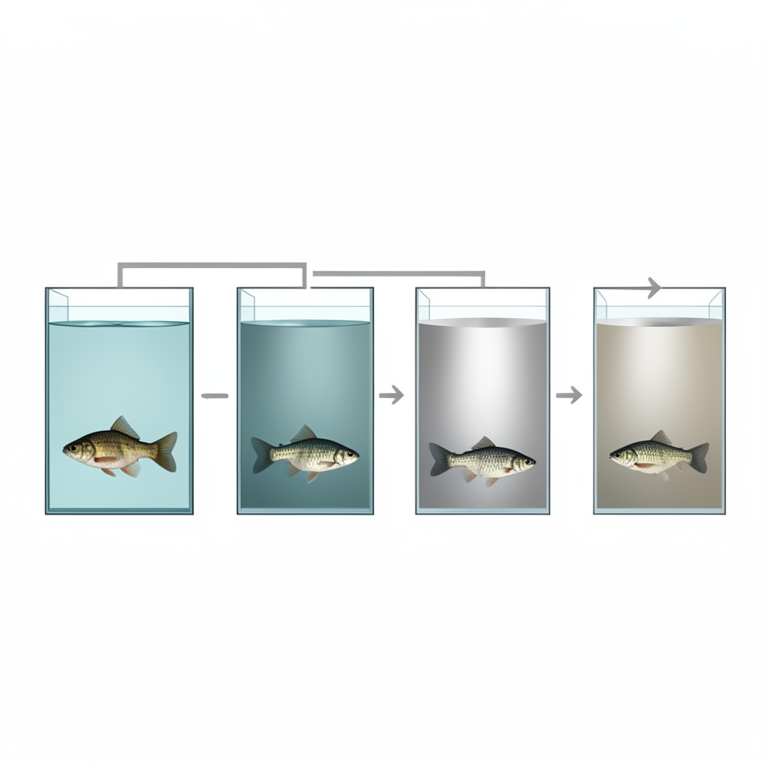
Experimental Setup Overview
AI-generated conceptual illustration (Labeo rohita fish in glass aquariums). It explains the idea and does not show study data.
View source evidence ↗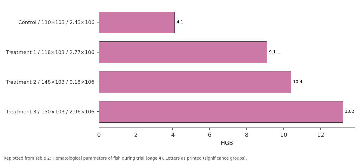
HGB: Control / 110×103 /... vs Treatment 1 / 118×103 /... vs Treatment 2 / 148×103 /... vs Treatment 3 / 150×103 /...
Replotted from Table 2 (page 4) of the original paper. Every bar is the paper's own table value.
View source evidence ↗Figures & tables
From the original paper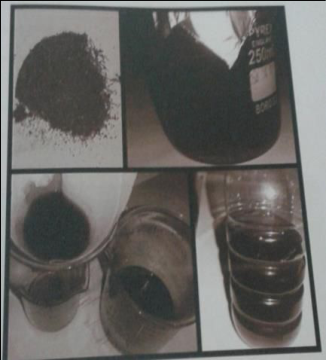 Figure 1
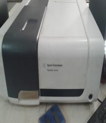 Figure 2
 Table 1
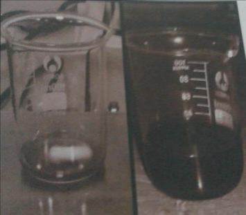 Figure 3
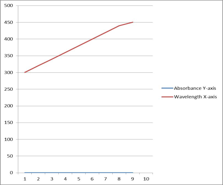 Figure 4
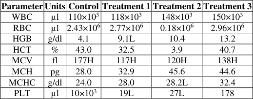 Table 2
Introduction
Nanoparticles are particles that have one dimension that is 100 nm or less in size. Nanoparticles are increasingly being used, or being evaluated for use, in many fields. Silver (Ag) is one of the most commonly used nanoparticles due to its bactericide effect [1] . The annual production of AgNPs is estimated at around 500 t worldwide and grows systematically [2] . The estimated production of Nano-enabled products has risen from less than 10 tons per year in 2011 [3] to 300 tons per year in 2015 [4] . Products based on Ag nanoparticles (Ag-NPs), such as odor-resistant textiles, food packaging, cosmetics, household appliances and medical devices may release Ag particles (nanoparticles or aggregates) or Ag ions via effluent discharge into the aquatic environment [5] . Fisheries and aquaculture contribute over 40% to the world's reported finfish production from less than 0.01% of the total volume of water on earth. These fisheries provide food for billions and livelihoods for millions of people worldwide [6] . Palmitic acid is the most abundant fatty acid in all species ranging from 32% to 46%. Although these fish contained reasonable amounts of essential PUFA such as docosahexaenoic, eicosapentaenoic and arachidonic acids, Labeo rohita contained the highest amounts of PUFA and protein. These fish contained appreciable levels of Omega-6 PUFA suggesting that these fish especially L. rohita could be used as a source of healthy diet for humans. These findings may benefit the fishing industry, nutritionists and researchers who are striving to improve the nutritive value, processing and marketing of selected fish species [7] . The present study aimed to determine the acute toxicity and evaluate the effect of subacute concentrations of Ag-NPs on hematological and plasma biochemical indices of, Roho, Labeo rohita. The 24-, 48-, 72-and 96-h median lethal concentration values of silver Nanoparticle for Labeo rohita .
Material and method
The present research was performed at the research and training center, Department of fisheries and aquaculture, UVAS, Ravi Campus .Block C, Pattoki. For the preparation of silver nanoparticles, plant extract was used. Plant extract was prepared with water and ethanol solution. After filtration, phytochemical analysis was done. Silver metal solution was mixed with that guava plant extract in chemistry Department of UET, Lahore [8]. Nano synthesis was conformed from color variation of nanoparticle solution from light yellow to brown and dark brown. Their absorbance was checked on UV-Vis spectroscopy and different peaks were observed. Particle size was check on particle analyzer. And ranged between 1-100 Nanometer in their size. Their relating functional groups were also checked from FTIR. Labeo rohita was used as an experimental animal
Severe toxicity valuation of silver nanoparticle (AgNps)
Severe toxicity influence of silver nanoparticles was examined on (Labeo rohita). All experiments performed for a time period of 4 weeks and silver nanoparticles were introduced to fish at three different concentration (5,10 and 20 mg/L) designed as treatment no. 1, treatment no. 2 and treatment no 3. Fourth one is control concentration which is without nanoparticles. Controls are compared with treatments.8 fishes were kept in each group. Control group was carried in tap water with free of any addition of nanoparticles, while treatment groups were revealed to the doses of 5, 10 and 20 mg/L of nano silver solution, for a period of 96-hours.
Water assumed rank parameter
Water assumed parameters like dissolved oxygen, pH, temperature, total hardness and electrical conductivity were measured on daily basis. The procedure of determining these parameter are given below by following the method of (A.P.H.A, 2005). DO meter was calibrated and probe was dipped in water to measure temperature and dissolved oxygen of aquarium water. Reading was noted when needle became stable on screen in mg/L. pH is a -log of H ion concentration in a medium. pH was measured by pH meter. In 50 ml of water sample, 1-2 drops of sodium-potassium titrate solution were added and mix well: 1 ml Nessler's reagent was added for the development of color and kept in stand for 15 minutes. Concentration was measured by spectrophotometer. Standards and sample were run at 420 nm for 1 cm light path. Calibration curve was formed at the same temperature and reaction time used for samples. Concentration readings were measured against a reagent blank and parallel checks were run frequently against standards in the nitrogen range of samples. It is an ability to conduct electric current or ionic concentration in water. It indicates the amount of ionize able substance (phosphate, nitrate etc.) dissolved in water. Calibrate the conductivity meter. Dip the probe of the meter in water and note the reading on the screen. Electrical conductivity is measured in µs/cm. Add 1 ml of ammonia buffer and 5 drops of indicator to 50 ml of water in a conical flask. The color of water sample will turn wine red. Titrate against EDTA solution, until a blue color appears. Note the end point.
Sample Collection
The experimental fish species Labeo rohita (100 g ± 10g , 6cm± 2cm) was picked from fisheries research ponds University of Veterinary and Animal Sciences Ravi Campus Pattoki and Fish were transferred to aquaculture and research center of University of Veterinary and Animal sciences and acclimated to laboratory condition for 2 weeks. Fishes were parceled in 4 glass tank (500L). The experimental aquaria were aerated and fish in holding tank equipped with semi static flow by aerated water system for at least 2 week for acclimatization to the lab before accomplishment of nanoparticles trails. The laboratory temperature was in between 29-30 0 C, pH 7.79 + 0.50, dissolved oxygen 7.93 + 0.25 mg/L and total hardness 298+2.35 mg/L as CaCO3 and normal illumination (approx. 12 hours light and 12 hours dark) was conserved during the whole experimental period. Water pH, temperature and dissolved oxygen were determined by Wagtech portable pH/temp meter and oxygen meter. Water total hardness was determined using portable photometer with commercial kits provided by the manufacturer (Wagtech Portable Photometer 7100, Berkshire, UK).
Statistical analysis
Data was subjected to ANOVA for its statistical significance among treatments. Mean values were compared to assess their intensity of significance among treatment groups through Duncan's multiple range test. Probability level was set at <0.05. The SAS (statistical analysis software) version 2005 was used for all statistical analysis due to its wider application.
Result
The experiment was conducted in department of fisheries and aquaculture, University of veterinary and animal sciences. During this research trial, Labeo rohita were grown under controlled condition with three different concentration of silver nanoparticles viz. treatment no 1 with 5 mg/L, treatment no 2 with 10 mg/L, treatment no 3 with 20 mg/L and control group without silver nanoparticles (AgNPs) for a period of 4 weeks. There was no statistical difference at the start of experiment between control and experimental groups. After 4 week of trial there was statistical difference in growth parameters, hematological parameter
Preparation of silver nanoparticles
For the preparation of silver nanoparticles, plant extract was prepared with water and ethanol solution. After filtration, photochemical analysis was done. Silver metal solution was mixed with that guava plant extract in chemistry Department of UET, Lahore [8] . Nano synthesis was conformed from color variation of nanoparticle solution from light yellow to brown and dark brown. Their absorbance was checked on UV-Vis spectroscopy and a different peak was observed. Particle size was check on particle analyzer. And range between 1-100 nanometers in their size. Their relating functional groups were also checked from FTIR.
Physiochemical parameters
Physiochemical parameters of water include temperature, pH, dissolved oxygen (mg/L), electrical conductivity (µs/cm), ammonia (mg/l) and carbon dioxide (mg/L). These water quality parameters helps to standardize the water that is at which fish can grow faster and better .During the experimental period physiochemical analysis of water was checked on daily basis. The data obtained during experiment is shown in Table below. Table 4.1 Show all the physiochemical parameters of water. All the physiochemical parameter provides significant result except ammonia. Ammonia show non-significant results, maximum value recorded in treatment no 3due to light concentration of silver nanoparticles (20mg/L).
Hematology
The number of the WBCs blood cell was higher in treatment no.3 than the control group. Hemoglobin (Hb), Hematocrit (Hct) and RBCs were high in the treatment no 3 as compared to the control group. The MCV in the treatment group's increase as the dose of nanoparticles increased. The values of MCV, MCHC and PLT were also high in treatment no.3 group than control
Discussion
In the current study, the experiment was conducted in department of fisheries and aquaculture, university of veterinary and animal sciences, Ravi campus, Block C, Pattoki. During this research trial Labeo rohita were grown under controlled conditions with three different concentrations of silver nanoparticles viz. Treatment 1 with 5 mg/l, treatment 2 with 10 mg/l and treatment 3 with 20 mg/l and control without silver nanoparticles (AgNPs) for a period of 4 weeks. There was no statistical difference at the start of experiment between control and experimental groups. After 4 weeks of trial there were statistical differences in physiochemical parameters, hematological parameters. The use of Nano silver is growing exponentially because of its strong antimicrobial properties. Ag-NPs may be released into aquatic environments from factory waste discharges, through leaks or spills during transportation and via materials containing Ag-NPs, while only little information is available on their possible eco toxicological effects [9] . The preparation silver of nanoparticle from guava plant extract is linked with green synthesis of nanoparticles. The aim of the present research was work to synthesize nanoparticle from natural components. The Psidium guajava is well-known medicinal plant and leaves of it are widely used in the Ayurveda system of medicine as well as in folk medicine [10] . Guava leaf extract is used as astringent, anodyne, febrifuge, antispasmodic, tonic to treat wounds, ulcer, cholera, diarrhea, vomiting, nephritis, toothache, gum inflammation, etc [11] . Psidium guajava leaf contains leucocyanidin, flavonoids which includes quercetin, quercetin-3-O-α-l-arabinofuranoside, quercetin-3-O-β-darabinopyranoside, quercetin 3-O-β-d-galactoside (hyperin), quercetin-3-O-α-l-arabinofuranoside (avicularin), quercetin 3-O-β-d-glycoside (isoquercetin), plant sterol, carotenes, tannins, saponins, vitamin C and B6, carbohydrates and diethoxy alkenes as plasma compounds. It is reported that flavonoids [12] present in leaf extract are responsible for reducing Ag + ions and capping for Ag nanoparticles. In this study, some of the flavonoids present in leaf extract may act as reducing and/or capping agent to synthesize Ag nanoparticles. Physiochemical parameter of water play important role in biology and physiology of fish. All the physiochemical parameters like temperature, dissolved oxygen, pH, total hardness, dissolved solids, ammonia and electrical conductivity were continuously measured on daily basis throughout whole study period of trial. The results of our study are like [13] where they reported that pH values were in between 6.62-7.85 which is optimum for the poly culture pond [14] reported the temperature ranges from (18.3-37.8 o C) which is larger than variation found in our study. Silver nanoparticles attract much attention due to expected toxicity. It can cause damage to the brain, liver, and stem cells in the human body. Thus, instead of using human, it is preferable to use animal models in toxicological studies. Among all models, fish is most dominantly used in toxicological studies. This review was published by [15] showed that Ag-NPs pose toxicity to all the life stages of fish. In chronic toxicity studies regarding hematological analysis, level of hemoglobin initially increased significantly and then decreased. Similar trends were recorded in RBC count. It was probably due to elevated levels of metabolic rate under stress condition [16] . Secondly, the flow of RBCs also increases in blood stream due to hypoxia and dehydration. As Ag-NPs produces hypoxic condition and increase the alkalinity, the kidney sensors detect this condition and increase RBCs movement in the blood flow. [17] of our study are like [16] in our study during control the hemoglobin level was 4.1 g/dl. It was normal range of hemoglobin but in treatment no1, treatment no 2 and treatment no 3 its level increase respectively 9.1 g/dl, 10.4 g/dl and 13.2 g/dl. These result shows that the toxicity of silver nanoparticles increase hemoglobin level in fish. Further, MCH and MCHC values were significantly different from control and decreased with increase in concentrations of Ag-NPs. This lowering of values were due to RBCs counts and hematocrit reduction, which itself was reduced due to deformation or damage to RBCs [18] . Many investigators reported decrease in the level of MCH and MCHC in freshwater fish exposed to metals and nanoparticles [19] the result of [18] and the results of [19] does not match with our result the reason was that they use fresh water. We also use fresh water but here water salinity is higher than normal fresh water due to this our results does not match with both of them. Overall changes in blood parameters were due to the reaction of defense against toxicity through the mechanism of the erythropoiesis [20] . There was also a significant difference in neutrophils of all treatments compared to control. Neutrophils increased in number because Ag-NPs increased the infection and damage in tissues [21] . This leads to neutrophils [22] . The degree of elevation of neutrophils represents the infection severity. Many fluctuations were also seen in a number of monocytes relatively in short response of respiratory burst. This change in monocytes might be due to disease condition or hematological tissue dysfunction [23] . The results of [21,22] and [23] like as our results according to them number of neutrophil increase due to the effect of silver nanoparticles. Neutrophil is a type of WBC cell. Our result also shows that the number of white blood cell WBC increases with the increase of silver nanoparticles. In control the number of WBC is 110×10 3 which was normal. But in treatment no 1, treatment no 2 and treatment no 3 the number white blood cell increases respectively 118×10 3, 148×10 3 and 150×10 3 . These result showed that the number of white blood cell increased with the increase of silver nanoparticles. Because silver nanoparticle causes toxicity and white blood cells cause immunity due to this number white blood cell increases with the increase of silver nanoparticles.
Conclusion
In conclusion, our results indicate that inducing stress due to subacute exposure to Ag-NPs (Nanocid) causes erythrocyte reduction, hematological disturbances and leukocytosis in Labeo rohita. Based on the results of this study, it is suggested that industrial and commercial applications of Ag-NPs should be more carefully and thoroughly assessed as to their potential toxic effects to the aquatic environment and fish. We concluded that silver nanoparticles affect the histological, biochemical and hematological parameters of fish. And these parameters are most important in every type of living animal's health
Figures and tables from the paper
| Parameters | Control | Treatment # 1 | Treatment # 2 | Treatment # 3 |
|---|---|---|---|---|
| Temperature(OC) | 29.63 ±0.03a | 30.10±o.95a | 29.87±0.85a | 30.02±0.95a |
| Ph | 7.50±0.01a | 7.43±0.28a | 7.39±0.02a | 7.50±0.01a |
| Total hardness (mg/l) | 200±0.02a | 230±0.03a | 250±0.05a | 230±0.03a |
| Ammonia(mg/l) | 0.31±0.01c | 1.65±0.02b | 1.87±0.03b | 2.17±0.02a |
| Dissolved oxygen(mg/l) | 5.72±0.04a | 5.28±0.86a | 5.14±0.46a | 5.36±0.13a |
| Electrical conductivity(µs/cm) | 2198.28± 148.37a | 2214.86± 247.02a | 2218.17± 226.87a | 2176.06± 201.76a |
Table text extracted from the original PDF; superscript letters are shown inline. The original table image is below.
View original table image
| Parameter | Units | Control | Treatment 1 | Treatment 2 | Treatment 3 |
|---|---|---|---|---|---|
| WBC | µl | 110×103 | 118×103 | 148×103 | 150×103 |
| RBC | µl | 2.43×10⁶ | 2.77×10⁶ | 0.18×10⁶ | 2.96×10⁶ |
| HGB | g/dl | 4.1 | 9.1L | 10.4 | 13.2 |
| HCT | % | 43.0 | 32.5 | 3.9 | 40.7 |
| MCV | fl | 177H | 117H | 120H | 138H |
| MCH | pg | 28.0 | 32.9 | 45.6 | 44.6 |
| MCHC | g/dl | 24.0 | 28.0 | 28.2L | 32.4 |
| PLT | µl | 10×103 | 19L | 27L | 178 |
Table text extracted from the original PDF; superscript letters are shown inline. The original table image is below.
View original table image
References
23 in the original paper- Farmen E, Mikkelsen HN, Evensen O, Einset J, Heier LS, Rosseland BO, et al. Acute and sub-lethal effects in juvenile Atlantic salmon exposed to low mg/L concentrations of Ag nanoparticles. Aquat Toxicol. 2012; 108:78-84.
- Fabrega J, Luoma SN, Tyler CR, Galloway TS, Lead JR. Silver nanoparticles: behavior and effects in the aquatic environment. Environ Int. 2011; 37:517-531.
- Hendren CO, Mesnard X, Droge J, Wiesner MR. Estimating production data for five engineered nanomaterials as a basis for exposure assessment. Environ Sci Technol. 2011; 45:2562-2569.
- King SM., Jarvie HP, Bowes MJ, Gozzard E, Lawlor AJ, Lawrence MJ. Exploring controls on the fate of PVPcapped silver nanoparticles in primary wastewater treatment. Environ Sci. Nano. 2015; (2):177-190.
- Benn TM, Westerhoff P. Nanoparticle silver released into water from commercially available sock fabrics. Environ Sci Tech. 2008; 42:4133-4139.
- Abigail J, Steven J, Cooke, Andrew MD, Shannon DB, David BB, et al. Social, economic, and environmental importance of inland fish and fisheries. Envir Rev. 2016; 24(2):115-121
- Farhat J, Abdul SC. Chemical compositions and fatty acid profiles of three fresh water fish species. F Chem. 2011; 125(3):991-996.
- Allafchian AR, Majidan Z, Lelbeigi V, Tabrizichi M. A novel method for the determination of three volatile organic componds in exhaled breath by solid-phase microextraction ion mobility spectrometry. Analytical bio analytical chem. 2016; 408(3):839-847.
- Ko¨hler AR, Som C, Helland A, Gottschalk F. Studying the potential release of carbon nanotubes throughout the application life cycle. J Cleaner Prod. 2008; 16:927-937.
- Gutierrez RMP, Mitchell S, Solis RV. Psidium guajava: a review of its traditional uses, phytochemistry and pharmacology. J Ethnopharmacol. 2008; 177:1-27.
- Ojewole JA. Hypoglycemic and hypotensive effects of Psidium guajava Linn. (Myrtaceae) leaf aqueous extract. Meth Find Exp Clin Pharm. 2005; 27:689-695.
- Sahadevan R, Sivakumar P, Karthika P, Sivakumar Muralidharan NG, Devendran P. Biosynthesis of silver nanoparticles from active compounds Quacetin -3-O-Bd-galactopyranoside containing plant extract and its antifungal application. Asian J Pham Clin Res. 2013; 6:76-79.
- Hossain MY, Begum M, Ahmed ZF, Hoque MA, Wahab MA. A study on the effects of iso-phosphorus fertilizers on plankton in fish ponds. South Pac Stud. 2006; 26:101110.
- Begum M, Hossain MY, Wahab MA, Kohinoor AHM. Effects of iso-phosphorus fertilizers on water quality and biological productivity in fish pond. J Aqua Trop. 2003; 18:1-12.
- Khan MS, Jabeen F, Qureshi NA, Asghar MS, Shakeel M, Noureen A. Toxicity of silver nanoparticles in fish: a critical review. J Bio Environ Sci. 2015; 6(5):211-227.
- Dobsikova R, Svobodova Z, Blahova J, Modra H, Velisek J. Stress response to long distance transportation of common carp (Cyprinus carpio L.). Acta Vet Brno. 2006; 75(3):437-448.
- Giulio DRT, Hinton DE. Integration of Human Health and Ecological Risk Assessment. The tox of fish Crc, 2008. Press. doi: 10.1201/9780203647295.
- Imani M, Halimi M, Khara H. Effects of silver nanoparticles (AgNPs) on hematological parameters of rainbow trout, Oncorhynchus mykiss. Comp Clin Pathol. 2015; 24(3):491-495.
- Vutukuru S. Acute effects of hexavalent chromium on survival, oxygen consumption, hematological parameters and some biochemical profiles of the Indian major carp, Labeo rohita. Int J Envir Res Pub Health. 2005; 2(3):456-462.
- Vinodhini R, Narayanan M. Bioaccumulation of heavy metals in organs of fresh water fish Cyprinus (common carp). Int J Env Sci. Tech. 2008; 5(2):179-182.
- Williams KM, Gokulan K, Cerniglia CE, Khare S. Size and dose dependent effects of silver nanoparticle exposure on intestinal permeability in an in vitro model of the human gut epithelium. J Nano biotec. 2016; 14(1):62.
- Soares T, Ribeiro D, Proença C, Chisté RC, Fernandes E, Freitas M. Size-dependent cytotoxicity of silver nanoparticles in human neutrophils assessed by multiple analytical approaches. Life Sci. 2016; 145:247-254.
- Bairuty AGA, Shaw BJ, Handy RD, Henry TB. Histopathological effects of waterborne copper nanoparticles and copper sulphate on the organs of rainbow trout (Oncorhynchus mykiss). Aqu Toxicol. 2013; 126:104-115.
Cited by 25
From OpenAlex, OpenCitations and reference lists read by Scinovex
- γ-aminobutyric acid (GABA) strengthened nutrient accumulation, defense metabolism, growth and yield traits against salt and endoplasmic reticulum stress conditions in wheat plants ↗
Sarika Kumari, Faroza Nazir, Apoorva Singh, Hyda Haroon, Nahida Rashid Khan, Rudra Narayan Sahoo et al. · Plant and Soil · 2024
❝ 26 - Salt-Tolerant Plant Growth-Promoting Bacteria (ST-PGPB): An Effective Strategy for Sustainable Food Production ↗
Syeda Tahseen Zahra, Mohsin Tariq, Muhammad Abdullah, Muhammad Kaleem Ullah, Abdul Rafay Rafiq, Aisha Siddique et al. · Current Microbiology · 2024
❝ 25 - Abiotic Stress Sensitivity and Adaptation in Field Crops ↗
Sukamal Sarkar, Saikat Saha, Saswati Ghosh, Sujay Kumar Paul, Saikat Dey, Debojyoti Moulick et al. · Springer eBooks · 2023
❝ 23 - Effect of Zinc Oxide Nanoparticles and Soil Amendments on Wheat Yield, Physiological Attributes and Soil Properties Grown in the Saline – Sodic Soil ↗
Mahmoud El‐Sharkawy, Esawy Kasem Mahmoud, Mohamed Abd Elaziz, Tamer H. Khalifa · Communications in Soil Science and Plant Analysis · 2022
Soil degradation with salinity and sodicity has become a major issue in Egypt causing a decline in soil quality and crop productivity in these soils. The addition of gypsum (G), rice straw compost (C) and foliar application of ZnO nanoparticles (Z) as foliar application and their combinations to improve soil quality and physiology of wheat plants in saline-sodic soil is…
❝ 21 - Thymbra spicata extract and arbuscular mycorrhizae improved the morphophysiological traits, biochemical properties, and essential oil content and composition of Rosemary (Rosmarinus officinalis L.) under salinity stress ↗
Mojtaba Solgi, Majid Bagnazari, Meisam Mohammadi, Afsaneh Azizi · BMC Plant Biology · 2025
BACKGROUND: Enhancing the content of essential oils and valuable secondary metabolites is a primary goal for medicinal plant breeders. In this study, the effects of Thymbra spicata extract at concentrations of 0% (C), 10% (TS1), and 20% (TS2), along with mycorrhizal fungus (MF) biofertilizer at a rate of 50 g/2.5 kg of soil, were evaluated on the growth, photosynthetic pigments,…
❝ 18PDF - Plants Response to Different Abiotic Stresses ↗
Sami Jan, M. Ashraf Bhat, Akshay Kumar, M. Altaf Wani, Farooq Ahmad Bhat, Raihana Habib Kanth et al. · Journal of Plant Diseases and Protection · 2025
❝ 16 - Development and evaluation of barley mini-core collection for salinity tolerance and identification of novel haplotypic variants for HvRAF ↗
Sonia Sonia, Vikender Kaur, Shashank Kumar Yadav, Sunder Singh Arya, Jayaraman Aravind, Sherry Rachel Jacob et al. · Plant and Soil · 2023
❝ 11 - The synergistic effect of biochar and poly(2-ethyl-2-oxazoline)/poly(2-hydroxyethylmethacarylate)/chitosan) hydrogels on saline soil properties and carrot productivity ↗
Sayed A. Abdeen, Hassan H. H. Hefni, Ahmed Awadallah‐F, Nasser R. Abd El‐Rahman · Chemical and Biological Technologies in Agriculture · 2023
Abstract Background Soil salinity is one of the most important factors limiting crop production. Furthermore, with the increasing population and saline soil worldwide there is no choice but to utilize saline soil to increase the agricultural regions. Therefore, to improve carrot productivity under saline conditions, it is necessary to provide good management such as applying hydrogels and biochar for improving…
❝ 9PDF - Application of zinc nanoparticles as seed priming agent improves growth and yield of wheat seedlings grown under salinity stress by enhanced antioxidants activities and gas exchange attributes ↗
Sara Zafar, Shahbaz Khan, Danish Ibrar, Muhammad Kamran Khan, Zuhair Hasnain, Kashf Mehmood et al. · Cereal Research Communications · 2024
❝ 8 - Evaluation of morphological and biochemical variations in peas under two widespread abiotic stresses ↗
Muhammad Farooq, Rafiq Ahmad, Muhammad Shahzad, Khalid Ahmad, Yasar Sajjad, Amjad Hassan et al. · Acta Physiologiae Plantarum · 2024
❝ 8PDF - Management of Salinity Stress by the Application of Trichoderma ↗
Nagamani Adusumilli, Sarojini Chakravarthy Kolli · Fungal biology · 2022
❝ 8 - Pomegranate and Salt Stress Responses—Assimilation Activities and Chlorophyll Fluorescence Performances: A Review ↗
Mehdi Khayyat, Mohammadreza Vazifeshenas, Mohadese Akbari · Applied Fruit Science · 2024
❝ 6 - Salinity and Its Impact on Sustainable Crop Production ↗
Hassan Auda Awaad · Earth and environmental sciences library · 2023
❝ 6 - Effect of Microbial Consortium Vs. Perfected Chemical Fertilizers for Sustainable Crop Growth ↗
Dinesh Kumar Maheshwari, Sandeep Kumar, Prashant Kumar, Sachin Kumar, Shrivardhan Dheeman · Microorganisms for sustainability · 2023
❝ 4 - Triticale (X Triticosecale Wittmack): Role and Responses Under Abiotic Stress ↗
Seyyed Hamid Reza Ramazani, Mohammad Zabet · Springer eBooks · 2022
❝ 4 - Adverse effects of sulfate-type saline-alkali water exposure on largemouth bass (Micropterus salmoides): insight into growth, tissue morphology damage, and liver inflammation ↗
Zhongzheng Xu, Xiaotian Zhang, Limin Ye, Ziyang Ren, Wenhao Huang, Chi Wang et al. · Aquaculture International · 2026
❝ 3 - Antiparasitic efficacy and safety evaluation of green-synthesized silver nanoparticles as sustainable nanotherapeutics against Argulus siamensis in Labeo rohita ↗
Pushpa Kumari, Saurav Kumar, Rajive Kumar Brahmchari, KV Rajendran, S.P. Shukla, Rupam Sharma et al. · Veterinary Parasitology · 2026
❝ 3 - Enhancing antioxidant enzyme activities in chrysanthemum plantlets (Chrysanthemum morifolium Ramat cv. 'Jimba') under salt stress: A response surface methodology approach evaluating NaCl and CeO2 nanoparticles interactions ↗
Do Manh Cuong, Nguyen Thi Nhu, Vũ Quốc Luận, Phan Phuoc Minh Hiep, Hoang Hai Dang, Bui Van The Vinh et al. · Plant Cell Tissue and Organ Culture (PCTOC) · 2025
❝ 3 - Biochemical, Physiological, and Molecular Mechanisms of Plant Adaptation to Salinity ↗
Deepu Pandita · Springer eBooks · 2023
❝ 3 - Biological agents and plant growth regulator promote rice growth and regenerative capacity ↗
Hang Zhou, Lang Liu, Jianqun Zhou, Aibin He, Zhaohui Wu · BMC Plant Biology · 2025
Against the backdrop of global population and food demand growth, the development of ratoon rice is conducive to increasing food production and ensuring food security. Applying agricultural biotechnology and plant growth regulator to ratoon rice production is a key measure to promote the development of green agriculture and address the challenges of global food security. A total of 4 treatments…
❝ 2PDF