
Research article · 2017 · Full text
International Journal of Veterinary Sciences and Animal HusbandryMorphological investigation of the dog (Canis familiaris) lacrimal drainage system
Volume 2(6) · Pages 42–50
Research areasNasolacrimal Duct Obstruction TreatmentsOcular Surface and Contact LensOcular Infections and Treatments
Abstract
Author-writtenTo demonstrate the histomorphological structure of the dog lacrimal drainage system, 15 adult healthy dogs (Canis familiaris) were used. The lacrimal drainage passages composed of the excretory ducts, lacrimal punctua, canaliculi, lacrimal sac, nasolacrimal duct and the nasolacrimal ostium. The nasolacrimal duct was divided into three distinct parts; the caudal "Osseous", middle "membranous" and rostral "cartilagenous" parts. Microscopically, the lacrimal puncta and canaliculi were lined with stratified squamous epithelium which was keratinized in the puncta. The epithelium of the lacrimal sac as well as the caudal and middle portions of the nasolacrimal duct were pesudostratified columnar epithelium without goblet cells. The epithelium changed to be a stratified squamous in the rostral portion of the nasolacrimal duct and became keratinized in the nasolacrimal ostium. The scanning electron microscopy showed that, the main opening of the excretory lacrimal ducts appeared heart shaped, while the opening of puncta was ovoid shaped. The internal surface of the nasolacrimal duct was smooth and showed a few sporadic pores and clefts. The nasolacrimal opening was a flask shaped. The normal histomorphological structure of the dog lacrimal drainage system was very significant to identify and diagnose any disorders in these structures in the same species and in experimental studies for human being.
Summary
Source-checkedPlain-language reading of the full paper, prepared with AI and checked against the text. Each point links to the authors' own words.
Why it matters
- Dogs are important laboratory animals for developing therapeutic drugs and studying diseases because of their similarities to humans. View evidence ↗
- The lacrimal drainage apparatus serves as the pathway that moves lacrimal fluid from the eye into the nasal cavity. View evidence ↗
- While the canine lacrimal gland is well-studied, there is limited research on the histomorphology of the canine lacrimal duct system. View evidence ↗
The question
- The study aimed to describe the morphology of the dog's lacrimal drainage passages to help identify and diagnose drainage disorders in dogs. View evidence ↗
What they did
- The researchers used 15 healthy adult dogs of both sexes, aged between 2 and 4 years. View evidence ↗
- A caliper was used to measure the dimensions of the lacrimal puncta, sac, nasolacrimal duct, and ostium. View evidence ↗
- Light microscopy was performed on specimens from various parts of the drainage system fixed in 10% neutral buffered formalin. View evidence ↗
- Scanning electron microscopy (SEM) was used to examine 1 mm³ specimens fixed in a glutaraldehyde and formaldehyde buffer. View evidence ↗
What they found
- The entire nasolacrimal duct had an average length of 8.43 ± 0.56 cm. View evidence ↗
- The dorsal puncta had an average diameter of 0.10 ± 0.003 cm, while the ventral puncta averaged 0.15 ± 0.05 cm. View evidence ↗
- The lacrimal sac and the caudal and middle parts of the nasolacrimal duct are lined with pseudostratified columnar epithelium that lacks goblet cells. View evidence ↗
- In the rostral portion of the nasolacrimal duct, the epithelium becomes stratified squamous and is keratinized with melanin granules at the ostium. View evidence ↗
What it means
- The authors conclude that understanding the histomorphology of the dog's lacrimal drainage system is essential for diagnosing canine disorders and supporting human experimental studies. View evidence ↗
- The authors state that further advanced studies are needed to understand the secretory functions of the nasolacrimal duct's lining epithelium. View evidence ↗
Visual summary
1 visualsIllustrations explain the paper; they do not show study data.
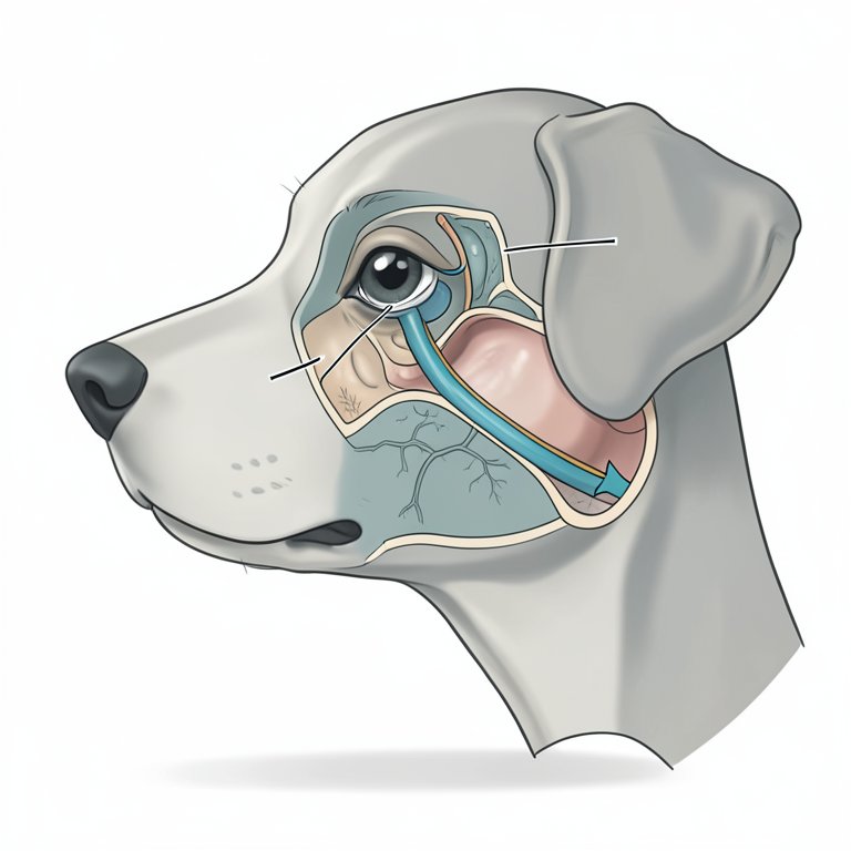
Canine Lacrimal Drainage Pathway
AI-generated conceptual illustration (dog (Canis familiaris) head and nasal cavity). It explains the idea and does not show study data.
View source evidence ↗Figures & tables
From the original paper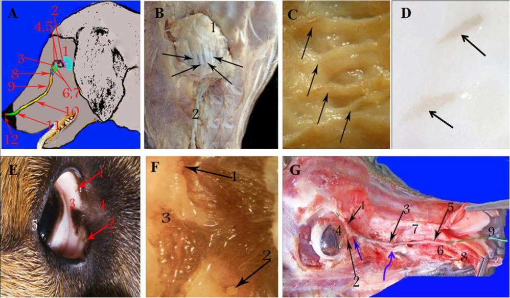 Figure 1
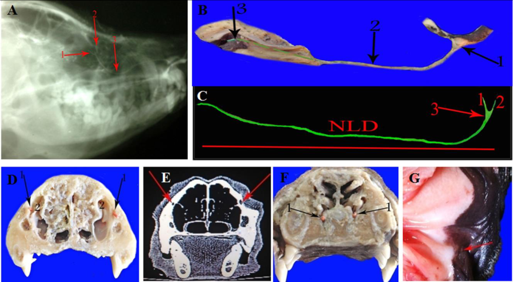 Figure 2
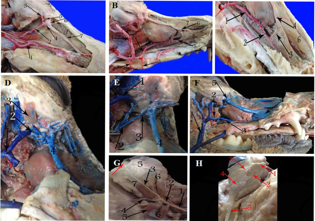 Figure 3
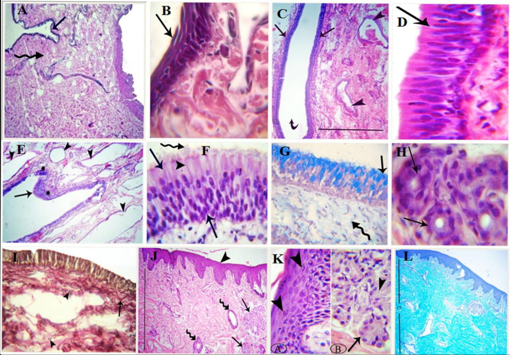 Figure 4
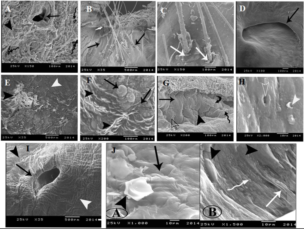 Figure 5
Introduction
Recently, dogs are considered as a vital laboratory animal due to the similarity between the human and dogs in certain diseases (Kijas et al., 2003) [20] also the canine model is used in developing the therapeutic drugs (Barabino and Dana 2004) [4] . The lacrimal drainage apparatus is a passage connects the eye with the nasal cavity; lacrimal fluid go through the punctum, passes in the lacrimal canaliculus and sac, to drain into the nasal cavity by the nasolacrimal duct. Microscopically, the nasolacrimal system of the human, rabbit, cat, deer, and pig lined with a pseudostratified columnar epithelium. Goblet cells are incorporated in the epithelium of human, rat, and cat. Whereas in rabbit, deer, and pig, the goblet cells are absent. The cavernous blood vessels system of lamina propria is present in human, rabbit, deer, and pig, but is deficient in rat and cat (Paulsen et al., 2002) [26] . The nasolacrimal duct system has been previously studied in human and numerous domestic animals (Jones, 1961; Gilanpour, 1979; Fernandez-Valencia and Pellico, 1990; Hurwitz, 1996; Paulsen et al., 1998; Shadkhast et al., 2008; Bigham and Shadkhast, 2009) [19,14,12,18,25,32,5] . Previous literatures revealed that the comparison of the rat and the human lacrimal excretory apparatus is impossible because the rat model possess significant histological variations. Despite anatomical differences of the nasolacrimal drainage passages, the rabbit model can be used in clinical applications in human patients (Frame and Burkat, 2009) [13] . Also Hirt et al. (2012) [17] explain that the canine and human lacrimal drainage system is equivalent; therefore the canine animal seems to be a suitable model for dry eye syndrome investigation. The morphology of the canine lacrimal gland is adequately studied, but few researches concern with the histomorphology of the canine lacrimal duct system. So this investigation was intended to demonstrate the morphology of the dog lacrimal drainage passages, which is important to recognize the canine lacrimal drainage disorders.
Materials and Methods
Materials and Methods
Animals
A total of 15 adult healthy dogs (Canis familiaris) of both sexes, aged 2-4 years were used in this investigation. They were obtained from the vicinity of the Faculty of Veterinary Medicine in Sharkia Governorate, Egypt. These animals were handled in accordance to the guidelines of Zagazig University. For sedation and anesthesia, the dogs were injected intramuscular by of xylazine (1 mg/kg) and ketamine (10 mg/kg) (Hall et al., 2001) [15] .
Macroscopical examination
For radiography, the nasolacrimal ostium of two anesthetized animals was cannulated and 5ml of a sodium and meglumine diatriozoate mixture (Urographin 76%) was injected into the nasolacrimal duct in normograde fashion. Dorsal and lateral radiographic view was taken (70 KV and 10mAs). The fresh heads of the same animals were used for Computed Tomography (CT), images were achieved by CT Optima 680, 16 Mulitsclices apparatus in the AL-Rhma center at Mahal ElKobra, Al Gharbiyah Governorate, Egypt. Five heads were delicately dissected for studying the shape, position, relations, parts of the lacrimal drainage system and their nerve supply. The measurements of the lacrimal punctua, lacrimal sac, nasolacrimal duct, nasolacrimal ostium and the distance of the latter from the nostril were made by using of a Caliper. The obtained dimensions were statistically evaluated by the SPSS software program (version 16.0; Chicago, USA) for calculation the mean ± standard error (SE). One frozen formalinized head of dog was cross sectioned at the level of the first molar and canine upper cheek teeth in dog, for description of the topography and relations of the nasolacrimal duct in situ. The nasolacrimal ostium of a fresh dog head was cannulated and injected with latex colored green for making cast of the whole length of the nasolacrimal duct. For studying the blood supply of the lacrimal drainage system, four heads were perfusing with normal saline in the right and left common carotid arteries. Two of them were injected by gum milk (Latex 60%) colored red through the right and left common carotid arteries and the others were injected by gum milk colored blue through the jugular vein. Then the heads were fixed in 10% formalin, 3% glycerine and 1% thymol solution and dissected cautiously.
Microscopical examination 2.3.1 Light Microscopy
Specimens from the lacrimal puncta, lacrimal caruncle, lacrimal sac, nasolacrimal duct (caudal, middle and rostral) and nasolacrimal ostium of one fresh head were fixed in 10% neutral buffered formalin for 24 h. The specimens were processed to obtain paraffin sections and stained with hematoxylin and eosin (H&E), Crossman's trichrome, Combination of Van Gieson's and Weighert's elastic and Alcian blue (pH:2.5) stains (Bancroft and Gamble, 2008) [3] .
Scanning Electron Microscopy
Specimens of each part of the lacrimal drainage system (1 mm³) of one fresh head were fixed in a buffered GA/FA (1% glutaraldehyde and 10 % formaldehyde in 0.1 M Phosphate buffer at pH. 7.4 and 4ºC) for two hours. Then, the specimens prepared to be examined by a JSM-35 (JEOL, CO, USA) scanning electron microscope with at 25KV voltage, in Faculty of Agriculture, El-Mansoura University, Egypt. The nomenclature used following Nomina Anatomica Vetrineria (2012) [24] .
Results
Results
Macroscopic findings
The lacrimal drainage apparatus composed of an orbital portion and a nasal cavity portion. The orbital lacrimal part consisted of the excretory ducts, lacrimal punctua, canaliculi and lacrimal sac. The nasal lacrimal part consisted of the nasolacrimal duct and the nasolacrimal ostium (Fig. 1A).
The excretory ducts of the lacrimal gland (Ductuli excretorii)
Three to four small excretory ducts emerged ventrally from the lacrimal gland. They passed in parallel manner to empty dorsolaterally in the conjunctival fornix of the upper eyelid. The lacrimal fluids accumulated and mixed with the secretions of the accessory glands in the conjunctival sac of the upper eyelid (Fig. 1B-D).
The lacrimal caruncle (caruncula lacrimalis)
The lacrimal caruncle was a triangular or ovoid body present in the most medial aspect of the palpebral fissure at the angle of the eye. It was yellow or dark brown in color and had protruded hairs (Fig. 1E andF). The average length and width of the lacrimal caruncle was about 0.20 ±0.06 cm and 0.30 ±0.08 cm, respectively. The tears collected around the lacrimal caruncle in the lacrimal lake in the medial angle anterior to the third eyelid.
The lacrimal puncta and canaliculi (Punctum lacrimale et Canaliculus lacrimalis)
The two dorsal and ventral lacrimal puncta were oval-like openings on the inner surface of the upper and lower eyelids at the medial canthus of the eye, at the mucocutaneous junction (Fig. 1E andF). The average diameter of the dorsal and ventral puncta was approximately 0.10 ± 0.003 cm and 0.15 ± 0.05 cm, respectively. The dorsal and ventral lacrimal puncta continued as the dorsal and ventral canaliculi. The latter were passed parallel to the eye lid margin dorsally and ventrally till joined the lacrimal sac.
The lacrimal sac (Saccus lacrimalis)
The lacrimal sac was the dilated beginning part of the nasolacrimal duct. It situated alongside the anteriomedial aspect of the orbit in a bony depression (fossa sacci lacrimalis) of the lacrimal bone. The lacrimal sac lay completely below the lacrimal caruncle. It measure approximately 0.34 ± 0.06 cm in length, and 0.137 ± 0.07 cm in width. The lacrimal sac was continued as the nasolacrimal duct (Fig. 1G).
The nasolacrimal duct and its opening (Ductus nasolacrimalis et Ostium Ductus Nasolacrimalis)
The nasolacrimal duct was divided into three distinct parts; the caudal "Osseous", middle "membranous" and rostral "cartilagenous" parts. The nasolacrimal duct was arched and passed in the bony lacrimal canal of the lacrimal and maxillary bones as osseous portion. It crossed the maxillary sinus and after the exit of the duct from the canal, it was covered only by the nasal mucosa as a membranous portion. It passed on the nasal surface of the maxilla in a straight way till the level of the canine tooth. Rostrally, the nasolacrimal duct ran deep to the ventral lateral nasal cartilage as a cartilaginous portion (Figs. 1G and2A-F). In about 10 % of dogs, the nasolacrimal duct had accessory openings in the medial wall of the duct at the level of the upper canine tooth. The duct terminated beneath the alar fold and opened on the ventrolateral part of the nasal vestibule at the union of the nasal mucosa and the skin (Figs. 1G, 2B andG). The average length of the entire nasolacrimal duct was about 8.43±0.56cm. The diameter of the duct opening was 0. 24 ± 0.08 cm and it placed away from the dorsal angle of the nostril by about 0.52 ± 0.11 cm.
Blood and nerve supply
The arterial blood supply of the lacrimal canaliculi, lacrimal sac and the caudal part of the nasolacrimal duct were by the medial superior palpebral, third palpebral and medial inferior palpebral arteries, respectively. These arteries were branches of the malar artery. While, the rostral portion of the nasolacrimal duct was supplied by the lateral nasal artery of the sphenopalatine artery. The malar and sphenopalatine arteries were originated from the infraorbital artery (Fig. 3A, B andC). The venous blood of the lower eyelid with its punctum was drained by the inferior palpebral vein. The lacrimal caruncule, dorsal and ventral puncta were drained by the angular oculi vein. The superior eyelid with its associated punctum was drained by the medial superior palpebral vein of the angular oculi vein. The venous blood of the lacrimal sac and caudal part the nasolacrimal duct was received by the malar vein which poured into the ventral external ophthalmic vein. However, the rostral part of the nasolacrimal duct was drained by several rami of the sphenopalatine vein. The inferior palpebral and angular oculi veins were terminated in the facial vein. While, the ventral external ophthalmic vein was the ventral part of the ophthalmic plexus and joined with the deep facial vein at the ventromedial aspect of the eye. The sphenopalatine vein was the terminal branch of the deep facial vein (Fig. 3 D, E andF). The nasociliary nerve arose from of the ophthalmic nerve and divided into the ethmoidal and the infratrochlear nerves under the medial rectus muscle of the eye. The infratrochlear nerve attained the medial angle of the eye and distributed in the lacrimal caruncule, lacrimal sac and nasolacrimal duct (Fig. 3G andH).
Microscopic findings 3.3.1 Light Microscopical examinations
The lacrimal puncta and canaliculi were lined with stratified squamous epithelium which was keratinized in the puncta. The underlying connective tissue layer consisted mainly of collagen fibers with a few amount of elastic fibers (Fig. 4 A andB). The lining epithelium of the lacrimal sac was the same as the caudal and middle portions of the nasolacrimal duct. The caudal portion had a wide lumen that composed of a pesudostratified columnar epithelium without goblet cells.
The connective tissue layer was rich with venous sinuses (Fig. 4C andD). The middle part of the nasolacrimal duct had a folding surface and lined with the same epithelium of the caudal part of the duct but with pale eosinophilic cytoplasm and clear acidophilic brush border. Lymphocytic aggregations were observed in the connective tissue layer underneath the epithelium of this portion (Fig. 4E andF). The epithelial layer was strongly reacted with alcian blue stain, although moderate reaction was noticed in the lamina propria (Fig. 4G). The lamina propria was composed mainly of collagen fibers with some elastic fibers, venous plexus, many blood vessels and seromucous glands. The latter glands had a narrow lumen and lined with a single layer of low cuboidal cells (Fig. 4H andI). The epithelium of the rostral portion of the nasolacrimal duct changed to be a stratified squamous and became keratinized with obvious melanin granules in the nasolacrimal ostium. The connective tissue layer was dense irregular collagenous with dispersed elastic fibers and contained aggregation of seromucous glandular acini and their ducts (Fig. 4J, K andL).
Scanning electron microscopical examinations
The main opening of the excretory ducts of the lacrimal gland was heart shaped (lotus appearance) while, the small openings were oval in shape (Fig. 5A). The surface of the oral part of the lacrimal caruncle was smooth, convex and containing many hairs follicles which nearly equal height and with different directions (Fig. 5B andC). The opening of the lacrimal puncta appeared ovoid shaped with narrow concave dorsal boundary and ventral broad flat edge. The internal surface of the opening was smooth (Fig. 5D). The surface of the lacrimal sac had two different zones; light and dark. The light one was irregular foamy and the dark was rough irregularly poreless surface (Fig. 5E). The wall of sac was appeared in form of elongated intermingle irregular lines with bulging (circular) areas (Fig. 5F). The internal surface of the nasolacrimal duct was smooth and characterized by presence of few sporadic pores and clefts. The shape of the pores ranged from medium sized oval openings to small ones with lateral lips. The lumen was covered with salty material, desquamated epithelium and mucus discharge (Fig. 5G andH). The nasolacrimal opening was a flask shaped and surrounded either with low smooth even or high fissure uneven surfaces (Fig. 5I). The lumen of the opening contains many discharge and secretions. The internal wall of the opening had a sandy appearance and varied between concave and convex areas with sporadic opening and transverse ridges (Fig. 5J).
Discussion
Our finding revealed that, the dog lacrimal drainage system composed of the excretory ducts, lacrimal puncta, lacrimal canaliculi, lacrimal sac, nasolacrimal duct and opening of nasolacrimal duct. These finding were agreement with Roy (1979) [29] in dog and Daryuos and Ahmed (2012) [6] in Awasi sheep and Black goat. Our results reported three to four small lacrimal excretory ducts, while Roy (1979) [29] in dog recorded the presence of three to five small secretory ducts. On the other hand Alsafy (2010) [2] in goat found only two excretory ducts. Furthermore, Henker et al. (2013) [16] in porcine observed that the excretory ducts were seven in number. Lacrimal puncta were situated on mucocutaneous junction of the of the eyelid margin near the medial canthus of the eye of dog. These findings were confirmed the previous study in sheep (El-Bhery, 2015) [10] , sheep and goat (Pugh and Baird, 2002) [28] , goat (Shadkhast et al., 2008 and Alsafy, 2010) [32,2] , Awasi sheep and Black goat (Daryuos and Ahmed, 2012) [6] and in Lori Sheep (Abbasi et al., 2014) [1] . Unlike, Saber and Makady (1987) [30] ; Sadegh et al. (2007) [31] and Alsafy (2010) [2] detected that, lacrimal puncta were absent in camel. In dog and cats Roy (1979) [29] ; Kohler (2008) [21] and Dyce et al. (2010) [9] declared that, the presence of one or both lacrimal puncta and in some dogs it may be smaller or absent. However Liebich and Konig (2004) [23] in domestic animals noticed somewhat large easily cannulated lacrimal puncta. The dorsal and ventral lacrimal puncta continued as dorsal and ventral lacrimal canaliculi, which converged to join the lacrimal sac. These investigations were agreement with Roy (1979) [29] ; Kohler (2008) [21] ; Dyce et al. (2010) [9] in dog and cats, El-Bhery (2015) [10] in sheep, Pugh and Baird (2002) [28] in sheep and goat, Shadkhast et al. (2008) [32] and Alsafy (2010) [2] in goat. Conflict to Daryuos and Ahmed (2012) [6] in Awasi sheep and Black goat, there were paired canacliculi for dorsal and ventral lacrimal puncta that had the same length in the same animals. The canacliculi were separated via a small bony substance. Regarding the course of the osseous and membranous portions of the dog nasolacrimal duct, it was similar to the previous investigations of Roy (1979) [29] ; Liebich and Konig (2004) [23] and Dyce et al. (2010) [9] in dog. While Gilanpour (1979) [14] and Shadkhast et al. (2008) [32] in goat reported that, the middle portion of the nasolacrimal duct was appearing crossing the ventral conchal crest. In humans and dogs the distal part of the duct was situated underneath the lower turbinate (Hirt et al., 2012) [17] . Furthermore, Roy (1979) [29] ; Liebich and Konig (2004) [23] ; Dyce et al. (2010) [9] and Hirt et al. (2012) [17] in dog stated that, a number of accessory openings located in the nasolacrimal duct at the level of the upper canine tooth. So, the inconstant connection between the duct and the nasal cavity moistened its mucosa. Concerning the blood supply of lacrimal puncta and lacrimal sac, it originated from the medial superior palpebral artery of the malar artery. The caudal part of nasolacrimal duct was supplied by the medial inferior palpebral artery while its rostral part was nourished by the lateral nasal branch of the sphenopalatine artery. These results were going in hand with Evans and Christensen (1979) [11] in dog and Suzuki and Okuda (1991) [33] in goat. The venous blood from the lacrimal puncta and lacrimal caruncle was drained by the angular oculi vein, While that of the lacrimal sac and caudal part of nasolacrimal duct poured into the ventral external ophthalmic vein. Although, the sphenopalatine vein received the venous drainage from the rostral part of the nasolacrimal duct. The same finding was recorded by popesko and Ghoshal (1982) in dog. The lacrimal caruncle, lacrimal sac and nasolacrimal duct in dog were supplied by the infratrochlear nerve. These results were in accordance with Liebich and Konig (2004) [23] in domestic mammals. Our results clarified that, the lacrimal canaliculi of dog were lined with stratified squamous epithelium, which came in accordance with that of Dellmann (1998) [7] in domestic animals. While, the sheep lacrimal canaliculi were lined with stratified cuboidal epithelium with goblet cells (Diesem, 1975) [8] . However, Tanenbaum and Mccord (2013) [34] in human stated that, the lining epithelium of the lacrimal canaliculi was pseudostratified columnar. Moreover, Kominami et al. (2000) [22] in human described the epithelium of the lacrimal canaliculi was non keratinized and without goblet cells. The substantia propria of the human lacrimal canaliculi was composed of dense elastic tissue (Tanenbaum and Mccord, 2013) [34] . The dog lacrimal sac was lined with pesudostratified columnar epithelium, the result was coinciding with Tanenbaum and Mccord (2013) [34] in human. These results disagreed with Diesem (1975) [8] in sheep who reported that, the lacrimal sac was lined with stratified cuboidal epithelium incorporated with goblet cells. Regarding to the lamina epithelialis of the caudal and middle portions of the nasolacrimal duct of dog lined with a pesudostratified columnar epithelium. The similar results were described by Paulsen et al. (2002) [26] in the human, rabbit, cat, deer, and pig. While in rat, the same authors demonstrate the epithelium was multilayered consisted of larger squamous upper cell layers over numerous cuboidal cells layers. The epithelium of cat was incorporated with solitary goblet cells although mucous glands were integrated in the epithelium of human and rat. Additionally Diesem (1975) [8] in sheep showed that, the epithelial lining the duct distal to the sac became thicker. Dellmann (1998) [7] in animals explained that, the duct had a stratified columnar epithelium with goblet cells. Also our results described that, the epithelium of the cranial portion of the nasolacrimal duct became stratified squamous which was keratinized with obvious melanin granules in the nasolacrimal ostium. The lamina propria of the dog nasolacrimal duct was characterized by presence of venous plexuses, lymphocytic infiltration and seromucous glands. This result was somewhat goes in hand with Diesem (1975) [8] in sheep; Dellmann (1998) [7] in animals; Thale et al. (1998) [35] and Tanenbaum and Mccord (2013) [34] in human. On the other hand Paulsen et al. (2002) [26] described the lamina propria of the human nasolacrimal system was divided into two strata: connective tissue layer including elastic fibers with lymphatic cells and over loaded venous plexus layer. A surrounding venous plexus was also recognized in rabbit, deer, and pig, however was lacked in rat and cat. So, the absorptive actions of experimental substances were high in rabbit and impossible in rat. Also the rabbit nasolacrimal system was homologous with that of human, except the distribution of goblet cells in the epithelium. In this study, the lamina propria contained mucous glands especially in the middle and cranial parts of the nasolacrimal duct of dog. Whereas in human and pig, the lamina propria was integrated with seromucous glands. In pigs, these glands were present along the entire nasolacrimal duct, but in the human few of the glands were found only in the lacrimal sac (Paulsen et al., 2002) [26] . It supposed that the glands and accessory openings of the nasolacrimal duct moistened the nasal mucosa. In regarding the scanning electron microscopy, in present study the main opening of the excretory lacrimal ducts appeared heart shaped, while the opening of puncta was ovoid shaped. The internal surface of the nasolacrimal duct was smooth and showed a few sporadic pores and clefts. The nasolacrimal opening was a flask shaped. These results were not recorded in the available literatures. However, Paulsen et al. (2002) [26] found that the epithelium of the human and rabbit nasolacrimal passages possessed microvilli. Also, the surface epithelial cells of the human and canine lacrimal sac and nasolacrimal duct showed microcilia. So, the similarity of the canine and human in the lacrimal sac and nasolacrimal duct epithelium and microvilli, could believed that the canine epithelium shared in the reabsorption of tear liquid substances (Hirt et al., 2012) [17] .
Conclusion
This study was demonstrating the histomorphology of the dog lacrimal drainage system, which is very significant to identify and diagnose any disorders in these structures in the same species and for experimental studies in human being. Advanced studies on the secretory functions of the lining epithelium the nasolacrimal ducts was necessary, because the mucosal mucus secretion comprising of various carbohydrates, trefoil factor (TFF) peptides, antimicrobial peptides and mucosal associated lymphoid tissue (MALT) which has an important immune defense action.
Figures and tables from the paper
References
35 in the original paper- Abbasi M, Karimi H, Gharzi A. Preliminary Anatomical and Histological Study of Lacrimal Gland in Lori Sheep. J Veterinary Sci. 2014; 5:154.
- Alsafy MM. Comparative Morphological Studies on the Lacrimal Apparatus of One Humped Camel, Goat, and Donkey. Journal of Applied Biological Sciences. 2010; 10:224-230.
- Bancroft JD, Gamble M. Theory and practice of histological techniques, 6th Ed. Churchill Livingstone, Edinburgh, London, 2008.
- Barabino S, Dana MR. Animal models of dry eye: a critical assessment of opportunities and limitations. Invest Ophthalmol Vis Sci. 2004; 45:1641-1646.
- Bigham AS, Shadkhast M. Lacrimal apparatus of Iranian river buffalos (bubalis bubalis): Anatomical study. Vet. Scan. 2009; 4(1):24-28
- Daryuos MM, Ahmed NS. Comparative Morphological and Morphometrical study of Lacrimal apparatus of Awasi sheep and Black goat. Qadisiya J of Vet. Medical Sci. 2012; 11(1):123-133.
- Dellmann HD. Eye. In Dellmann H.G. Eurell J. A.: in Textbook of the veterinary histology. 5th ed. Williams & Wilkins Company. 1998; I:333-344.
- Diesem C. Equine sense organs and common integument. Ruminant sense organs and common integument. Ruminant osteology. In Getty R.; Sisson and Grossman's: The Anatomy of the Domestic Animals. 5th ed. Volume I. W.B. Saunders Co. Philadelphia and London. 1975; 783:703-719, 1180-1203.
- Dyce KM, Sack WO, Wensing CJG. Textbook of Veterinary Anatomy. 4 th Edition. W.B. Saunders Company, Philadelphia. 2010, 268-318, 374-398, 501527.
- El-Bhery EI. Morphological studies on the lacrimal apparatus in some farm animals. PhD thesis, Zagazig University, 2015.
- Evans HE, Christensen G. Miller's anatomy of the dog, 2 nd Edition. W.B. Saunderscompany, Philadelphia, London, Toronto, 1979, 654-677.
- Fernandez-Valencia R, Pellico LG. Functional anatomy of the human saccus lacrimalis. Acta Anat. 1990; 139:5459.
- Frame NJ, Burkat CN. Identifying an Appropriate Animal Model for the Nasolacrimal Drainage System. Ophthalmic Plastic & Reconstructive Surgery. 2009; 25(5):354-358.
- Gilanpour H. Anatomic and radiographic studies of the lacrimal drainage system in sheep (Ovis aries). Am. J Vet. Res. 1979; 40:1177-1179.
- Hall LW, Clarke KW, Trim CM. Veterinary Anaesthesia, 10th ed. WB Saunders, Harcourt Publishers Limited, 2001.
- Henker R, Scholz M, Gaffling S, Asano N, Hampel U, Garreis F et al. Morphological Features of the Porcine Lacrimal Gland and Its Compatibility for Human Lacrimal Gland Xenografting. PLoS ONE. 2013; 8(9).
- Hirt R, Tektas OY, Carrington SD, Arnett R, Patrick FE, Knipping S et al. Comparative Anatomy of the Human and Canine Efferent Tear Duct System, Impact of Mucin MUC5AC on Lacrimal Drainage. Current Eye Research. 2012; 37(11):961-970.
- Hurwitz JJ. Physiology of the lacrimal drainage system. In: The Lacrimal System, Lippincott-Raven, Philadelphia and New York, 1996, 23-28.
- Jones LT. An anatomical approach to problems of the eyelids and lacrimal apparatus. Archs Opthalmol. 1961; 66:111-115.
- Kijas JW, Miller BJ, Pearce-Kelling SE, Aguirre GD, Acland GM. Canine models of ocular disease: outcross breedings define a dominant disorder present in the English mastiff and bull mastiff dog breeds. J Hered. 2003; 94:27-30.
- Kohler RPE. Experimental anatomical and histological study of the canine lacrimal drainage system. TOU 3 - 4010., PhD. Veterinaire Diplome D'etat, 2008.
- Kominami P, Yasutaka S, Taninguchi Y, Shinohara H. Anatomy and Histology of the Lacrimal Fluid Drainage System.Okajimas Folia Anat. JPN. 2000; 77(5):155-160.
- Liebich HG, Konig HE. Eye (organum visus); in Veterinary Anatomy of Domestic Mammals Textbook and colour Atlas. Schattauer, Stuttgart and NewYork, 2004, 547-564.
- Nomina Anatomica Veterinaria, 5th ed, prepared by the International Committe on Veterinary Gross Anatomical Nomenclature (ICVGAN) and authorized by the General assembly of the World Association of Veterinary Anatomists (W.A.V.A.), konxville, T.N (USA). Published by the Editorial Committee, Hannover (Germany), Columbia, MO (U.S.A.), Ghent (Belgium) and Sapporo (Japan), 2012.
- Paulsen F, Thale A, Kohla G, Schauer R, Rochels R, Parwaresch R et al. Functional anatomy of human lacrimal duct epithelium. Anat Embryo. 1998; 198:1-12.
- Paulsen FP, Föge M, Thale AB, Tillmann BN, Mentlein R. Animal model for the absorption of lipophilic substances from tear fluid by the epithelium of the nasolacrimal ducts. Invest. Ophthalmol. Vis. Sci. 2002; 43:3137-3143.
- Popesko P, Ghoshal NG. Veins of the head and neck and thoracic wall and thoracic cavity in Ghoshal, N.G; Koch, T.K. and popesko, P. The venous drainage of the domastic animals. 1st ed. Saunders company, Philadelphia, London, Toronto, Sydney, 1981; I:11-95.
- Pugh DG, Baird N. Sheep and Goat Medicine, 2 nd Edition. Elsevier health sciences, 2002, 366-431.
- Roy VH. The eye, in Miller's anatomy of the dog; by Evans, H.E. and Christensen, G.C. 2 nd Edition. W.B. Saunders Company, Philadelphia, London and Toronto. (1979):1103-1105.
- Saber AS, Makady FM. Anatomical and clinical studies on the lacrimal system in camel (Camelus dromedarius). Assiut. Vet. Med. J. 1987; 19:17-21.
- Sadegh BA, Shadkhast M, Sharifi S, Mohammadnia A. Lacrimal Apparatus System in One-humped Camel of Iran (Camelus dromedarius): Anatomical and Radiological Study. Iranian Journal of Veterinary surgery. 2007; 29 (5):75-80.
- Shadkhast M, Bigham AS, Vajdi N, Shafiei Z. Lacrimal apparatus system in goat (Capra aegagrus hircus): Anatomical and radiological study. Asian Journal of Animal and Veterinary Advances. 2008; 3(6):457-460.
- Suzuki Y, Okuda H. On the a. malaris of the goat (Capra hircus). Okajimas Folia Anat Jpn. 1991; 6(6):405-415.
- Tanenbaum M, Mccord JR. Lacrimal Drainage System. In Duane T.D. Tasman W. Jaeger, E A.Duane's Ophthalmology. Ch. 13. ed. Philadelphia: Lippincott Williams & Wilkins, 2013, 6.
- Thale A, Paulsen F, Rochels R, Tillmann B. Functional anatomy of the human efferent tear ducts: a new theory of tear outflow mechanism. Graefe's Archiv of Clinical and Experimental Ophthalmology. 1998; 236(9):674-678.