
Research article · 2019 · Full text
International Journal of Agriculture and Food ScienceIsolation of DNA from medicinal plants
Volume 1(1) · Pages 14–19 · DOI: 10.33545/2664844x.2019.v1.i1a.3 ↗
Research areasMedicinal Plants and NeuroprotectionGABA and Rice Research
Abstract
Author-writtenA laboratory experiment was carried out during summer session of 2012-13 at Department of Biochemistry and Molecular Biology, MGM College of Agricultural Biotechnology, Aurangabad, to study the isolation of DNA from medicinal plants without using liquid nitrogen. Five Different medicinal plants were used for isolation of DNA. The Tris (hydroxymethyl) amino methane based extraction buffer with different concentrations of PVP was found effective in isolation of DNA for various molecular biology applications. DNA quality was determined by the ratio of absorbance at 260 nm and 280 nm. It was found that the isolated DNA showed ratio in between 1.4 to 1.8. Thus present protocol can be used for isolation of DNA from medicinal plants (Neem (Azadirachta indica), Bel (Aegle marmelos), Brahmi (Bacopa monnieri), Tulsi (Ocimum sanctum) and Sandalwood (Santalum album) for various molecular biology applications. The highest purified DNA was used for restriction digestion and PCR studies. It was confirmed that the extracted DNA was suitable for PCR and Restriction digestion.
Summary
Source-checkedPlain-language reading of the full paper, prepared with AI and checked against the text. Each point links to the authors' own words.
Why it matters
- Traditional medicines are widely used, with approximately 60% of the global population utilizing them alongside other medical treatments. View evidence ↗
- Extracting usable DNA from medicinal plants is difficult because they contain high levels of secondary metabolites, such as polyphenols and polysaccharides, which interfere with molecular biology research. View evidence ↗
- While commercial DNA isolation kits exist, their high cost per sample makes routine large-scale genomic applications economically difficult. View evidence ↗
The question
- The study aimed to develop a method for isolating DNA from medicinal plants that does not require the use of liquid nitrogen. View evidence ↗
What they did
- The researchers used leaf tissue from five medicinal plants: Neem, Bel, Brahmi, Tulsi, and Sandalwood. View evidence ↗
- DNA purity was determined using a UV-VIS Spectrophotometer by calculating the ratio of absorbance at 260nm and 280nm, where 1.8 is considered ideal. View evidence ↗
- To test the DNA's suitability for downstream applications, the researchers performed restriction digestion using the EcoRI enzyme. View evidence ↗
What they found
- For Neem, Tulsi, Bel, and Sandalwood, the highest DNA yield and purity were achieved using a 1.5% PVP concentration. View evidence ↗
- At 1.5% PVP, the maximum DNA yields were 1086 µg/g for Neem, 1025 µg/g for Bel, 1016 µg/g for Tulsi, and 993 µg/g for Sandalwood. View evidence ↗
- Brahmi required a higher PVP concentration of 2.5% to reach its maximum DNA yield of 1025 µg/g, likely due to its higher concentration of secondary metabolites. View evidence ↗
- The purity ratios (A260/A280) of the isolated DNA ranged from 1.4 to 1.8. View evidence ↗
- The isolated DNA was successfully amplified using a RAPD primer, confirming its suitability for PCR studies. View evidence ↗
What it means
- The authors conclude that this method is a cost-effective and safe alternative to commercial kits, making it accessible for laboratories with limited technology. View evidence ↗
- The authors suggest that this protocol can be adapted for other related plant species with minor modifications. View evidence ↗
- The authors note that the quantity of isolated DNA depends on several variables, including the plant species, genomic size, cell size at different physiological phases, and the ratio of tissue to extraction buffer. View evidence ↗
Visual summary
2 visualsIllustrations explain the paper; charts are replotted from the paper's own table values.
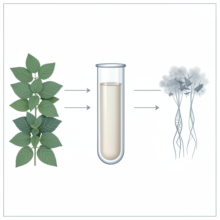
PVP Concentration Effect
AI-generated conceptual illustration (Medicinal plant leaves (Neem, Tulsi, Bel, Sandalwood, and Brahmi)). It explains the idea and does not show study data.
View source evidence ↗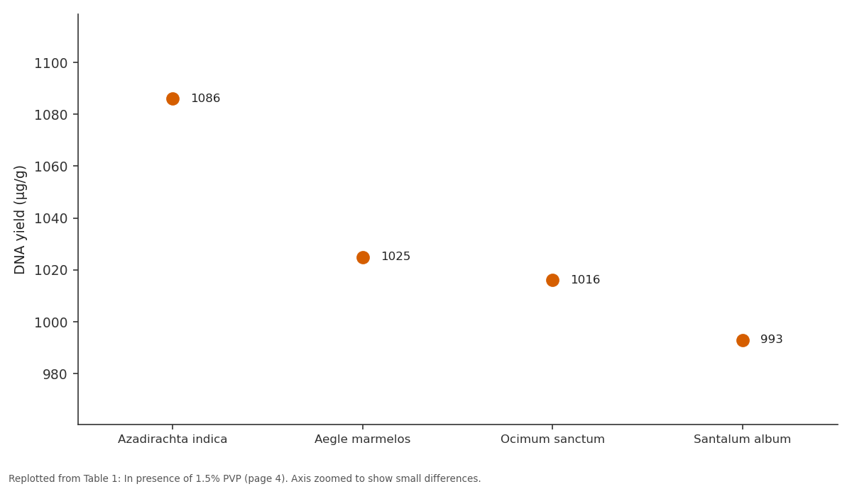
DNA yield (µg/g): Azadirachta indica vs Aegle marmelos vs Ocimum sanctum vs Santalum album
Replotted from Table 1 (page 4) of the original paper. Every bar is the paper's own table value.
View source evidence ↗Figures & tables
From the original paper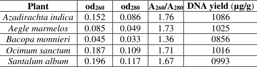 Table 1
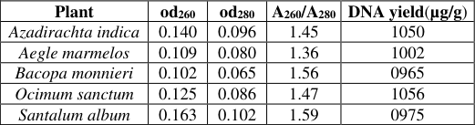 Table 2
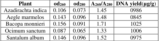 Table 3
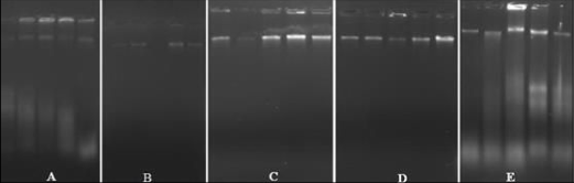 Figure 1
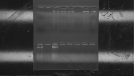 Figure 2
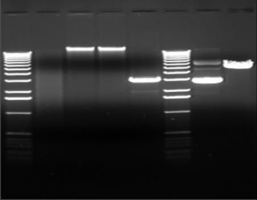 Figure 3
Introduction
Traditional medicines are used by about 60 per cent of the world's population along with other medicine. These are not only used for primary health care not just in rural areas in developing countries, but also in developed countries as well where modern medicines are predominantly used. While the traditional medicines are derived from medicinal plants, minerals, and organic matter, the herbal drugs are prepared from medicinal plants only. Use of plants as a source of medicine has been inherited and is an important component of the health care system in India. In the Indian systems of medicine, most practitioners formulate and dispense their own recipes; hence this requires proper documentation and research. In western world also, the use of herbal medicines is steadily growing with approximately 40 per cent of population reporting use of herb to treat medical illnesses within the past year. Public, academic and government interest in traditional medicines is growing exponentially due to the increased incidence of the adverse drug reactions and economic burden of the modern system of medicine. There are about 45,000 plant species in India, with concentrated hot spots in the region of Eastern Himalayas, Western Ghats and Andaman & Nicobar Island. The officially documented plants with medicinal potential are 3000 but traditional practitioners use more than 6000. India is the largest producer of medicinal herbs and is appropriately called the botanical garden of the world. There are currently about 250 000 registered medical practitioners of the Ayurvedic system (total for all traditional systems: approximately 291 000), as compared to about 700,000 of the modern medicine system. In rural India, 70 per cent of the population is dependent on the traditional system of medicine, the Ayurveda. It is estimated that about 80,000 species of plants are utilized in some form or other by the different systems of Indian medicine.
Plants have been studied on the basis of clearly defined biological parameters like rasa (taste), vipaka (metabolic property), guna (quality), prabhava (biological effect) and virya (potency). The codified traditions have about 25,000 plant drug formulations that have emerged from such studies. In addition to this, over 50,000 formulations are believed to be existing in the folk and tribal traditions. All these point to the deep passion for an exhaustive knowledge about medicinal plants that have existed in this land from time immemorial effects of the adverse drug reactions and cost factor of the modern system of medicine. (http://www.nmpb.nic.com) Medicinal plants have played a key role in world health. Medicinal plant species has a valuable economic importance because of its usage as pharmaceuticals, nutritional, as well as its use in popular medication. For DNA-based techniques, Nano gram quantities of the purified DNA are requisite to amplify and yield sufficient amounts of PCR products. Medicinal utilization and conservation has attracted global attention (Parrotta, 2001). Neem (Azadirachta indica) originates from northeast India. It is also known as Margosa or the Persian lilac. In Indonesia, this plant is referred to as Mimba. In India this plant is referred to as the village pharmacy because of its ability to cure many disorders ranging from bad teeth and bed bugs to ulcers and malaria. Neem is of particular interest to the field of dentistry for it has a long history treating teeth and gum problems. Various studies have shown that Neem can inhibit the growth of Streptococcus mutans and can be used in mouth rinses to reduce periodontal diseases Tulsi (Ocimum sanctum) is a small plant, sub-shrub which has multiple uses. Ayurveda mentions the importance of medicinal uses of it. The leaves are quite effective for the ulcer and infections in the mouth. A few leaves chewed will cure these conditions. The herb is useful in teeth disorders. Its leaves, dried in the sun and powdered, can be used for brushing teeth. It can also be mixed with mustered oil to make a paste and used as toothpaste. This is very good for maintaining dental health counteracting bad breath and for massaging the gums. It is also useful in pyorrhea and other gum disorders. The antiinflammatory and anti-infectious properties of Tulsi make it a powerful treatment for gum disease Brahmi (Bacopa monnieri) is also known as "Medhya rasayanas" in ayurveda as it increases mental clarity and brain stimulating action (Bhattacharya and Ghosal 1998). The medicinal properties of Bacopa monnieri responsible for improving memory-related function have been attributed to the presence of different types of saponins such as Bacosides A, B, C, and D called the "memory chemicals" (Rastogi, 1994). Bacopa monnieri also contains variety of medically active substances i.e. stigma sterol, sapogenins and flavonoids. Other compounds are D-mannitol, betulic acid, beta-sisterol, octacosane, nicotine and amino acid. It also possesses anti-inflammatory, analgesic, antipyretic, epilepsy, insanity, anticancer and antioxidant activities (Satyavati, 1976). It also used for the treatment of asthma, water retention and blood clearing. In Pakistan, the herbal drugs, Brahmi-buti, is used to treat skin diseases, leprosy, epilepsy, eczema, asthama, hoarseness of the voice, and diseases of the nervous system (Shakoor, 1994). They had tested previously established DNA isolation protocols but these methods resulted in DNA with lot of impurities and not very suitable for RAPD analysis Therefore, we report here a total genomic DNA isolation protocol derived from a method originally developed for other plants. The isolated DNA would be suitable for further downstream applications. Medicinal plants contain high levels of polysaccharides, polyphenols, several pigments, and other secondary metabolites, which makes DNA unusable for downstream work in molecular biology research (Wen and Deng, 2002). Polyphenols as powerful oxidizing agents can decrease the yield and purity of extracted DNA. Polysaccharides make DNA viscous, glue-like and non-amplifiable in PCR by inhibiting Taq polymerase enzyme activity and also interfere with accurate DNA digestion (Porebski et al., 1997) [9] . A good isolation protocol should be simple, rapid and efficient, yielding appreciable amounts of high-quality DNA suitable for molecular analysis. Different methods for DNA extraction have effectively been applied for many plant species. DNA has been treated with DNase-free ribonuclease A, since large amounts of RNA in the sample can chelate Mg 2+ and reduce yield in PCR. It also involves successive long-term RNAse treatment with all steps carried out at room temperature. RNase treatment degrades RNA into small ribonucleosides that do not contaminate the DNA preparation and yields RNA-free pure DNA (Jena et al., 2010). At present, there are several DNA isolation kits, but the main problem with these commercially available kits is their high cost per sample (Ahmed et al., 2009). Because most of the available procedures are based on the use of commercial kits, routine DNA extraction is economically difficult for large scale genomic applications. Therefore, to solve the problem of DNA isolation from medicinal plant the experiment entitled "Isolation of DNA from medicinal plant" has been planned during academic year Nov 2012 to April 2013 in MGM College of Agricultural Biotechnology, Aurangabad with following objectives. 1. Standardization of protocol for isolation of DNA. 2. To quantify the yield of isolated DNA.
3. To study possibilities of isolated DNA for PCR study.
Review of Literature
An attempts has been made in this chapter to review and classify the work done in past on this aspect of present investigation by eminent scientists in India and abroad. Anna et al. (2001) [1] carried out an experiment and successfully isolated DNA from leaves of milfoil (Achillea millefolium L.) and Siberian ginserg (Eleutherococcus senticosus) suitable for molecular biology application. Nagarajan (2005) [7] developed a modified CTAB based protocol for DNA isolation from leaf tissue of Phyllanthus emblica and obtained high quantity genomic DNA. They have obtained yield of DNA ranged from 1-2 μg/μl per gram of the leaf tissue and the purity (ratio) was between 1.6-1.7 indicating minimal levels of contaminating metabolites. Padmalatha et al. (2005) [8] optimized DNA isolation and PCR based protocol for RAPD analysis of selected medicinal and aromatic plants of conservation concern from Peninsular India containing high levels of polysaccharides, polyphenols and secondary metabolites. The method involves a modified CTAB extraction employing polyvinyl pyrrolidone while grinding, an overnight RNAse treatment with all steps carried out at room temp. These techniques are ideal for isolation of DNA from different plant species and the DNA isolated was used for randomly amplified polymorphic DNA (RAPD) analysis. They isolated DNA from eight different medicinal plants for RAPD analysis. Khan et al. (2007) [6] demonstrated the protocol for isolated genomic DNA from dry and fresh roots of medicinal plant suitable for RAPD and restriction digestion. The research work carried out at Centre for transgenic plant development, Department of Biotechnology, Jamia Hamdard, New Delhi. Pandey et al. (2008) [10] [11] conducted research to study the isolated genomic DNA from medicinal plant without using liquid nitrogen and obtained DNA quantity and quality comparable those isolated with liquid nitrogen. DNA isolated by this method was used for various molecular biology applications. Adhikari et al. (2011) [3] has been observed to create interference with DNA isolation procedure and downstream reaction, such as DNA amplification, restrictions and cloning and to prepare a simplified high yielding miniprep genomic DNA extraction protocol for medicinal plant. They have developed the protocol was to make this techniques readily available in low facility laboratories and to minimize the duration of plant DNA isolation. Alatar et al. (2012) [2] [4] improvement the protocol for DNA isolation from the medicinal plant of lemon balm (Melissa officinalis). They are used PVP to overcome the problem of polyphenols. DNA yields ranged from 10-20 μg (in 100-μL elution volumes) from all plant material evaluated. The DNA was obtained free of any contaminating proteins, polysaccharides and colored pigments. Sahare et al. (2012) [12] conducted the experiment for the Isolation of Genomic DNA from Medicinal Plants Producing Large Amount of Secondary Metabolites; the experiment was carried out at Department of Botany, Rashtrasant Tukadoji Maharaj University, Nagpur.
Materials and Method
The detail of materials was used and a method was adopted for conducting the present investigation is described in this chapter under appropriate heads. Added 1ul of a 10µg/ml stock solution of RNAse A to DNA. Incubated at 37 • C then added 1/10 volume of 3M Sodium acetate and 2 volumes of Isopropanol to the DNA containing solution then incubated on ice for 10 min, and centrifuged at maximum speed for 5 min at room temperature, to pallet the DNA. Then discarded the alcohol, pallet was washed with 70% ethanol and dried the DNA, and dissolved in dH 2 O. (Ref. Adhikari, S. and Ghosh, T. 2011) [3] 3.3. Quantification of yield and purity of DNA using A260/A280 by UV-VIS Spectrophotometer 1. The isolated DNA was quantified by its absorbance at 260 nm and 280 nm, the ideal value for A 260 /A 280 is 1.8, if the ratio decreases it represented protein contamination and if it increases RNA contamination. 2. The spectrophotometer (UV-1800, schimatzu) has an inbuilt program for quantification of DNA yield and purity. 3. The milli-Q water was used for absorbance blank of both cuvets, then one of the cuvet is replaced by solution contaminant DNA and readings were recorded. (Ref. Alatar, A. and Mahmoud, M. 2012.) [2] 3.4. Restriction digestion of purified DNA fragment 1. The reaction mixture was prepared by mixing 20µl of DNA sample ,10 µl of reaction mixture, 3µl of restriction enzymes (EcoRI) and final volume was maintain 50µl using sterile milli-Q water. 2. The mixture was incubated at 37 0 C for 45 min and placed on ice to stop the reaction. The digestion was confirmed by agarose gel electrophoresis. (Ref. Ghaffariyan, S. and Mohammadi, S. 2012.) [4] 3.5. RAPD PCR Analysis of DNA 1. The DNA was used to perform PCR with RAPD marker.
Collection of
2. The 25µl of master mix supplied by Genei TM was mixed with 1µl of DNA sample, the PCR was carried out using applied Bio Student PCR machine. The result was analyzed using agarose gel electrophoresis. 3. Master mixture for one reaction was consist of following components 10X assay buffer dNTP mix Taq DNA polymerase Genomic DNA Primer MgCl 2 Final volume adjusted using double distilled water.
PCR amplification
The DNA was amplified using thermal cycler. It involves following steps- Initial Denaturation at 95 0 c for 3 min. Denaturation at 94 0 c for 1 min. Primer annealing at 52 0 c for 1 min. Extension at 72 0 c for 2 min. Went to step 2 for repeated cycles. Final extension at 72 0 c for 3 min. Hold at 4 0 (Ref. Sharma, P. and Joshi, N. 2010.) [11] 3.6. Agarose Gel Electrophoresis 1. Gel electrophoresis is a widely used technique for the analysis of nucleic acids. Every molecular biology research laboratory routinely uses agarose gel electrophoresis for the preparation and analysis of DNA.
Agarose gel electrophoresis consist of following components-
Agarose gel electrophoresis consist of following components-
Material
Agarose, TAE Buffer, 6X Sample Loading Buffer, DNA ladder standard, Electrophoresis chamber, Power supply, Gel casting tray and combs, DNA stain, Ethidium bromide, Gloves, Pipette and tips Protocol Measured 1.25 g Agarose powder and added it to a 500 ml flask Added 125 ml TAE Buffer to the flask. (The total gel volume well vary depended on the size of the casting tray) Melted the agarose in a microwave or hot water bath until the solution becomes clear. (if used a microwave, heat the solution for several short intervals -did not let the solution boil for long periods as it may boil out of the flask). The solution cold to about 50-55°C, swirling the flask occasionally to cool evenly. Seal the ends of the casting tray with two layers of tape. Placed the combs in the gel casting tray. Pour the melted agarose solution into the casting tray and let cool until it is solid (it should appear milky white). Carefully pull out the combs and removed the tape. Place the gel in the electrophoresis chamber. Added enough TAE Buffer so that there is about 2-3 mm of buffer over the gel.
3. Product was separated on agarose gel by electrophoresis. It was visualized in gel documentation system after staining with fluorescent dye and photo was saved for further analysis by using appropriate software. (Ref. Anna, M. and Laura, Z. 2001.) [9]
Results and Discussion
The data on yield and purity of DNA as influenced by different extraction method is given in Table 1 (1.5 % PVP), Table 2 (2% PVP) and Table 3 (2.5% PVP). 2011) [3] and Sahare et al. (2012) [12] . Nevertheless, the quantity of isolated DNA has been dependent on the species, the genomic size, the size of cell at different phonological phases of the plant, the amount and type of tissue, the proportion between high quantities of tissue and volume of the extraction buffers used in three different concentration of PVP, and the effect of some substances in the extraction buffer that maintain the stability of pure DNA The concentration of the PVP in the solution and order of addition of the chemicals could be vital, as it has been already reported that adding SDS after 1 h of incubation in extraction buffer, instead of adding it directly to buffer, could improve efficiency ,this was confirmed in this experiment. In present study, standard protocol was modified by using SDS as a detergent with NaCl for solubilization of cell membrane and release cell contents. PVP was used in different concentration (1.5%, 2.0%, and 2.
| Plant | od260 | od280 | A260/A280 | DNA yield(µg/g) |
|---|---|---|---|---|
| Azadirachta indica | 0.106 | 0.073 | 1.45 | 0986 |
| Aegle marmelos | 0.143 | 0.096 | 1.48 | 0845 |
| Bacopa monnieri | 0.156 | 0.091 | 1.71 | 1025 |
| Ocimum sanctum | 0.087 | 0.065 | 1.33 | 1006 |
| Santalum album | 0.146 | 0.096 | 1.52 | 0975 |
Table text extracted from the original PDF; superscript letters are shown inline. The original table image is below.
View original table image
| Plant | od260 | od280 | A260/A280 | DNA yield(µg/g) |
|---|---|---|---|---|
| Azadirachta indica | 0.140 | 0.096 | 1.45 | 1050 |
| Aegle marmelos | 0.109 | 0.080 | 1.36 | 1002 |
| Bacopa monnieri | 0.102 | 0.065 | 1.56 | 0965 |
| Ocimum sanctum | 0.125 | 0.086 | 1.47 | 1056 |
| Santalum album | 0.163 | 0.102 | 1.59 | 0975 |
Table text extracted from the original PDF; superscript letters are shown inline. The original table image is below.
View original table image
| Plant | od260 | od280 | A260/A280 | DNA yield (µg/g) |
|---|---|---|---|---|
| Azadirachta indica | 0.152 | 0.086 | 1.76 | 1086 |
| Aegle marmelos | 0.085 | 0.049 | 1.73 | 1025 |
| Bacopa monnieri | 0.045 | 0.033 | 1.36 | 0856 |
| Ocimum sanctum | 0.187 | 0.109 | 1.71 | 1016 |
| Santalum album | 0.196 | 0.117 | 1.67 | 0993 |
Table text extracted from the original PDF; superscript letters are shown inline. The original table image is below.
View original table image
Effect of Different extraction methods on Yield and purity of DNA
Effect of Different extraction methods on Yield and purity of DNA
Restriction digestion of purified DNA fragment
The extracted DNA which was of highest purity and yield was subjected for restriction digestion analysis. The restriction digestion was done by using EcoR1 restriction enzyme. The purity and clean nature of DNA samples could be confirmed through complete digestion EcoRI (3U/µg DNA). This indicated that the isolated DNA was amenable to further processing in various molecular biology applications as well as DNA fingerprinting. The clear bands were not visible but DNA was restricted by enzymes and it formed a smear on. 8% of Agarose gel.
RAPD PCR Analysis of DNA
The DNA was used to perform PCR with RAPD marker. The 25µl of master mix supplied by Genei TM was mixed with 1µl of DNA sample, the PCR was carried out using applied Bio Student PCR machine. The result was analyzed using agarose gel electrophoresis.
The good utility of the DNA in PCR amplification for RAPD primer (5'GCACGCCGGA3') and with preparation of all the medicinal plant species tested. Only one single RAPD primer was used and it was tested that whether the DNA quality is suitable for RAPD PCR or not. It is found that the DNA got random amplification using PCR and the DNA quality is suitable for the further PCR studies.
Summary and Conclusion
From the above results is clear that PVP concentrations (1.5%, 2.0%, 2.5%) changes affect the yield and purity of DNA. The good quality, intact DNA of high molecular weight was obtained in larger quantities from the five different medicinal plant employed in the study. Nevertheless, the quantity of isolated DNA has been dependent on the species, the genomic size, the size of cell at different physiological phases of the plant, the amount and type of tissue, the proportion between high quantities of tissue and volume of the extraction buffers used, and the effect of some substances in the extraction buffer that maintain the stability of pure DNA.
In the present study, the maximum yield 1086 µg/g obtained from Azadirachta indica as compared to the tissue sample of other medicinal plant. In the present study, most of the concerns have been addressed in DNA extraction protocol for plants rich secondary metabolites or polysaccharides derivatives. The present method required small amount of tissues for successful extraction and large numbers of samples could be processed in parallel. The method performed with chloroform/phenol to purify DNA rather than more expensive cesium chloride or CTAB used in other method. It needed about 1 h to prepare DNA for any molecular biology application and there was no need of liquid nitrogen during crushing the plant material. This method did not require expensive and highly hazardous reagent. It could perform even in low technology laboratories. The quality and quantity of DNA extracted by this method were high enough to perform thousands of PCR-based reaction and could also be used in other DNA manipulation techniques. The protocol could be further extended to related species with relatively minor modification. The aim to develop this protocol was to make this technique readily available in facility laboratories and to minimize the cost of DNA extraction by using commercially available kits. Above all, a high level of genomic DNA with fair quality suitable for PCR-based marker and other genomic studies has successfully been extracted.
Figures and tables from the paper
References
12 in the original paper- Anna M, Laura Z. DNA Isolation Methods for Medicinal and Aromatic Plants. Plant Molecular Biology Reporter. 2001; 19(5):273.
- Alatar A, Mahmoud M. Simple and rapid protocol for the isolation of PCR-amplifiable DNA from medicinal plants. Genetics Molecular Research. 2012; 11(1):348-354.
- Adhikari S, Ghosh T. A simplified high yielding miniprep genomic DNA Extraction protocol for medicinal plant. Indian Journal of Biotechnology. 2011; 11(8):337-340.
- Ghaffariyan S, Mohammadi S. DNA isolation protocol for the medicinal plant lemon balm (Melissa officinalis). Genetics Molecular Research. 2012; 11 (2): 1049-1057.
- Javornik B. Robust CTAB-activated charcoal protocol for plant DNA extraction. Acta Agricultural slovenica. 1987; 87(1):427-436.
- Khan S, Alam T. Protocol for isolation of genomic DNA from dry and fresh roots of medicinal plant suitable for RAPD and restriction digestion. African Journal of Biotechnology. 2007; 6(3):175-178.
- Nagarajan S, Kavitha M. Improved protocol for isolation of genomic DNA from leaf tissues of Phyllanthus emblica. Iranian journal of biotechnology. 2005; 9(4):56-98.
- Padmalatha K, Prasad M. Optimization of DNA isolation and PCR protocol for RAPD analysis of selected medicinal and aromatic plants of conservation concern Peninsular India. African Journal of Biotechnology. 2005; 5(3):230234.
- Porebski. Polyphenols as powerful oxidizing agents can decrease the yield and Purity of extracted DNA. Journal of Biotechnology. 1997; 6(3):175-178
- Pandey M, Changtragoon S. Isolation and characterization of microsatellite Markers in Indian neem (Azadirachta indica var. A. Juss) and cross-amplification in Thai neem (A. indica var. Siamensis valenton).Springer Science Business Media. 2008; 48(1):117-123.
- Sahare, P. and Srinivasu, T. 2012. Method for the Isolation of Genomic DNA from Medicinal Plants Producing Large Amount of Secondary Metabolites. The International Journal of Engineering and Science. 2010; 1(2):37-39.
- Tiwari K, Jadhav S. Modified CTAB Technique for isolation of DNA from some medicinal plants. Research Journal of Medicinal Plant. 2012; 6(5):65-73.