
Research article · 2017 · Full text
International Journal of Statistics and Applied MathematicsA scientific model of finite element technique to study spiral heat regulation in human limbs
Volume 2(3) · Pages 01–03
Research areasInfrared Thermography in MedicineThermoregulation and physiological responsesTextile materials and evaluations
Abstract
Author-writtenWe consider this paper as the one-dimensional bio-heat exchange condition with the perfusion of blood, reliant on temperature which oversees physiological heat dispersion issues relating to limbs and the organic properties are supposed to be distinctive along the radial direction. By methods for an extensive technique with metaphorical estimation, we acquire the correct analytical answer for inspect the heat circulation in the tissues. The dermal region is comprised of three layers, to be specific epidermis, dermis, and subcutaneous tissues. The model consolidates critical varieties of physical and physiological parameters like blood mass stream rate, rate of metabolic heat generation, and thermal conductivity in each layer. Numerical outcomes have been acquired for different instances of functional interest.
Summary
Source-checkedPlain-language reading of the full paper, prepared with AI and checked against the text. Each point links to the authors' own words.
Why it matters
- Heat transmission through human skin is a highly complex and unpredictable process. View evidence ↗
- One-dimensional models for heat distribution in limbs are particularly relevant at low environmental temperatures, where core temperatures of appendages can fluctuate significantly. View evidence ↗
- Temperature variations in limbs may occur because blood cools as it flows toward the extremities. View evidence ↗
The question
- The study aims to use a variational finite element approach to analyze how temperature is distributed across a normal cross-section of a human limb. View evidence ↗
What they did
- The model defines the dermal region as consisting of three specific layers: the epidermis, dermis, and subcutaneous tissues. View evidence ↗
- The model incorporates key physiological and physical parameters, including thermal conductivity, metabolic heat generation rates, and blood mass flow rates for each layer. View evidence ↗
- Numerical results were calculated for three different atmospheric temperature scenarios: 15°C, 23°C, and 33°C. View evidence ↗
- The researchers tested three different sets of values for the thickness of the layers in the skin and subcutaneous tissue (SST) region. View evidence ↗
What they found
- Nodal temperatures on the external surface and within each sub-district vary significantly based on changes in the rate of sweat dissipation and environmental temperatures. View evidence ↗
- The slope of temperature change is most pronounced in the epidermal sub-layer, as it is the layer directly exposed to environmental conditions. View evidence ↗
What it means
- The authors conclude that their extensive technique and metaphorical estimation provide a correct analytical answer for inspecting heat circulation in tissues. View evidence ↗
Visual summary
2 visualsIllustrations explain the paper; charts are replotted from the paper's own table values.
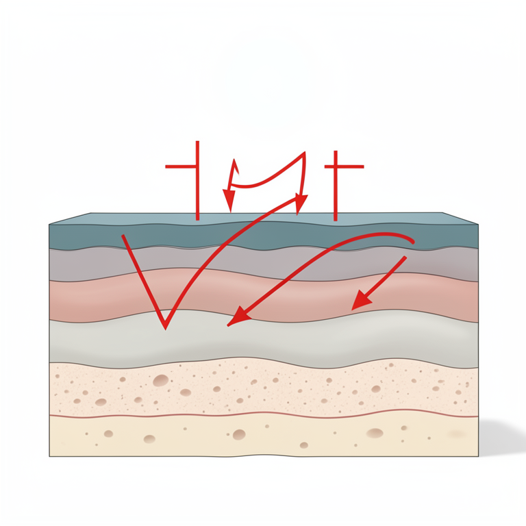
Conceptual Layering of Human Skin
AI-generated conceptual illustration (human skin cross-section). It explains the idea and does not show study data.
View source evidence ↗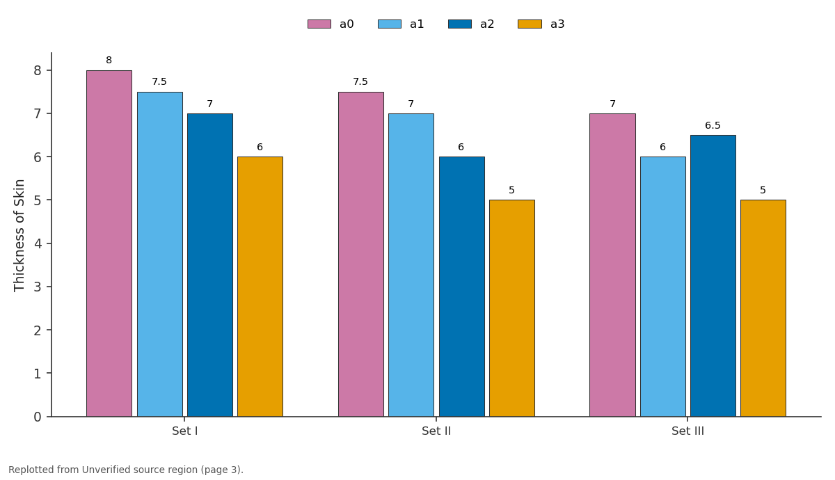
a0 and a1 and more: Set I vs Set II vs Set III
Replotted from a table (page 3) of the original paper. Every bar is the paper's own table value.
View source evidence ↗Introduction
The heat transmit methodology through human dermal locales is an extremely unpredictable phenomena because of various reasons. At lower environmental temperatures, the one dimensional models for heat distribution in limbs have wide degree as the center temperature of the human appendages fluctuates broadly at lower air temperatures. For falling climatic temperature, the body center therapists quickly and isotherm shells in the appendage change their separate position progressively. This might be because of the way that the blood vessel blood has chilled off while streaming towards the furthest points. Likewise the two inverse side of internal center of a human appendage might be at various temperature. This might be on the grounds that one side of the appendage contains real veins (corridors) with blood originating from fundamental trunk at body center temperature and the external side of the appendage contains veins with blood coming back from the furthest points at lower temperature. Perl [12] took collectively the forms of differential equations and Fick's perfusion principle with heat conduction and matter diffusion equations and metabolic term to get equation. He used equation to solve its simple cases by taking all parameters as constant throughout the region. Perl and Hirsch [7] used this equation to test the transient response for measuring local tissue blood flow on dog and rabbit kidney. Trezek and Cooper [1] computed thermal conductivity of tissue by taking all parameters as constant. Cooper and Trezek [2] obtain solution of equation in SST region by taking all parameters as constant. Patterson [6] made experimental attempts, to determine temperature profiles in skin and subcutaneous region. This paper employs a variational finite element approach to study the temperature distribution in a normal cross-sectional region of a limb. Due to unsymmetric situations of large blood vessels passing through the core of the limb the inter-face has angular variation. The peripheral part of limb is directly exposed to atmosphere. Different types of variations of parameters have been considered for different natural subregions such as stratum corneum, stratum germinativum, dermis and underlying tissue (Montagana, Jarrett and Gray) [5,4,3] . Finite element formulation provides necessary flexibility in taking care of different behavior of distinctly different subregions.
Material and Methods
The heat flow in SST region is given by the following partial differential equation
(2.1) Where T and Tb are Temperature and Body center temperature, S is Rate of metabolic warmth generation, mbis Blood mass stream rate in the tissue, is thickness of the tissue. Cb=Specific warmth of blood, K is warm conductivity of the tissue. Above condition has been adjusted and extensively used by Saxena [11] , Saxena and Arya [8] , Saxena and Bindra [9,10] in the warm investigation of human skin and subcutaneous tissue. In this, skin and fundamental tissues of round and hollow locales, for example, appendages of a human are partitioned into four annular layers. They can be considered as multi layered districts. Each layer has diverse physical and physiological properties. The external body surface is presented to the earth and warmth misfortune from the body surface happens because of conduction, convection, radiation and vanishing. Here we utilize for a human appendage with round symmetry. The properties and temperature conveyance are thought to be uniform along and Z headings. In this manner the condition diminishes in one dimensional precarious state case for each layer to the accompanying round and hollow from.
Where K (i) ,M i ,S i and T i denote the values of K, M, S and T in i the sub-region.
Boundary and Interface Conditions:
Inview of continuity of temperature and temperature gradient in various sublayers, the following boundary and interface conditions can be formulated (i) T (1) =T (2) at r= a 1
(iv) at r = a 2 (v) T (3) =T (4) at r = a 3
It is assumed that at the outermost layer (r = a 0 ) the heat is lost to the environment by conduction, convection, radiation and evaporation. Therefore at this layer we take
Where h is coefficient of convection,L is dormant warmth of dissipation and E is the Rate of sweat vanishing TA is the barometrical temperature.At the deepest layer the temperature will be same as that of the body center. Thus the limit conditions will beT4 =Tb atr= a4
Solution of the Problem
The variational form is defined in the region as I1, I2, I3 and I4 respectively for stratumcorneum, stratum germinativum, dermis and subdermal parts. Assigning the values to T as Ti (i = 0, 1, 2, 3, 4) called as nodal values. The distance between the nodal values is given by ai (I = 0, 1, 2, 3, 4) from the outermost layer of epidermis to innermost layer of subdermallayer. LetT (i)
I = f(T ,T ,T ,T ,T )
Now minimizing I with respect to parameters T ,T ,T ,T ,therefore
Finally taking La-place transform, we get four non homogenous simultaneous equations as
x T +y T +z T =n =n y T +z T +w T =n z T +w T =n solving for , , , by using matrix method, we get the values of , , , in the form of polynomials
Where x (p) is a polynomial of degree less than n that of Y (P)i.e.,(n-1)
The value of nodal temperature T T T T ,can be obtained by taking the inverse La-place transform T = e Therefore, we can see that nodal values are dependent on time.
Tables
The numerical result have been obtained with the help of following values -
Conclusion
Distinctive T(i) (i = 0, 1, 2, 3) versus time t for various thickness of skin and diverse estimations of climatic temperatures have been ascertained On looking at the nodal temperature on external surface and in each sub district, it is watched that these nodal temperatures differ extensively with the change in environmental temperatures and rate of sweat dissipation.
Clearly sharpness of slope is more set apart in the epidermal sub-layer which is specifically presented to condition. In addition blood compliment in the dermal and sub dermal sub layers.
References
10 in the original paper- GRAY H. Text Book of Gray's Anatomy, Longmans Press, 1973.
- JARRETr A. The Physiology and Pathophysiology of Skin, Academic Press, 1973.
- MONTAGNA W. The Structure and Function of Skin, Academic Press, 1962.
- Patterson AM. Measurement of temperature profiles in human Skin, S. Afr J Sci. 1976; 72:78-79.
- Perl W, Hirsch RL. Local blood flow in kidney tissue by heat clearance method. J Theoret Biol. 1966; 10(2)251280.
- SAXENA VP, ARYA D. Exact Solution of the Temperature Distribution Problem in Epidermis and Dermis Regions ofHuman Body, Proc. VNM, Medical and Biological Engineering, Sweden, 1981, 364-366.
- SAXENA VP, BINDRA JS. Steady State Temperature Distribution in Dermal Regions of Human Body with Variable Blood Flow, Perspiration and Self-Controlled Metabolic Heat Generation, J Pure Appl Math. 1984; 15(1):31-42.
- SAXENA VP, BINDRA JS. Quadratic Shape Functions in Variational Finite Element Approach to Heat Distribution in Cutaneous and Subcutaneous Tissues, Ind. J. pure Appl Math L. 1987; 8(9):846-855.
- SAXENA VP. Temperature Distribution in Human Skin and Subdermal Tissues, 1, Theo. Biol, 1983; 102:277286.
- Perl W. Heat and matter distribution in body tissue and determination of tissue blood flow by local clearance methods. Journal of theoretical biology. 1962; 2(3):201235.