
Research article · 2018 · Full text
International Journal of Veterinary Sciences and Animal HusbandryClinical management of puerperal uterine infection in a cow caused by Edwardsiella tarda
Volume 3(4) · Pages 01–02
Research areasFish Ecology and Management StudiesReproductive Physiology in LivestockMilk Quality and Mastitis in Dairy Cows
Abstract
Author-writtenEdwardsiella tarda infection, an uncommon pathogen mostly associated with pet reptiles and the ingestion of raw fish. Present case report envisages the successful management of Edwardsiella tarda isolated from the uterine discharge of a Jersey cow 3 weeks after parturition. This may be the first report of puerperal uterine infection caused by E. tarda in a cow.
Summary
Source-checkedPlain-language reading of the full paper, prepared with AI and checked against the text. Each point links to the authors' own words.
Why it matters
- Edwardsiella tarda is a gram-negative bacillus commonly found in aquatic environments, primarily affecting farmed and feral fish. View evidence ↗
- In humans and other hosts, E. tarda infections can lead to severe septicemia and lethality, with gastrointestinal issues often appearing in immunocompromised individuals. View evidence ↗
The question
- The study aims to report and describe the successful clinical management of a puerperal uterine infection caused by E. tarda in a Jersey cow. View evidence ↗
What they did
- The subject was a 6-year-old pluriparous Jersey cow presenting with pus-mixed uterine discharge three weeks after giving birth. View evidence ↗
- Diagnosis involved collecting uterine discharge, streaking it on McConkey’s Lactose Agar and XLD agar, and performing biochemical tests including catalase, oxidase, and indole production. View evidence ↗
- A culture sensitivity test was performed to determine which antibacterial drugs were effective against the isolated pathogen. View evidence ↗
- The cow was treated with Ciprofloxacin injections at a dosage of 5mg/kg body weight for a duration of 5 days. View evidence ↗
- Treatment success was evaluated based on the return of estrus after 3 months and successful conception upon the first insemination. View evidence ↗
What they found
- The isolated pathogen was identified as E. tarda, characterized as gram-negative motile short rods that produced small, grayish-white colonies on XLD agar and tested positive for Indole and H2S production. View evidence ↗
- The pathogen was sensitive to Levofloxacin, Ciprofloxacin, and Ceftriaxone, but completely resistant to Penicillin, Amoxicillin, Streptomycin, Cloxacillin, Ampicillin, and Cephalexin. View evidence ↗
- The treatment was successful, as the cow returned to estrus after 3 months and conceived following the first insemination. View evidence ↗
What it means
- The authors conclude that this case may represent the first reported instance of a puerperal uterine infection caused by E. tarda in a cow. View evidence ↗
- The authors note that while the cause of enteric disease from E. tarda in bovines is known, the specific source of the uterine infection in this case remains unknown. View evidence ↗
Visual summary
1 visualsIllustrations explain the paper; they do not show study data.
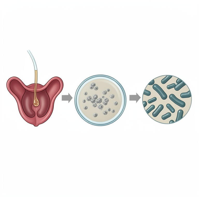
Diagnostic Workflow
AI-generated conceptual illustration (cow uterine discharge and Edwardsiella tarda bacteria). It explains the idea and does not show study data.
View source evidence ↗Figures & tables
From the original paper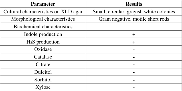 Table 1
Introduction
Edwardsiella tarda, a gram-negative bacillus, is widely distributed in the aquatic environment which mainly affects feral and farmed fish (Mikamo et al. 2003) [3]. The infection is associated with severe septicemia and lethality in a broad host range, which includes humans and gastrointestinal disorders are mostly observed in immunocompromised hosts (Kebede et al. 2016) [1]. E. tarda has caused septicemia, meningitis, peritonitis, osteomyelitis, tubo-ovarian abscess, and liver abscesses in fish and humans (Tamada et al. 2009) [4], but no reports of puerperal uterine infection in domesticated large animals have been reported to date. Case details A pluriparous Jersey cow aged 6 years was presented in Teaching Veterinary Clinical Complex, CSKHPKV, Palampur with a history of pus mixed uterine discharge at 3 weeks after parturition.
Cow also had a history of post-partum uterine prolapse. For diagnostic purpose, the uterine discharge was collected in a sterile vial and streaked on McConkey’s Lactose Agar for identification. Thereafter, purified colonies were streaked on Xylose Lysine Deoxycholate (XLD) agar and various biochemical tests like catalase, oxidase, Indole production, Hydrogen sulphide, Citrate utilization and motility tests (Table 1) were done to confirm the presence of Edwardsiella tarda. Table 1: Phenotypic and biochemical characteristics of Edwardsiella tarda Parameter
| Parameter | Results |
|---|---|
| Cultural characteristics on XLD agar | Small, circular, grayish white colonies |
| Morphological characteristics | Gram negative, motile short rods |
| Biochemical characteristics | |
| Indole production | + |
| H2S production | + |
| Oxidase | - |
| Catalase | - |
| Citrate | - |
| Dulcitol | - |
| Sorbitol | - |
| Xylose | - |
Table text extracted from the original PDF; superscript letters are shown inline. The original table image is below.
View original table image
Results
Cultural characteristics on XLD agar Small, circular, grayish white colonies Morphological characteristics Gram negative, motile short rods Biochemical characteristics Indole production + H2S production + Oxidase - Catalase - Citrate - Dulcitol - Sorbitol - Xylose - VET 2018; 3(4): 01-02 Akshay Sharma PhD Scholar, Department of Veterinary Gynecology and Obstetrics, College of Veterinary and Animal Sciences, Himachal Pradesh Agricultural University, Palampur, Himachal Pradesh, India Madhumeet Singh Professor and Head, Department of Veterinary Gynecology and Obstetrics, College of Veterinary and Animal Sciences, Himachal Pradesh Agricultural University, Palampur, Himachal Pradesh, India Pravesh Kumar Assistant Professor, Department of Veterinary Gynecology and Obstetrics, College of Veterinary and Animal Sciences, Himachal Pradesh Agricultural University, Palampur, Himachal Pradesh, India Nirdeshika Chaudhary Veterinary Officer, Department of Veterinary Gynecology and Obstetrics, College of Veterinary and Animal Sciences, Himachal Pradesh Agricultural University, Palampur, Himachal Pradesh, India Correspondence Akshay Sharma PhD Scholar, Department of Veterinary Gynecology and Obstetrics, College of Veterinary and Animal Sciences, Himachal Pradesh Agricultural University, Palampur, Himachal Pradesh, India Culture sensitivity test was also done for the pathogen against some anti-bacterial drugs. Levofloxacin, Ciprofloxacin and Ceftriaxone were found sensitive to the pathogen whereas Penicillin, Amoxicillin, Streptomycin, Cloxacillin, Ampicillin, Cephalexin were completely resistant. As a part of treatment, Inj. Ciprofloxacin @ 5mg/kg body weight (C- Flox Power; Intas Pharmaceuticals Ltd.) was administered for 5 days to the cow.
Success of the treatment was judged on the basis of estrus (after 3 months) and conception with first insemination.
Discussion
The genus Edwardsiella is genetically distinct from other members of the Enterobacteriaceae. Three species of the genus Edwardsiella are recognized; E. hoshinae. E. ictaluri, and E. tarda. E. tarda is the most common species and was first described in 1965.
Although it exists in a variety of environmental and animal sources, E. tarda is predominantly found in freshwater and fish (Kebede et al. 2016) [1]. It mainly causes gastroenteritis with acute watery diarrhea with a dysentery-like course (Leung et al. 2012) [2]. Uterine infection in the immediate postpartum period may have predisposed to the development of clinical disease due to immunosuppression (Mikamo et al. 2003) [3]. E. tarda can cause enteric disease in the bovine species due to consumption of contaminated products but source of uterine infection is still unknown.
References
4 in the original paper- Kebede B, Habtamu T. Isolation and Identification of Edwardsiella tarda from Lake Zeway and Langano, Southern Oromia, Ethiopia. Fisheries and Aquaculture Journal, 2016; 7(4):1-6.
- Leung KY, Siame BA, Tenkink BJ, Noort RJ, Mok YK. Edwadsiella tarda-Virulence mechanisms of an emerging gastroenteritis pathogen. Microbes and Infection. 2012; 14:26-34.
- Mikamo H, Ninomiya M, Sawamur H, Tamaya T. Puerperal intrauterine infection caused by Edwardsiella tarda. Journal of Infection and Chemotherapy. 2003; 9:341-343.
- Tamada T, Koganemaru H, Matsumoto K, Hitomi S. Urosepsis caused by Edwardsiella tarda. Journals of Infection and Chemotherapy. 2009; 15:191-194.