
Research article · 2019 · Full text
International Journal of Veterinary Sciences and Animal HusbandryEffects of deluteinization on gestation and progesterone concentration in the viviparous Mexican lizard Sceloporus grammicus (Sauria: phrynosomatidae)
Volume 4(6) · Pages 22–28
Research areasTurtle Biology and ConservationSperm and Testicular FunctionVeterinary Medicine and Surgery
Abstract
Author-writtenAim: The corpus luteum is a transitory endocrine gland present in all vertebrate gravid females. Several authors have assigned a central role to the capacity to produce progesterone in egg retention during gestation and in the evolution of reptilian viviparity. However, its role in maintaining gestation in viviparous reptiles is currently under discussion. Here, we describe the effects of lutectomy on progesterone concentration and the maintenance of gestation in the temperate Mexican viviparous lizard Sceloporus grammicus. Materials and Methods: For this purpose, sixteen pregnant females were assigned to three different groups: lutectomy, sham lutectomy, or non-surgical treatment, the latter used as a control group. Blood samples were obtained before surgery, as well as 24 hours and 8 weeks after the procedure to determine the effects of lutectomy on the concentration of progesterone and the maintenance of gestation. Results: We found a significant increase in the plasma concentration of progesterone 24 h after surgery (lutectomized and sham lutectomized lizards); however, these progesterone levels dropped significantly after 8 weeks. There were no significant changes in this regard in the control group at equivalent times when compared with the other groups. Normal parturition and no abortion events were observed in any of the study groups. Conclusion: These observations indicate that the corpus luteum, although capable of progesterone production, is not necessary for the maintenance of gestation nor for parturition. Moreover, these results suggest the existence of a secondary source of progesterone capable of maintaining gestation.
Summary
Source-checkedPlain-language reading of the full paper, prepared with AI and checked against the text. Each point links to the authors' own words.
Why it matters
- Progesterone production is widely considered to play a central role in egg retention during gestation and the evolution of viviparity in reptiles. View evidence ↗
- Despite its known role in oviparous species, it remains unclear whether the corpus luteum (CL) is necessary for maintaining gestation in viviparous lizards. View evidence ↗
The question
- The study aimed to determine how the surgical removal of the corpus luteum (lutectomy) affects progesterone levels and the maintenance of pregnancy in the Mexican lizard Sceloporus grammicus. View evidence ↗
What they did
- Sixteen pregnant female lizards were randomly divided into three groups: lutectomy (n=7), sham lutectomy (n=5), and a non-surgical control group (n=6). View evidence ↗
- Blood samples were collected before surgery, 24 hours after, and 8 weeks after the procedure to measure progesterone concentrations. View evidence ↗
- Plasma progesterone (P4) concentrations were determined using a radioimmunoassay (RIA) on samples stored at -40°C. View evidence ↗
- In the lutectomy group, all corpora lutea were surgically removed from the ovaries. View evidence ↗
What they found
- Lutectomy did not cause any abortions; all lutectomized lizards experienced normal parturition between March and May. View evidence ↗
- In lutectomized lizards, plasma progesterone significantly increased from 0.438 ± 0.09 ng/ml before surgery to 2.29 ± 0.63 ng/ml 24 hours after surgery. View evidence ↗
- Progesterone levels in lutectomized lizards dropped significantly to 0.16 ± 0.03 ng/ml eight weeks after the procedure. View evidence ↗
- Sham-lutectomized lizards also showed a significant increase in progesterone 24 hours after surgery (1.11 ± 0.32 ng/ml from 0.55 ± 0.18 ng/ml), followed by a significant decrease at 8 weeks (0.34 ± 0.12 ng/ml). View evidence ↗
- Control lizards showed no significant changes in progesterone concentrations across the measured time points. View evidence ↗
- Histochemical analysis showed that the corpus luteum was positive for ∆ 5–43β-HSD activity during early and middle pregnancy, particularly in the luteal cell mass. View evidence ↗
- Atretic vitellogenic follicles (AVF) showed low ∆ 5–43β-HSD activity compared to luteal tissue, suggesting they contribute minimally to progesterone production. View evidence ↗
What it means
- The authors conclude that while the corpus luteum can produce progesterone, it is not essential for maintaining pregnancy or for the process of parturition in S. grammicus. View evidence ↗
- The results suggest the existence of a secondary organ, possibly the adrenal gland, capable of producing progesterone to maintain gestation. View evidence ↗
- The authors note that it is unknown if the chorioallantoic placenta (CAP) synthesizes progesterone in this species, though they cannot rule out its participation in hormone production or metabolism. View evidence ↗
Visual summary
1 visualsIllustrations explain the paper; they do not show study data.
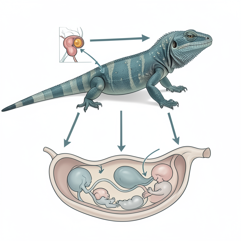
Conceptual Model of Progesterone Sources
AI-generated conceptual illustration (Mexican lizard Sceloporus grammicus). It explains the idea and does not show study data.
View source evidence ↗Figures & tables
From the original paper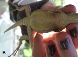 Figure 1
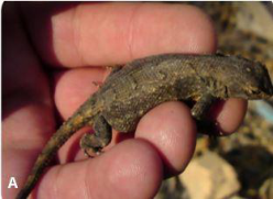 Figure 1
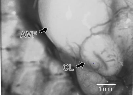 Figure 2
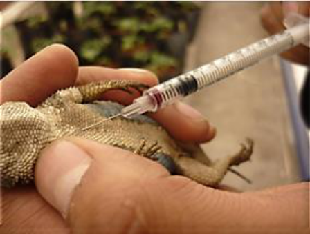 Figure 3
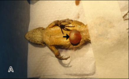 Figure 4
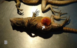 Figure 4
 Table 1
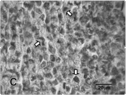 Figure 5
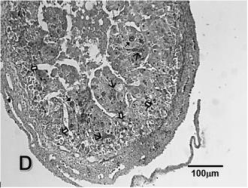 Figure 5
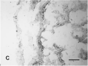 Figure 6
Introduction
The goal of this study was to determine the effects of lutectomy on the maintenance of gestation and progesterone plasma concentrations in the viviparous temperate lizard Sceloporus gammicus. The corpus luteum (CL) is a transitory endocrine gland present in vertebrate gravid females (Xavier, 1987) [39] . Several authors concur in that embryo retention in gravid reptiles is controlled by the capacity of CL to produce progesterone (P 4 ) (reviewed in Xavier, 1987 [39] , Albergotti and Guillette, 2011) [1] . In oviparous lizards, there is a positive correlation between luteal function and longtime of the egg retention (Jones and Guillette, 1982) [22] since luteal regression occurs just before (Sceloporus anneus, Guillette and Jones, 1985) [16] , or immediately after oviposition (Calotes versicolor, Varma 1970; Uromastick hardwicki, Arslan et al., 1978) [37, 4] . Further, the surgical removal of the luteal tissue causes premature oviposition and diminished P 4 levels (Sceloporus undulatus, Roth et al., 1973; Cnemidophorus uniparens, Cuellar, 1979) [34,8] . However, viviparity requires that the period of the egg retention in utero be extended until the embryonic development is complete (Guillette, 1993) [18] . Moreover, there is no direct correlation between luteolysis and parturition in viviparous lizard's species, since luteal regression can occur at any time during pregnancy and differs among species Jones and Guillette, 1982; Xavier, 1987) [22,39] . Further, the extirpation of either the ovaries or the CL in gestating viviparous lizards have different effects according to species and pregnancy stage. In this regard, abortion can be induced as a result of lutectomy or ovariectomy if performed during early pregnancy in Xantusia vigilis (Yaron, 1972) [40] and Sceloporus jarrovi (Guillette, 1987) [17] . However, the same treatment in Lacerta vivipara (Panigel, 1956) [30] , Sceloporus cyanogenys (Lien and Callard, 1968, Callard et al., 1972) [24,6] , and Barisia imbricata imbricata (Martínez-Torres et al., 2010) [28] does not result in abortion, but cause abnormal parturition (stillborn and delayed parturition). In other species, such as Mabuya carinata (Sekharappa and Devaraj-Sarkar, 1978) [35] and Chalcides ocellatus (Badir, 1968) [5] , the extirpation of the luteal tissue or ovariectomy had no effect in gestation length or parturition. These data highlights that, at present, it is unclear if the CL participates in maintaining gestation in viviparous lizards. [31,19] . Males begin their gametogenesis in May or June, with a maximal testicular volume reached in July, August, or September (according to the region they inhabit) (Guillette and Casas, 1980; Jiménez-Cruz et al., 2005; Ramirez-Bautista et al., 2005) [14,32] ; whereas in the females the vitellogenesis occurs between May-September, ovulation in October-November and the young birth in April, May or July (Ramirez-Bautista et al [21,32] . The reproductive ecology of this species has been studied extensively ( [21,14] , as well as histology follicular development, CL and placenta the S. grammicus; however, their reproductive endocrinology remains unapproached.
Materials and Methods
Materials and Methods
Experimental design
We collected adult females (snout-vent length: 59.0 6.0 mm; body weight: 7.3 ± 2.1 g) (Fig. 1
Experimental procedure
One week after capture, the specimens assigned to either the lutectomy or sham lutectomy groups were anaesthetized with sodium pentobarbital (16.0 g/10 g body weight) via IP; afterwards, a ventrolateral incision was performed. In the females subjected to lutectomy, the ovaries were exposed, all CL were surgically removed from each ovary and its total number registered. Additionally, two atretic vitellogenic follicles (AVF) were obtained from 3 females (Fig. 2). One CL and one AVF from each female were fixed in 10% buffered formalin and processed for routine histological methods; the remaining samples were embedded in tissue-teck and immediately frozen in a mixture of dry ice and acetone and stored at -40 ºC until the time for histochemistry analysis of ∆ 54 3-hydroxyl steroid dehydrogenase (∆ 5-4 3-HSD) (Levy et al., 1959) [23] . The specimens subjected to sham lutectomy were treated identically; however, the CL and AVF were not removed and only their number was registered. In addition, the embryo development stage was determined according to the development table for Lacerta vivipara (Dufaure and Hubert, 1961) [11] . Once the surgical procedure was completed, the lizards were sutured and each female deposited in individual terrariums within our laboratory, with free access to food and water (the diet included Tenebrio mealworms, Galleria mellonela wax worms, Achaeta spp. domestic crickets, and grasshoppers) during the following three days to allow for their recovery. On the day of surgery, each lizard received an injection of 5,000 IU of penicillin G, and of 500 μg streptomycin sulfate (IM) in the following two days. Lizards of the control group did not receive surgical treatment but were administered the antibiotics treatment regardless. After the recuperation period, all females were housed individually in terrariums (30 cm × 50 cm × 30 cm) and kept in the greenhouse of the UMF-FES Iztacala UNAM throughout the pregnancy period. All lizards had unrestricted access to water and food (mealworms, grasshoppers and crickets) and were kept under natural temperature and photoperiod throughout the experiment. The terrariums were scrutinized daily from the day of surgery until parturition to detect early or late embryo expulsion, or nonviable eggs. The dates of abortion, expulsion of non-viable eggs, and parturition, as well as the number of offspring, including live neonates, stillborn embryos or nonviable eggs, were registered. We also recorded whether parturition was A normal or abnormal and each lizard was dissected the following day after labor for to confirm CL absence in the ovaries, as well as the lack of embryos or eggs in the uterus of lutectomized females. The uteri of sham-lutectomized specimens were analyzed in the same manner; the number of CL was recorded, in addition to the state of the uterus, in the control specimens as well.
Histology
Previously fixed CL and AVF were processed according to routine histological technique and embedded in paraffin. Serial sections (7 m thick) were obtained and stained using Harris hematoxylin and eosin method (Luna, 1968) [26] . and observed under the microscope to determine the histological characteristics for CL and AVF and development stage for CL according to Martínez-Torres et al., (2003) [27] .
Histochemistry
Corpora lutea and AVF samples embedded in tissue-tek were sectioned (12 µm thick) in a cryostat and processed according to Levy et al., (1959) [23] . The freshly frozen sections were placed in medium containing 40 mg nicotine adenine dinucleotide and 20 mg nitro blue tetrazolium dissolved in 40 ml phosphate-buffered solution, pH 7.4 and incubated at 37 ºC for 1 h. As a substrate, we used 2 mg dehydroepiandrosterone (3b-hydroxyandrost-5-en-20-one) dissolved in 0.5 ml N, Ndimethylformamide. Control sections of mice ovaries and kidneys were incubated in a medium with or without substrate. All reagents were provided by Sigma Chemical.
Effect of lutectomy on progesterone concentration
One day before surgery (lutectomy or sham lutectomy), a blood sample of 80 ± 10 μl was obtained from each female by intracardiac puncture with a heparinised syringe. Blood samples were also obtained 24 hours and eight weeks after surgery. Control females were bled in similar manner as the experimental specimens. The blood samples were centrifuged immediately after collection (6000 rpm/1 min), and the plasma decanted and stored at -40°C until the P 4 radioimmunoassay (RIA) was performed (Martínez-Torres et al., 2003) [27] . All aliquots were obtained between 9:00 a.m. and 11:00 a.m. Plasma P 4 concentration was determined using a commercial kit (Coat-A-Count Progesterone, Diagnostic Products Corporation, Ca 90045). The assay was performed without duplicates, using 50 μl of plasma without prior dilution or extraction. 125 I-labelled P 4 was supplied as a reactive tracer. We used a P 4 specific antiserum, finding a steroid cross-reaction (relative to P 4 , 100%) with androstenediol (non-detectable), corticosterone (0.9%), cortisol (0.03%), 11deoxicorticoesterone (2.2%), 20-dihydroprogesterone (0.2%), oestradiol (non-detectable), 17 hydroxyprogesterone (3.4%), 5b-pregnan-3a-ol-20-one (0.05%), 5a-pregnan-3, 20dione (3.2%), pregnenolone (0.1%), and testosterone (0.1%). Inter-and intra-assay coefficients of variation were of 7.9 and 7.6%, respectively. The sensitivity of the assay was of 0.02 ng/ml. The values were obtained using the GAMBYT software. All experimental procedures were approved by the FES Iztacala-UNAM Bioethical Committee.
Statistical analysis
To determine the difference in litter size and number of live offspring, as well as to compare the number of CL among the groups, we used a one-way ANOVA. A two-way ANOVA for repeated measures was used to determine any significant changes in P 4 concentrations. Post-hoc tests were carried out using the Holm-Sidak method to determine differences; the significance threshold was set at p< 0.05 (Ambrose and Ambrose, 1987). All statistical analyses were performed with the Sigma Stat software (version 3.5 for Windows).
Results
The eggs of specimens subjected to lutectomy during early pregnancy (two females collected in early November) presented a "cicatricela" (Fig. 4A), and, according to Dufaure and Huber (1961) [11] , these embryos were between stages 4-7.
In the remaining lizards, surgery was performed during the second part of gestation, when the embryos were in stages 29 (two lizards) and 32 (one lizard) (Fig. 4B) (according to Dufaure and Hubert, 1961) [11] .
Effects of lutectomy on the maintenance of gestation
We observed no abortion events in any of the lutectomized lizards; parturition was normal and occurred from the first week in March through April and May. A similar situation was observed in sham-lutectomized and control females. Further, we did not observe any significant difference in the number of neonates among groups (Table 1).
| Treatment | Number of CL (means ± SD) | Number young (means ± SD) | Progesterone* (means ± SEM) | — | — |
|---|---|---|---|---|---|
| BS | 24h AS | 8 wAS | |||
| Lutectomy (n = 7) | 4.8 ± 2.2 | 4.8 ± 2.4 | 0.44 ± 0.09a | 2.29 ± 0.6b | 0.16 ± 0.03c |
| Sham Lutectomy (n = 5) | 4.5 ± 1.9 | 4.5. ± 2.8 | 0.55 ± 0.18d | 1.11 ± 0.3e | 0.34± 0.12f |
| Intact (n = 6) | 5.2 ± 1.6 | 5.6 ± 1.9 | 0.34 ± 0.13g | 0.62 ± =0.3h | 0.36± 0.13i |
Table text extracted from the original PDF; superscript letters are shown inline. The original table image is below.
View original table image
Histology
The CL samples obtained during late October and early November showed a histological appearance corresponding to stage I of luteal development (Fig. 5A). This stage is characterized by an irregular cavity filled with erythrocytes and surrounded by the luteal cell mass (LCM) (Fig. 5A). The latter showed oval or rounded cells with acidophilic cytoplasm, a vesicular nucleus, a nucleolus (Fig. 5B), and a few cells with condensed nucleus. The CL obtained from lutectomized specimens during the second half of gestation showed typical histological characteristics of luteal tissue in regression. The luteal cell mass of CL obtained from females with embryos in stage 29 showed cells with slightly shrunken nuclei and without nucleolus, corresponding with stage I of regression (Fig. 5C), whereas CL from females with embryos in stage 32 showed CL in stage II of regression (cells with shrunken nuclei and connective tissue trabeculae). In the AVF, the theca contains many fibroblasts, blood vessels and acidophilic intercellular material. The oocyte lacks zona pellucida, and has been invaded by follicular cells, the cytoplasm presents some blood vessels and scanty vitelline residuals (Fig. 5D)
Histochemistry
All CL obtained from early and middle pregnancy stages were positive. The highest activity of ∆ 5-4 3-HSD was observed in the luteal cell mass, whereas lower activity was observed in the thecal tissue (Fig. 6A). Atretic vitellogenic follicles also showed low activity in both theca (Fig. 6B) and the oocyte cytoplasm (Fig. 6C), however, the formazan granules were observed mainly in the theca. 100X.
Effects of lutectomy on progesterone concentration
The changes in plasma P 4 concentration in S. grammicus females (lutectomized, sham lutectomy, and control lizards) are shown in Table 1. We found a significant increment in plasma P 4 concentration in lutectomized lizards 24 h after surgery [F (4, 28) = 9.43, p< 0.05]. The values obtained previous to lutectomy were of 0.438 ± 0.09 ng/ml, whereas after surgery these were of 2.29 ± 0.63 ng/ml. However, the levels of this hormone diminished significantly eight weeks after lutectomy (0.16 ± 0.03 ng/ml) [F (4, 28) = 9.43, p< 0.05].
Regarding sham-lutectomized lizards, we also found a significant increment of P 4 concentration in plasma [F (4, 28) = 7.57, p< 0.05] (previous to surgery: 0.55 ± 0.18 ng/ml; 24 h after surgery: 1.11 ± 0.32 ng/ml). A significant decrement (p< 0.05) in P 4 levels was observed at 8 weeks after surgery as well (0.34 ± 0.12 ng/ml). In contrast, the control specimens did not show any significant changes in P 4 concentration in the evaluated time points (previous to surgery: 0.33 ± 0.13 ng; 24 hr. after surgery: 0.62 ± 0.36 ng/ml). 5.2 ± 1.6 5.6 ± 1.9 0.34 ± 0.13 g 0.62 ± =0.3 h 0.36± 0.13 i *ng/ml SD = standard deviation SEM = standard error medium BS = before surgery AS = after surgery wAS = weeks after surgery n = number of females used. b = the means was significantly higher (at p< 0.05) with respect to remaining times of pregnancy examined (whiting to the same group and among groups). e = the mean was significantly higher (at p< 0.05) with respect to remaining times of pregnancy examined (whiting to the same group).
Discussion
Previous studies have evidenced the role of the luteal tissue as a major source of gestational P 4 in all reptiles (reviewed in Yaron, 1985; Xavier, 1987) [41,39] . Since gestation is supported by this hormone, several authors have assigned a central role to this gland in the maintenance of the gravidity period (Yaron, 1985; Guillette, 1987; Callard et al., 1992) [41,17] . However, the role of the CL in the maintenance of gestation is yet in dispute since surgical deluteinization causes different effects according to species and gestation stage in which the surgical procedure is performed (Guillette, 1987) [17] . In this study, we found evidence that the CL is not essential for the maintenance of gestation in S. grammicus as lutectomy did not result in abortion. In this species, luteal development occurs during the first third of gestation whereas luteolysis occurs during the last two (Villagrán-Santa Cruz, 1989) [38] , where luteal regression occurs gradually and ends around the time of parturition. In our study, we found that the luteal tissue of this species is positive for ∆ 5-4 3-HSD activity in both early and middle stages of gestation, when the CL are in development and regressive stages, respectively. These observations suggest that, same as in other studied lizards (reviewed in (Xavier, 1987) [39] , the CL of S. grammicus is capable of producing P 4 throughout gestation. Several authors have reported that both in viviparous lizards (Tiliqua rugosa, (Fergusson and Bradshaw, 1991; Barisia imbricata, Martínez-Torres et al., 2010) [12,28] and in snakes (Thamnophis elegans, (Highfill and Mead, 1975) [20] lutectomy results in a decreased concentration of P 4 , either immediately (e.g. 24 h after surgery) or in the long term (T. rugose, Fergusson and Bradshaw, 1991; T. elegans, Highfill and Mead, 1975; B. i. imbricata, Martínez-Torres et al., 2010) [12,20,28] . In contrast, we observed that in S. grammicus there is a significant increment (around five-fold when compared to the observed values prior to CL extirpation) of this hormone in the plasma of all lutectomized lizards 24 h after surgery. However, the plasma concentration of P4 decreased significantly after 8 weeks and fell below the levels observed prior to surgery, although not below the detection limit. In sham lutectomized lizards, we observed that the concentration of P 4 increased significantly as well; however, this increment was below the observed in lutectomized females. In control females, P 4 concentration did not change significantly in any of the evaluated time points. These results make clear that in S. grammicus, as previously suggested for other viviparous lizards (Fergusson and Bradshaw, 1991; Martínez-Torres et al., 2010) [12,28] , exist a secondary source of this steroid capable to maintenance of gestation in absence of CL. Some authors have suggested that AVF may be an important ovarian source of P 4 during gestation (Guillette et al., 1981; Villagrán-Santa Cruz, 1989) [15] [38] . Further, Villagrán-Santa Cruz (1989) [38] suggested that the AVF may be capable of P 4 production since it possesses a glandular appearance, proposing that these structures could assist the CL in the synthesis of this hormone. In our study, we found that the AVF obtained from lutectomized lizards showed low ∆ 5-4 3-HSD activity when compared with the luteal tissue. These observations suggest that in this species the contribution of AVF towards P 4 production during gestation is minimal; therefore, we consider that the AVF does not participate in maintaining gestation in this lizard. On the other hand, Guillette et al., (1981) [15] have proposed that the chorioallantoic placenta (CAP) could be a secondary source of P 4 in S. jarrovi, since the presence of this placental tissue coincides with CL regression. Painter and More (2005) [29] demonstrated that the CAP is capable of metabolizing steroids in this lizard. According to Villagrán-Santa Cruz (1989) [38] , S. grammicus develops the CAP during the last stage of gestation (from stage 30 of embryonic development). In our study, we performed the lutectomy procedure during early and middle gestation stages, when the CAP would not have formed yet. Although it is unknown if the CAP is capable of synthetizing P 4 in this species, we do not discard the possibility that the placenta may participate in the production or metabolism of this hormone, in similar manner to S. jarrovi (Painter and More, 2005) [29] and Chalcides chalcides (Guarino et al., 1998) [13] . In some species of viviparous lizards, such as Xantusia vigilis, (Yaron, 1972) [40] and Sceloporus jarrovi (Guillette 1987) [17] , lutectomy causes abortion when performed during early pregnancy. However, the same treatment did not provoke abortion in either Lacerta vivipara (Panigel, 1956) [30] , Sceloporus cyanogenys (Lien and Callard, 1968; Callard et al., 1972) [24] , or B. i. imbricata (Martínez-Torres et al., 2010) [28] ; however, it did cause abnormal parturition (stillborn young and delayed parturition). In other species, such as Mabuya carinata (Sekharappa and Debaraj-Sarkar, 1978) [35] and Chalcides ocellatus (Badir, 1968) [5] , neither the extirpation of luteal tissue nor ovariectomy affected the length of gestation or parturition. In S. grammicus, we did not observe abortion or abnormal parturition in lutectomized lizards. In a previous paper, Martínez-Torres, et al., (2010) [28] proposed that although the reptilian luteal tissue is capable of producing P 4 , their relevance in the maintenance of gestation in the viviparous saurians has been overshadowed by the emergence of other structures capable of P 4 production, such as the adrenal gland (AG). Moreover, there is evidence on the capacity of the AG to produce P 4 in several viviparous squamata species, such as Lacerta vivipara, (Dauphin-Villemant and Xavier, 1985 [9] and Dauphin-Villemant et al., 1990), Thamnophis elegans, (High fill and Mead, 1975) [20] , and that it could be, therefore, participating in maintaining gestation in the absence of CL (T. rugosa, Fergusson and Bradshaw, 1991; T. elegans, Highfill and Mead, 1975; B. i. imbricata, Martínez-Torres et al., 2010) [12, 20, 28] , this is supported by the fact that the levels of plasma P 4 concentration never fell below the limit of detection despite being lutectomized. In contrast, adrenalectomy results in decreased plasma P 4 concentration, at non-detectable levels, in non-pregnant T. elegans snakes (Highfill and Mead, 1975) [20] . These evidences support the idea that the AG actively participates in P 4 production and maintaining gestation in viviparous lizards. Moreover, we consider that these observations demonstrate the necessity of evaluating the role of the AG in maintaining gestation in viviparous squamata.
Conclusion
1. Although the CL of S. grammicus is capable of producing P 4 , it is not necessary for maintaining gestation or parturition. 2. There is a plausible possibility of a 'secondary' organ capable of producing P 4 and maintaining gestation. 3. It is necessary to re-evaluate the participation of the corpus luteum in maintaining gestation in viviparous saurians.
Conflict of interest
The authors declare that there is no conflict of interest that could affect the integrity of the currently reported results. The corresponding author asserts that the co-authors have read the final manuscript, agree with its contents and consent to joining the list of authors of this paper.
Acknowledgement
AcknowledgmentsThe authors thank the staff of the Laboratorio de Microscopía FES Iztacala UNAM, Martha Martínez-Cisneros for her technical assistance and Norma B. Cruz by the careful revision of manuscript. This manuscript was carried out in part during a sabbatical semester 2019-1 of MMT.
Figures and tables from the paper
References
41 in the original paper- Albergotti LC, LJ Jr Guillette. Viviparity in Reptiles: Evolution and Reproductive Endocrinology, in: Norris D.O., Lopez K.H. (Eds.). Hormones and Reproduction in vertebrates. Amsterdan, Boston, Heidelberg, London, New York, Oxford, Paris, San Diego, San Francisco, Singapure, Sidney, Tokyo, Academic Press, 2011, 247275.
- Ambrose HW, Ambrose KP. A handbook of biological investigation. Hunter Textbooks Inc., Winston-Salem, North Carolina, 1987, 208.
- Arévalo E, Porter CA, González A, Mendoza F, Camarillo JL, Sites JW Jr. Population cytogenetics and evolution of the Sceloporus grammicus complex (Iguanidae) in central México. Herpetol Monogr. 1991; 5:79-15.
- Arslan M, Zaidi P, Lobo J, Zaidi AA, Qazi MH. Steroids levels preovulatory and gravid lizards (Uromastix hardwicki). Gen Comp Endocrinol. 1978; 34:300-303.
- Badir N. Structure and function of corpus luteum during gestation in the viviparous lizard Chalcides ocellatus. Anat Anz. 1968; 122S:1-10.
- Callard IP, CG Bayne, McConnell WF. Hormones and reproduction in the female lizard Sceloporus cyanogenys. Gen Comp Endocrinol. 1972; 18:175-194.
- Callard IP, Fileti LA, Perez LE, Sorbera LA, Giannoukus G, Klosterman LL et al. Role of the corpus luteum and progesterone in the evolution of vertebrate viviparity. Am Zool. 1992; 32:264-275.
- Cuellar HS. Disruption of gestation and egg shelling in deluteinized oviparous whiptail lizards Cnemidophorus uniparens (Reptilia, Teiidae). Gen Comp Endocrinol. 1979; 39:150-157.
- Dauphin-Villemant C, Xavier F. In vitro steroid biosynthesis by adrenal gland of the female Lacerta vivipara Jacquin: The metabolism of exogenous precursors. Gen Comp Endocrinol. 1985; 58:1-9.
- Dauphin-Villemant C, Leboulenger F, Vaudry H. Adrenal activity in the female lizard Lacerta vivipara Jacquin during artificial hiberntion. Gen Comp Endocrinol. 1990; 79:201-214.
- Dufaure JP, J. Hubert. Table de développement du lézard vivipare: Lacerta (Zootoca) vivipara Jacquin. Archiv Anat Microscop Morphol Exp. 1961; 50:309-328.
- Fergusson B, Bradshaw SD. Plasma arginine vasotocin, progesterone and luteal development during pregnancy in the viviparous lizard Tiliqua rugosa. Gen Comp Endocrinol. 1991; 82:140-151.
- Guarino FM, Paulesu L, Cardone A, Bellini L, Ghiara G, Angelini F. Endocrine activity of the corpus luteum and placenta during pregnancy in Chalcides chalcides (Reptilia, Squamata). Gen Comp Endocrinol. 1998; 111:261-270.
- Guillette LJ Jr, Casas-Andreu G. Fall reproductive activity in the high-altitude Mexican lizard Sceloporus grammicus microlepidotus. J Herpetol. 1980; 14:143-147.
- Guillette LJ Jr, Spielvogel S, More FL. Luteal development, placentation and plasma progesterone concentration in the viviparous lizard Sceloporus jarrovi. Gen Comp Endocrinol. 1981; 43:20-29.
- Guillette LJ Jr., Jones RE. Ovarian, oviductal, and placental morphology of the reproductively bimodal lizard, Sceloporus aneus. J Morphol. 1985; 184:85-98.
- Guillette LJ Jr. The evolution of viviparity in fishes, amphibians and reptiles: An endocrine approach, in: Norris DO, Jones RE. (Eds.), Hormones and Reproduction in Fishes, Amphibians, and Reptiles. New York and London, Plenum Press, 1987, 523-562.
- Guillette LJ Jr. Evolution of viviparity in Lizards. Bioscience. 1993; 43:742-751.
- Hernández-Salinas U, Ramírez-Bautista A, LeyteManrique A, Smith GR. Reproduction and sexual dimorphism in two populations of Sceloporus grammicus (Sauria: Phrynosomatidae) from Hidalgo, México. Herpetologica. 2010; 66:12-22.
- Highfill DR, Mead RA. Sources and levels of progesterone during pregnancy in the garter snake, Thamnophis elegans. Gen Comp Endocrinol. 1975; 27:389-400.
- Jiménez-Cruz E, Ramírez-Bautista A, Marshall JC, Lizana-Avia M, Nieto-Montes de Oca A. Reproductive cycle of Sceloporus grammicus (Squamata: Phrynosomatidae) from Teotihuacán, México. Southwest Nat. 2005; 50:178-187.
- Jones RE, JL Jr. Guillette. Hormonal control of oviposition and parturition in lizards. Herpetologica. 1982; 38:80-93.
- Levy H, Deane HW, Rubin BL. Visualization of steroid Δ 5 , 3-ol-dehydrogenase activity in tissues of intact and hypophysecomized rats. Endocrinol. 1959; 65:932-943.
- Lien JP, Callard IP. The effect of gonadectomy and hormone replacement therapy on reproduction and the reproductive tract of the ovo-viviparous lizard, Sceloporus cyanogenys. Va J Sci. 1968; 19:179.
- Lozano A, Ramírez-Bautista A, Uribe MC. Oogenesis and ovarian histology in two populations of the viviparous lizard Sceloporus grammicus (Squamata: Phrynosomatidae) from the central Mexican Plateau. J Morphol. 2014; 275:949-60.
- Luna GL. Manual of Histological Staining Methods of the Armed Forces Institute of Pathology. McGraw-Hill Book Co, 1968, 237.
- Martínez-Torres M, Hernández-Caballero ME, ÁlvarezRodríguez C, Luis-Díaz JA, Ortiz-López G. Luteal development and progesterone levels during pregnancy of the viviparous temperate lizard Barisia imbricata imbricate (Reptilia, Anguidae). Gen Comp Endocrinol. 2003; 132:55-65.
- Martínez-Torres M, Hernández-Caballero ME, Luis-Díaz JA, Ortiz-López G, Cárdenas-León M, Moreno-Fierros L. Effects of lutectomy in early pregnancy on the maintenance of gestation and plasma progesterone concentrations in the viviparous temperate lizard Barisia imbricata. Rep Biol Endocrinol. 2010; 8:19.
- Painter DL, Moore MC. Steroid hormone metabolism by the chorioallantoic placenta of the mountain spiny lizard Sceloporus jarrovii as a possible mechanism for buffering maternal-fetal hormone exchange. Physiol Biochem Zool. 2005; 78:364-372.
- Panigel M. Contribution a l'ètude de l'ovoviviparité chez les reptiles: gestation et parturition chez le lézard vivipare Zootoca vivipara. Ann Sci Nat Zool Biol Anim. 1956; 18:569-668.
- Ramírez-Bautista A, Jiménez-Cruz E, Marshall JC. Comparative life history for populations of the Sceloporus grammicus (Squamata: Phrynosomatidae). West N Am Nat. 2004; 64:175-83.
- from Pachuca, Hidalgo, México. Acta Zool. Scinica. 2005; 51:998-1005.
- Ramírez-Bautista A, Stephenson BP, Lozano A, UribeRodríguez H, Leyte-Manrique A. Atypical reproductive cycles in a population of Sceloporus grammicus (Squamata: Phrynosomatidae) from the Mexican Plateau. Ecol Evol. 2012; 2:1903-1913.
- Roth JJ, Jones RE, Gerrard AM. Corpora lutea and oviposition in the lizard Sceloporus undulatus. Gen Comp Endocrinol. 1973; 21:569-572.
- Sekharappa BM, Devaraj-Sarkar HB. Role of the corpora lutea in the skink Mabuya carinata (Schn.). Indian J Exp Biol. 1978; 16:1097-1098.
- IUCN. Guidelines for appropriate uses of IUCN red list data. Version 3.0, 2018. [Adopted by the IUCN Red List Committee].
- Varma SK. Morphology of ovarian changes in the garden lizard, Calotes versicolor. J Morphol. 1970; 131:195-209.
- Villagrán-Santa Cruz M. Desarrollo embrionario placentación y su relación con el cuerpo lúteo y la atresia folicular en Sceloporus mucronatus y Sceloporus grammicus, (Sauria, Iguanidae). Ph D Thesis Facultad de Ciencias, UNAM, 1989, 132.
- Xavier F. Functional morphology and regulation in the corpus luteum, in: Norris DO, Jones RE (Eds.), Hormones and Reproduction in Fishes, Amphibians and Reptiles. New York and London. Plenum Press, 1987, 241-281.
- Yaron Z. Effects of ovariectomy and steroid replacement on the genital tract of the viviparous lizard, Xantusia vigilis. J Morphol. 1972; 136:313-325.
- Yaron Z. Reptilian placentation and gestation: structure, function and endocrine control, in: Gans C, Billet F. (Eds.), Biology of the Reptilia, New York, Wiley. 1985; 15:528-603.