
Research article · 2019 · Full text
International Journal of Veterinary Sciences and Animal HusbandryHistology of the male gonad of adult Khaki Campbell duck (Anas platyrhynchos domesticus) in Bangladesh
Volume 4(4) · Pages 36–39
Research areasSperm and Testicular FunctionLivestock and Poultry ManagementComparative Animal Anatomy Studies
Abstract
Author-writtenThe current research was designed to investigate the histological description of testes of adult (6 months old) Khaki Campbell duck in Bangladesh. The study was conducted in the Department of Anatomy & Histology, Bangladesh Agricultural University, Mymensingh. The experimental Khaki Campbell ducks (Anas platyrhynchos domesticus) were collected from poultry farm of Bangladesh Agricultural University, Mymensingh, having apparently good health and devoid of any external deformities. The testes were collected immediately after ethical killing of the birds for histological observations. The collected tissue samples were then processed and stained with Hematoxylin & Eosin (H & E) stain for histological observations. Each testis was covered by a connective tissue capsule. There were no thick connective tissue stripes dividing the testis into lobules. The stroma of the testis possessed no mediastinum. Leydig cells were commonly found in groups within the connective tissue which filled the spaces between the seminiferous tubules. Germ cells and Sertoli cells constituent the main component of the seminiferous epithelium. This basic histological feature may help to explore the molecular changes of adult testes of Khaki Campbell duck.
Summary
Source-checkedPlain-language reading of the full paper, prepared with AI and checked against the text. Each point links to the authors' own words.
Why it matters
- Duck farming is expanding in Bangladesh as a way to diversify poultry farming and address shortages in animal protein through increased egg and meat production. View evidence ↗
- The Khaki Campbell duck breed is recognized globally as the highest producer of eggs. View evidence ↗
- Histological studies of the testes are essential for evaluating quantitative changes in testicular components and the functions of spermatogenesis. View evidence ↗
What they did
- The researchers used ten healthy 6-month-old Khaki Campbell ducks for the study. View evidence ↗
- Testes were fixed in 10% formalin for 24 hours, dehydrated through ascending alcohol grades, embedded in paraffin, sectioned at 5-7 µm, and stained with Hematoxylin and Eosin. View evidence ↗
What they found
- The testicular capsule consists of three layers: an outer thin tunica serosa, a thick tunica albuginea, and an innermost very thin tunica vasculosa. View evidence ↗
- The average thickness of the testicular capsule was measured at 51.12 ± 6.10 µm. View evidence ↗
- The parenchyma of the testis lacked thick connective tissue stripes that would divide it into lobules. View evidence ↗
- The seminiferous tubules had a mean length of 468.6 ± 166.91 µm and a mean width of 231.46 ± 41.68 µm. View evidence ↗
- Spermatogonia were located next to the basement membrane, while the largest spermatogenic cells, the primary spermatocytes, were adjacent to the spermatogonia. View evidence ↗
- The interstitial space contained compact triangular or quadrilateral tissue consisting of Leydig cells, fibroblasts, collagenous and reticular fibers, and blood and lymphatic vessels. View evidence ↗
- Sertoli cells were observed extending from the basement membrane to the tubular lumen, typically possessing oval nuclei. View evidence ↗
What it means
- The authors concluded that the histological structure of the adult Khaki Campbell duck testis is similar to that of other avian species. View evidence ↗
- The findings are expected to be useful for quantitative assessments of testicular function under different conditions and for exploring molecular changes in the breed. View evidence ↗
Visual summary
1 visualsIllustrations explain the paper; they do not show study data.
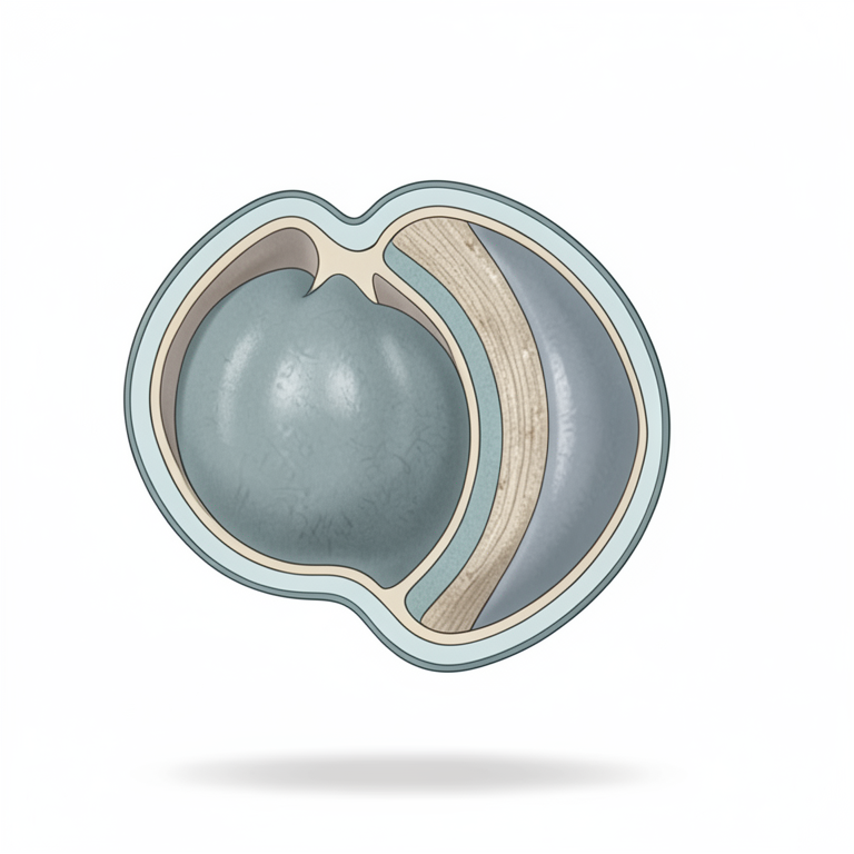
Layers of the Testicular Capsule
AI-generated conceptual illustration (adult Khaki Campbell duck testis). It explains the idea and does not show study data.
View source evidence ↗Figures & tables
From the original paper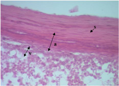 Figure 1
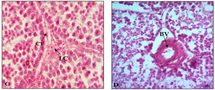 Figure 2
Introduction
Testes vary in size and structure depending on age in a particular species. In Bangladesh, the duck farming is growing faster. In near future, it will be a new venture of diversification of poultry farming to diverse the choice of taste and to strengthen the egg and meat production unit for fulfilling the shortage of animal protein demands. In Bangladesh, the available duck genotypes are Khaki Campbell, Indian Runner, Jinding, Pekin, Muscovy and Desi (Ahmed, 1986) [1] . Among the duck breeds Khaki Campbell is known to be the highest egg producer in the World. The preliminary studies showed that Khaki Campbell is medium sized egg laying duck having potentials to survive well and giving very good production (Pervin et al., 2013) [11] . At present, there is paucity of information on the general histology of testis of this breed of duck. It has been established that histological study of the testis of any breed or species is necessary in assessing and estimating quantitative changes in testicular components and spermatogenic functions. The present study is aimed with following objective: To provide the detailed knowledge on the histological features of testis of Khaki Campbell duck. To compare its histological features with other avian species.
Materials and Methods
Ten apparently healthy Khaki Campbell duck of 6 months of age were used in this study. The birds were sacrificed by severing common carotid artery. The birds were killed by dislocation of the neck vertebrate and the abdomen was immediately opened. The location of testes was examined prior to their removal from the abdominal cavity. Tissue samples for histological study were obtained from the testis. The testes of each bird were cut into thin slices, fixed in 10% formalin solution for 24hours, and then dehydrated by using a series of ascending grade of alcohol (70%, 80%, 95%, 100%, 100%, and 100%) solutions, cleared in xylene, embedded in paraffin wax, sectioned at 5-7 µm thickness and stained with Hematoxylin and Eosin.
Results and Discussion
Each testis was covered by testicular capsule. Histologically, the testicular capsule is composed of three main tissue layers: an outer, thin tunica serosa, a thick tunica albuginea and the innermost, very thin tunica vasculosa (Fig. 1.). The tunica serosa was the outermost layer and consists of simple squamous epithelium or mesothelium derived from the peritoneal lining of the structures within the abdominal cavity of the bird. The tunica albuginea forms the bulk of the capsule, and is composed of collagen, elastic fibers and abundant fibroblasts. The tunica albuginea was comprised of dense connective tissue which is consisting mainly of collagenous fibres. The tunica vasculosa comprised loose connective tissue, fibroblasts and blood vessels. The structure of the testicular capsule in Khaki Campbell ducks was similar to that of other birds reported in Sudanese duck by Aire (1997) [2] ; Jamieson (2007) [9] and Salwa et al., (2013) [14] ; in White Rooster by Razi et al., (2010) [12] .; in Muscovy duck by Gerzilov et al., (2016) [6] . In examined observation fields, there were no thick connective tissue stripes dividing the parenchyma of the testis into lobules. These findings were similar to those found in White Rooster by Razi et al., (2010) [12] .; in Muscovy duck by Gerzilov et al., (2016) [6] .; in Sudanese duck by Salwa et al., (2013) [14] .; in White Rooster by Razi et al., (2010) [12] .; in indigenous duck by Khadem (2014). The average thickness of testicular capsule was 51.12 ± 6.10 µm. The thickness of testicular capsule that was found in this study was lower than that has been by Aire and Ozegbe (2007) [3] in birds. The finding was similar to that has been reported for the domestic fowl (30-60 µm) by Hodges (1974) [8] . The reasons for this difference in testicular capsule thickness is not known, but could be due to fixatives and fixation methods used, as well as age of the subjects, species difference and possibly but not probably reproductive activity. The parenchyma of testis was consists of thousands of seminiferous tubules. The seminiferous tubules begin blindly, anastomose and form a complex tubular network. They were found to have a dense and compact organization within the testicular tissue (Fig. 2A.). They were lined internally by the germinal epithelium and surrounded externally by peritubular tissue. These observations were in agreement with Razi et al., (2010) in white rooster. In this study, the mean length of the seminiferous tubules was recorded as 468.6± 166.91 and mean width of the seminiferous tubules was 231.46 ± 41.68 µm. In available literature, there are no consistent data about the length and width of the seminiferous tubules of Khaki Campbell duck. Tingari et al., (1984) [16] reported in the adult chicken and duck, the mean diameter of the seminiferous tubules in the winter season about 126 and 124 µm and in the hot season they were 135 and 134µm, respectively. According to Jamieson (2007) [9] the diameter of the seminiferous tubule increases at active breeding period and decreases at nonbreeding period. These was also reported in domestic duck by Simoes et al., (2017) [15] , in domestic quail by Baraldi-artoni et al., (1997) [5] , in gander (Anser anser domesticus) by Gumulka and Rozenboim (2015) [7] . The large cells lying next to the basement membrane were the spermatogonia (Fig. 2B.).The largest of the spermatogenic cells were the primary spermatocytes which lie adjacent to the spermatogonia (Fig. 2B.). More than one generation was found. Their nuclei were darkly stained with the chromatin showing a net-like appearance. The secondary spermatocytes were smaller than primary spermatocytes and possessed darkly stained nuclei (Fig. 2B.).The early spermatids are round cells with pale nuclei that occur in clusters toward the lumen of the somniferous tubule. Late spermatids are characterized by small, oval to elongated, dark heads and long, faint tails that project into the lumen. Spermatozoa possessed small deeply staining head; their tails extend freely into the lumen of the seminiferous tubules ((Fig. 2B.). Similar findings were reported in domestic fowls by Hodge (1974) [8] ; in cockerel by Reviers and Williams (1984) [13] ; in general quail by Al-Shamary (2001) [4] ; in Sudanese duck by Salwa et al., (2013) [14] ; in Muscovy duck by Gerzilov et al., (2016) [6] ; in indigenous duck by Khadem (2014) [10] . The interstitial space contained interstitial tissue which was very compact, either triangular or quadrilateral in shape. The interstitial cells consisted ofleyding cells, fibroblast, collagenous fibres and reticular fibres, lymphatic vessels and blood vessels (Fig. 2D.). The shape of the leydig cells (Fig. 2C.) was highly variable and ranged from round, oval, polyhedral, triangular and flattened. Similarly, the shape of the nucleus also varied from round to flat. Sertoli cells extend from the basement membrane to the tubular lumen and posses oval nuclei but some of them look irregular in shape (Fig. 2B.). These results were in agreement with Salwa et al., (2013)
Conclusion
The result of the present study explored that the testisicular stroma possessed no mediastinum. Leydig cells were commonly found in groups within the connective tissue of the interstitial space. Germ cells and Sertoli cells were present within the seminiferous epithelium. In this study, it was revealed that there is a similarity between the histological structure of testes of adult Khaki Campbell duck and other avian species. These observations will be valuable in quantitative assessment of testicular structure and function under various physiological as well as pathological conditions and may also help to explore the molecular changes of adult testes of Khaki Campbell duck.
Figures and tables from the paper
References
16 in the original paper- Ahmed S. Duck production in Bangladesh. In "Duck Production Science and World Practice". Farrell, D. J. and P. Stapleton, (Ed.). University of New England, Armidale, Australia, 1986, 342-350.
- Aire TA. The structure of the interstitial tissue of the active and resting avian testis. Onderstepoort Journal of Veterinary Research. 1997; 64:291-299.
- Aire TA, Ozegbe PC. The testicular capsule and peritubular tissue of birds: morphometry, histology, ultrastructure and immunohistochemistry. Journal of Anatomy. 2007; 210:731-740.
- Al-Shamary BF. Anatomical and histological study of the male genital system and some ultrastructure studies of spermatogenesis in common quail (Coturnix C. coturnix L.) Msc thesis, College of Veterinary Medicine. University of Baghdad, Irak, 2001.
- Baradi-artoni SMB, Orsi AM, Lamano-carvalho TL, Vicentini CA, Stefanini MA. Seasonal morphology of the domestic quail (Coturnix coturnix japonica) testis. Anatomia Histollogia Embryologia journal. 1999; 28:217-220.
- Gerzilov V, Bochukov А, Penchev G, Petrov P. Testicular Development in the Muscovy duck (Cairina moschata Bulgarian). Journal of Veterinary Medicine. 2016; 19(1):8-18.
- Gumulka M, Rozenboim I. Effect of breeding stage and photoperiod on gonadal and serotonergic axes in domestic ganders. Journal of Poultry science. 2015; 00:19.
- Hodges RD. The histology of the fowl. 1st.ed. Academic press, London, U.K, 1974, 300-326.
- Jamieson B. Reproduction Biology and Phylogeny of Birds, part A, University of Queensland, Australia, 2007.
- Kadhem AZ. The cycle event of spermatogenesis and spermiogenesis in the testes of indigenous duck (Anas platyrhynchos). Bashra journal of veterinary research. 2014; 1(2).
- Pervin W, Chowdhury SD, Ali MA, Khan JU, Raha SK. Growth performance of indigenous (desi) ducklings receiving diets of varying nutrient concentrations. In: Proceedings of 8th Poultry Show and Seminar, World's Poultry Science Association, Bangladesh Branch. 2013, 45-50.
- Razi M, Hassanzadeh SH, Najafi GR, Feyzi S, Amin M, Moshtagion M et al. Histological and anatomical study of the White Rooster of testis, epididymis and ductus deferens. International Journal of Veterinary Research. 2010; 4(4):229-236.
- Reviers DM, Williams JB. Testis development and production of spermatozoa in the cockerel (Gallus domesticus). In: Reproductive biology of poultry. Cunningham, J., Lake, P.E., and Hewitt, D., eds. 1984, 183-202.
- Salwa I, Abdelgader E, Tingari DE, Mohamed A. Morphological Study of the Testis of Adult Sudanese Duck (Anas platyrhynchos) International Journal of Animal and Veterinary Advances. 2013; 5(3):103-107.
- Simoes K, Orsi AM, Schimming BC, Benetti EJ. Seasonal reproductive cycle and variability in plasma testosterone levels in the domestic duck (Anas platyrhynchos). Bioscience Journal. 2017; 33(1):153-164.
- Tingari MD, Ramos AS, Gaili ESE, Rahma BA, Saad AH. Morphology of the testis of the one-humped camel in relation to reproductive activity. Journal of Anatomy. 1984; 139:133-143.