
Research article · 2019 · Full text
International Journal of Veterinary Sciences and Animal HusbandryA rare case of subcutaneous emphysema in a 28 weeks old rooster
Volume 4(5) · Pages 23–25
Research areasPneumothorax, Barotrauma, EmphysemaPleural and Pulmonary DiseasesRespiratory Support and Mechanisms
Abstract
Author-writtenSubcutaneous emphysema with unknown etiology was diagnosed in a 28 weeks old rooster suffering from respiratory difficulties in a free-range flock situated in the region of Sidi Thabet (northeast of Tunisia). Clinical exams, done at the avian clinic of the National Veterinary Medicine School of Tunisia, revealed a high quantity of gas accumulation under skin. Gas was detected in whole body. Radiographic examination showed diffuse radio-transparent zones confirming the emphysema without other anatomical modifications in the different internal organs and skeleton. Simple use of needle puncture was able to relieve the condition and animal was treated with antibiotic injection during three days. Eventually, rooter was recovered.
Summary
Source-checkedPlain-language reading of the full paper, prepared with AI and checked against the text. Each point links to the authors' own words.
Why it matters
- Subcutaneous emphysema is a condition where gas accumulates under the skin; while common in mammals like dogs and horses, it is rare in poultry. View evidence ↗
- Air can enter the subcutaneous tissues through skin wounds or injuries to the respiratory tract, including the trachea, lungs, or air sacs. View evidence ↗
- Gas accumulation under the skin can also be caused by anaerobic bacteria associated with gangrenous inflammation. View evidence ↗
The question
- The study aims to describe the clinical presentation and therapeutic approach for a 28-week-old rooster with subcutaneous emphysema of unknown cause. View evidence ↗
What they did
- Clinical examinations were performed at the avian clinic of the National Veterinary Medicine School of Tunisia to assess gas accumulation. View evidence ↗
- Radiographic examinations were used to confirm the presence of emphysema and check for other anatomical abnormalities in the skeleton and internal organs. View evidence ↗
- The rooster received intramuscular injections of tetracycline at a dose of 25 mg/kg for three days to prevent bacterial infection. View evidence ↗
What they found
- The rooster presented with anorexia, respiratory difficulties, and subcutaneous gas accumulation that had persisted for four days. View evidence ↗
- Gas accumulation was generalized across the body, specifically affecting the head, neck, breast, right flank, and thigh. View evidence ↗
- Radiographs confirmed large radio-transparent zones in the head, neck, and right flank, but showed no evidence of lung damage or bone fractures. View evidence ↗
- The rooster recovered following the use of needle punctures to relieve the gas and a three-day course of antibiotic injections. View evidence ↗
What it means
- The authors concluded that the cause of the emphysema in this specific case remained unknown as no obvious triggers were identified. View evidence ↗
- The authors state that the prognosis depends on the severity of the emphysema and how quickly the intervention occurs. View evidence ↗
Visual summary
1 visualsIllustrations explain the paper; they do not show study data.
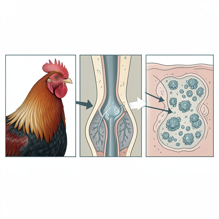
Potential Causes of Subcutaneous Emphysema
AI-generated conceptual illustration (rooster). It explains the idea and does not show study data.
View source evidence ↗Figures & tables
From the original paper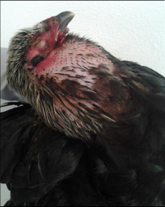 Figure 1
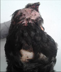 Figure 1
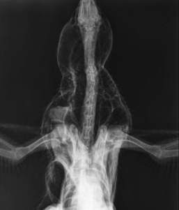 Figure 2
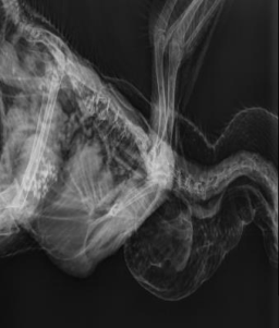 Figure 2
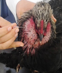 Figure 3
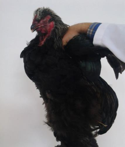 Figure 3
Introduction
Subcutaneous emphysema, more diagnosed in mammalian species (horse, dog), is rarely observed in poultry. This affection, characterized by the accumulation of gas under skin, is sometimes called "windpuff" (Riddle, 1997; Crespo and Shivaprasad, 2008) [1,5] . The air can penetrates in subcutaneous tissues through a skin wound or as the consequence of injure or defect in respiratory tract, such as air sacs, lungs or trachea. Fracture of pneumatic bones such as humerus, coracoid and sternum allows air to accumulate under the skin of aquatic or flying birds (Crespo and Shivaprasad, 2008) [1] . Accumulation of gas under the skin can also arise when some anaerobic bacteria implicated in gangrenous inflammation. In this condition, subcutaneous emphysema is usually associated to illness and loss of function of the part concerned. Infected tissue shows red, green or black discoloration associated to insensitivity. Affected bird is usually dying or dead (Miroslay and Nelly, 1950; Kamani et al., 2009) [4,5] . Spontaneous restoration of the emphysema is possible on 2-3 weeks, but risk of bacterial complication is frequent. Treatment of affected birds can also be occurred by puncturing the skin with a sharp instrument. This operation must be completed by antibiotic administration in order to prevent bacterial infection (Kamani et al., 2009; Devarathnam and Naveen, 2013) [2,4] .
Case history
A 28 weeks old rooster from a backyard flock of 20 birds was presented to the avian clinic, National Veterinary School of Tunisia. The main complaint was anorexia, respiratory difficulties and gas accumulation under the skin for the past four days. Rest of animals was healthy. Trapping of air stared from the neck, thoracic region, abdomen region and finally whole body.
Results and discussion
On special clinical examination puffy areas all over the body was observed with loss of weight and pale mucous membranes. No pain was observed while palpation of emphysema. No sign of wound or traumatic lesion or skin break was observed to suggest the point of entry of gas. Generalize gas accumulation under the skin was revealed especially, in the head, the neck, the breast, the right flank and thigh (Figures 1a &1b). This findings were in agree with results of other authors (Kamani et al., 2009; Devarathnam and Naveen, 2013; Reddy et al., 2013) [2,4,6] . A preliminary diagnosis of subcutaneous emphysema of unknown etiology was made. Radiographic investigation was decided in order to confirm clinical suspicion and to search other anatomical modification in relation with under skin gas accumulation. Radiographic features showed large radiotransparent zones in the head, the neck and right flank. No bone fracture or lung damage was found. (Figure 2a & 2b) Subcutaneous emphysema is rarely described in poultry. The precise etiology is unknown. In fact, many hypothetic causing factors were supposed. The etiology of a potential trauma may be mentioned in this context. Indeed, the rupture of the skin can occur and the accumulation of a large quantity of gas will lead to severe emphysema (Reddy et al., 2013) [6] . Subcutaneous emphysema may arise also after blunt trauma when the glottis is closed, or after a penetrating injury which allows entry of air into the tissues but prevents its escape. Air escape from spontaneous rupture of lung parenchyma or from erosive diseases of the lung. More frequently, fractures of pneumatic bones or defective epiphyses. However, idiopathic etiology is also documented. This condition may be observed in some diseases. Indeed, subcutaneous emphysema, particularly at the cervical region is commonly described in avian influenza infection. In fact, air escaping from damaged lung tissue, through the thoracic inlet and into under skin of the neck and back (Reddy et al., 2013) [6] . Differential diagnosis must be established with another condition which could cause swellings in birds, is generalized edema. In this case, edema usually involves most of the soft tissues of the body, and tends to accumulate mainly in dependent areas, while subcutaneous emphysema was usually localize to one part or side of the bird. Differentiation can be made in most case by meticulous clinical examination by pressing the swelling zone with finger and then releasing it. In this context, edema will usually show the imprint of the finger for at least several seconds, while emphysema, where the air is under pressure, the swollen zone regains immediately its shape and the finger leaves no impression. Prognosis is reserved among the importance of emphysema and the precocity of intervention. Birds can resume normal activity within three or four days of post therapy if the emphysema was detected early. Gochfeld (1974) [3] described spontaneous release without treatment, when bird develops moderate emphysema. However, the situation is deteriorating and the animal may die if the therapeutic intervention is too late and emphysema is very extensive. Puncture of several zones was done, after large disinfection of the skin, using sterile hypodermic needle, in order to evacuate the subcutaneous gas until considerable reduction (Figures 3a &3b). Then, animal was treated by intra-muscular injection of 25 mg/kg of tetracycline during three days to prevent bacterial complications and further gas entry. Reaccumulation of gas under the skin is frequent when emphysema is severe. Consequently, bird may be treated by making a little incision (1-2 mm) of the skin (Gochfeld, 1974) [3] . a.
Conclusion
This paper describes clinical and therapeutic approach of a rare condition of subcutaneous emphysema in backyard rooster. A ballooning of overall body was detected and differentiated from subcutaneous edema. The etiology of this emphysema was unknown in the absence of obvious causes.
Acknowledgement
Treatment, based on puncture of several zones with a sterile needle, was made and followed by intramuscular injection of a large specter antibiotic to prevent bacterial complications.
Figures and tables from the paper
References
5 in the original paper- Crespo R, Shivaprasad HL. Developmental, metabolic, and other noninfectious disorders. 2008. In: Diseases of poultry, 12th edition, Saif YM, Fadly AM, Glisson JR, McDougald LR, Nolan LK, Swayne DE. Blackwell publishing, Iwoa, Pages 1149-1195.
- Gochfeld M. Prevalence of subcutaneous emphysema in young Terns, Skimmers and Gulls. J. Wildl Dis. 1974; 10: 115-120.
- Kamani J, Tijjani A, Yidawi JP, Gana AL, Egwu OK, Gusi AM. Subcutaneous emphysema (windpuff) in a 13 weeks old pullet: Case report. Int. J. Poult. Sci. 2009; 8: 1121-1122.
- Miroslav R, Nelly RA. Anaerobic subcutaneous emphysema of poultry. Nature. 1950; 166: 442. https://www.nature.com/articles/166442a0
- Reddy SB, Sivajothi S, Reddy AP. Subcutaneous emphysema in a pullet. Int. J. Livestock Res. 2013; 3: 7780.
Cited by 1
From OpenAlex, OpenCitations and reference lists read by Scinovex
- Management and treatment procedure of windpuff caused by lice infestation in domestic pigeon: A case report ↗
Mishuk Shaha, Arjuman Lima, Ashutosh Das, Gous Miah, Sabuj Kanti Nath, Pranab Paul et al. · World Journal of Advanced Research and Reviews · 2021
A rare case of windpuff was diagnosed in 7 months old two domestic pigeons due to itching and scratching with inanimate objects caused by lice infestation reported in Begumganj upazila hospital, Noakhali district, Bangladesh. This is the first case of domestic pigeon recorded in Bangladesh and the aim of this case study is to determine the causal agent, treatment and…
❝ 0PDF