
Research article · 2018 · Full text
International Journal of Veterinary Sciences and Animal HusbandryEffects of autologous platelet-rich plasma on skin healing in dogs
Volume 3(2) · Pages 49–55
Research areasPeriodontal Regeneration and TreatmentsSexual function and dysfunction studiesGenital Health and Disease
Abstract
Author-writtenThis examination was intended to assess the part of Platelet-Rich Plasma (PRP) on recuperating of tentatively injured skin in ten grown-up canin, matured 2-3 years and measuring 25-30 kg. The creatures separated arbitrarily and similarly into (control and treatment gatherings). Four of 2×2 cm of full- thickness square cutaneous injuries was prompted on the two sides of the sidelong thoracic district of every creature under the impact of neighborhood analgesic continuing by xylazine hydrochloride as a narcotic. A couple of left injuries was dealt with by infusion with 5 mL of self-governing PRP (treatment gathering), 2 mm parallel to the injury edges and in the injury focus. While, the correct injury were infused by 5 mL of clean saline by a similar strategy (control gathering), transform metrical and histopathological assessments of wound recuperating process spoke to by percent of wound withdrawal, epithelialization and aggregate mending at 0, 3, 5, 7, 10, 13, 17, 20, 24 and 35days post-injuring. The morphometrical appearance of the injuries which treated with PRP, demonstrated that the compression, re-epithelialization and recuperating percent were statically critical (p<0.05) in examination with control wounds amid a month contemplate. In light of histopathological comes about, there was re- epithelialization of epidermis, with very cell granulation tissue, all around separated keratinocytes of epidermis with scar arrangement in the dermis of the segmented skin. We infer that neighborhood infusion of PRP prompts quicken and change of twisted mending in contrast with control wounds.
Summary
Source-checkedPlain-language reading of the full paper, prepared with AI and checked against the text. Each point links to the authors' own words.
Why it matters
- Wound healing is a complex, multifactorial process that involves the contraction and closure of a defect to rebuild a functional skin barrier. View evidence ↗
- A primary goal in treating various types of wounds is to improve the recovery of both the dermal and epidermal layers. View evidence ↗
- Platelet-rich plasma (PRP) is used as a combination treatment because it provides various cytokines essential for the natural healing process. View evidence ↗
The question
- The study aimed to evaluate the effect of Platelet-Rich Plasma (PRP) on the healing of experimentally induced skin injuries in ten adult dogs. View evidence ↗
What they did
- The experiment used ten adult male crossbreed dogs, aged 3.5 to 4 years and weighing between 18 and 25 kg. View evidence ↗
- Four full-thickness square skin wounds, each measuring 2x2 cm, were created on the dorsal thoracic sides of each dog, with two wounds per side separated by 10-15 cm. View evidence ↗
- Control wounds were treated with 5 mL of sterile saline, while treatment wounds received 5 mL of PRP, both administered 2 mm from the edges and within the wound bed. View evidence ↗
- Wound healing was assessed over a 35-day period, with measurements taken on days 0, 3, 7, 10, 14, 17, 21, 24, 28, and 35. View evidence ↗
What they found
- By day 35, the percentage of epithelialization was significantly higher in PRP-treated wounds (72.10%) compared to control wounds (31.1%). View evidence ↗
- Wound contraction was significantly faster in the PRP group starting from the first week, reaching 84.44% compared to 76.66% in the control group by the end of the study. View evidence ↗
- The total wound healing rate was highest in the PRP treatment group, reaching 89.44% by day 35. View evidence ↗
- Epidermal re-epithelialization began in the second week for the PRP treatment group, whereas it did not appear until the fourth week in the control group. View evidence ↗
- Histopathological analysis of the PRP treatment group at day 28 showed well-separated epidermal keratinocytes and scar formation in the dermis. View evidence ↗
What it means
- The authors conclude that PRP has a beneficial effect on skin wound healing by accelerating epithelialization and promoting effective wound contraction. View evidence ↗
- The study concludes that the local implantation of PRP is a safe and simple procedure. View evidence ↗
Visual summary
3 visualsIllustrations explain the paper; charts are replotted from the paper's own table values.
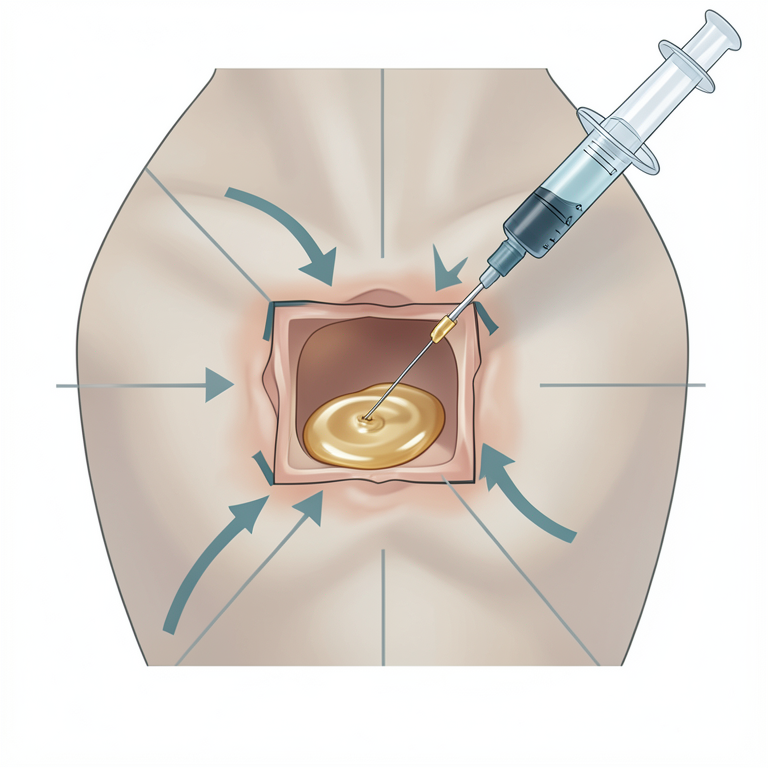
PRP Treatment Method
AI-generated conceptual illustration (dog thoracic skin with a square wound). It explains the idea and does not show study data.
View source evidence ↗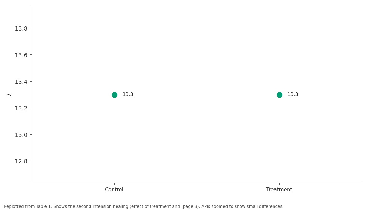
7: Control vs Treatment
Replotted from Table 1 (page 3) of the original paper. Every bar is the paper's own table value.
View source evidence ↗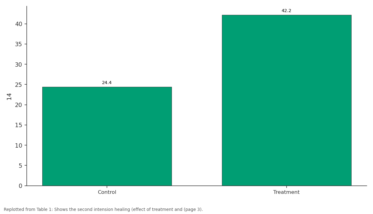
14: Control vs Treatment
Replotted from Table 1 (page 3) of the original paper. Every bar is the paper's own table value.
View source evidence ↗Figures & tables
From the original paper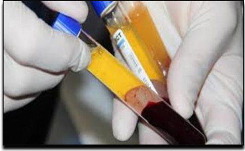 Figure 1
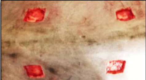 Figure 2
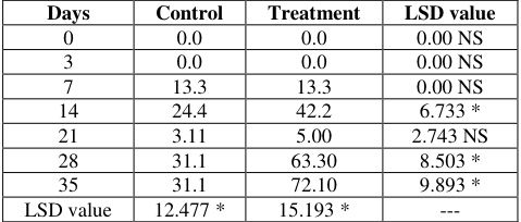 Table 1
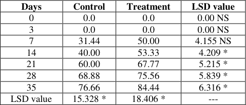 Table 2
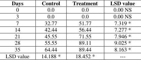 Table 3
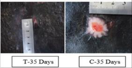 Figure 3
Introduction
Wound mending is complexes multifactorial process those outcomes in the constriction and conclusion of the imperfection and rebuilding of a useful boundary. This procedure happens as a grouping of occasions including hemostasis, incendiary cell invasion, tissue regrowth, and remodelin [9] . Wound recuperating scientists expect to see how an injury mending methodology can be prompted to repair the harmed tissues speedier and more efficiently [12] . Improvement of dermal and epidermal recovery is a critical objective for the treatment of a wide range of sorts of wounds [13] . Wound mending is directed by a few cell composes and by a course of peptides, for example, cytokines or development factors. Following damage, development factors emission by platelets and macrophages are actuated and provocative process which is required for recuperating is initiated [1] . Another region of tissue designing including bioactive atom based medicines has at present increased much consideration. A few examinations have demonstrated that cytokine treatment may quicken mending of tissues and particularly advance the repair of disabled injuries in the assortment of creatures. [14,29,30] , Goren lamentably, these decontaminated bioactive specialists has missed the mark regarding desires in clinical examinations, as it is right now obvious that no single exogenous operator can successfully intercede all parts of wound recuperating process, [34,[7][8][9] . This might be because of the way that cytokines work in show, [36] , both transiently and spatially [3] . So, the dynamic idea of wound repair process substantiates the requirement for cytokine blend treatments [30] In an exertion, to give this blend treatment, specialists have swung to Plateletrich plasma (PRP), which is a rich wellspring of various cytokines basic for common recuperating process. It is discharged from platelet α-granules at destinations of tissue damage, [31, 16] . These incorporate catecholamines, serotonin, adenosine triphosphate (ATP), egg whites, fibrinogen, osteonectin, osteocalcin and calcium particles.
Different coagulating factors and locally dynamic development factors, for example, platelet-determined development factor (PDGF), changing development factor-α (TGF-α), changing development factor-β (TGF-β), insulin-like development factor (IGF), fibroblast development factor (FGF), vascular endothelial development factor (VEGF) and epidermal development factor (EGF) likewise start wound mending, [5,29,7] Platelets additionally emit fibrin, fibronectin, and vitronectin, which give a framework to connective tissue and go about as grip particles for epithelial relocation [19,40,41] , these entire scopes of bioactive elements display in PRP gel appear to come about the foremost restorative preferred standpoint of autologous platelet-rich plasma over segregated decontaminated cytokines, quickening the recuperating arrangements keeping in mind the end goal to impersonate the regular procedure as physiologically as could be allowed [24] albeit a few in vitro contemplates have been done in this field, there are a few examinations in puppies assessing PRP adequacy amid cutaneous injury mending or potentially tissue recovery. There is just a solitary report outlining the adequacy of PRP gel in treatment of ceaseless injuries on the tail of a puppy [22] to the best of our insight, there are no past. Studies evaluating utilization of PRP in crisp injuries in puppies. In this, we planned to assess the impacts of topical use of PRP, coagulated with various activators. We evaluated in the case of expanding grouping of arbiters in the injury with PRP can improve recuperating rate and time required to accomplish sufficient tissue recovery [42] .
Materials and Methods
Preparation of platelet-rich plasma: Blood test 50 mL was gathered under a septic procedure from the jugular vein of each buck through a 21 check needle and stored in 3.2% sodium citrate tube with limit with respect to 10 mL. At that point, set in an axis at 3200 rpm for 15 min. Amid centrifugation, blood is isolated intothree distinctive divisions: platelet-poor plasma (unrivaled layer), white platelets (middle layer) and red platelets (sub-par layer). The main supernatant plasma division (half), contiguous the pooch, was gotten under aseptic conditions in a laminar stream chamber. This portion was centrifuged at 3200 rpm for another15 minutes keeping in mind the end goal to get two sections: the upper part which is platelet poor plasma PPP and the lower part was the PRP (25%). At that point the PRP was suctioned with another pipette and set in a sterile tube and initiated with calcium chloride (4.5 mEg/5 mL), utilizing 50 μL/mL of PRP, to give a gel network to a PRP to hold fast to the site of infusion [17] . A normal of 5ml of PRP was acquired from each 50 ml of entire blood. A small amount of 1ml of every creature was examined for platelet check. Upon the arrival of PRP acquisition, the platelets tally in PRP was 200.000 to 350.000 cells/μL.
Experimental animals and management:
Ten grown-up male crossbreed canines, 3.5 to 4 years old and weighing, around (18-25 kg), were utilized as a part of this examination. The mutts were housed in pet hotels, sustained by business delicate upkeep abstain from food once every day, and water was offered not indispensable.
Pre-operative considerations:
The dorsal surface of thoracic districts was sans cut hair and arranged aseptically for the injuring. Thirty minutes preceding injuring, Penicillin-Streptomycin was managed I.M., in a measurement of 20000 IU/kg and 10 mg/kg B.W., individually. The mutts were premedicated with xylazine-HCl (xylazine 2%, Alfasan International BV, Woerden, The Netherlands) (2 mg/kg) intramuscularly. This was trailed by acceptance of general anesthesia with intravenous infusion of the blend of ketamin-HCl (ketamin10%, Alfasan International BV) (10 mg/kg) and diazepam (0.5 mg/kg), and kept up by similar medications.
Technique of Wounding: Four square full-thickness skin wounds (2×2 cm) were made on the dorsal thoracic sides of every canine (two injuries on each side), 10-15cm separated (Fig. 1). The aggregate number of the injuries is (40 wounds). The creatures submitted to two equivalent gatherings, the control gathering and the treatment gatherings. All injuries were not sutured (opened injuries). The control wounds were infused by 5 mL of clean saline, 2 mm horizontal to the injury edges and inside the injury bed, instantly in the wake of injuring. While the treatment wounds were infused intradermal by 5 mL of PRP at various locales, by a similar procedure said for the control gathering. After finishing of wounds treatment in the two gatherings, the injuries were wrapped by utilization of a defensive, non-weight and nondisciple dressing. The wraps have been changed, two days interims and wounds were tenderly cleaned with bandage wipes absorbed typical saline, without debridement of the injury bed. In the vicinity of 26 and 30 kg, were utilized as a part of this investigation after the creatures were cured with penicillin-streptomycin, and profoundly quieted with acepromazin maleate (0.1mg/kg B.W), the surgical destinations were arranged aseptically for the injuring. Under nearby anesthesia with lidocain hydrochloride (2%), a four square full-thickness skin wounds (2×2)cm were made on twisted (two in each side) were made on each canine (n=40 wounds), two cranial and two caudal with a separation of (5-10)cm between every one.
Macroscopic evaluation of the wounds
The injuries were assessed more than 35 days time frame. At the days 0, 3, 7, 10, 14, 17, 21, 24, 28, and 35. Computerized photos were taken of all injuries after the territory had been painstakingly cleaned to imagine wound edge. The scab of each twisted was arefully expelled utilizing saline for better assessment of epithelialization and granulation tissue. Rulers were held vertically and on a level plane near the injury as a source of perspective. The region of the epithelialization and granulation tissue were measured for each twisted utilizing Sigma Scan programming Percentage of the injury compression, epithelialization and recuperating were computed for every twisted. The accompanying detail were utilized:
Wound Contraction 1. Twisted size at the day (x) mm2/twisted size at the day (0) mm2 × 100 = percent of the injury measure at the day (x) 2. 100percent of twisted size at day (x) = percent of wound constriction-Wound epithelialization: 3. Size of epithelialization territory at the day (x) mm2/size of the injury at the day (x) mm2 × 100 = percent of the epithelialization Wound Recuperating 1. Granulation tissue at the day (x) mm2/twisted size at the day (0) mm2 × 100 = 2. Percent of the non-mended territory to look at of the injury measure at the day (0) 3. 100 -percent of the non-recuperated zone to analyze of the injury estimate at the day (0) = percent of the mending.
Hydroxyprolin Measurement
At the days 0, 3, 7, 10, 14, 17, 21, 24, 28, and 35 after injuring, biopsies were taken from the focal point of each twisted utilizing 0.7 mm biopsy punch for hydroxyprolin estimation. Tissue tests for hydroxyprolin examine were washed with physiologic saline and dried in a 100°C stove for 72 hours. Hydroxyprolin levels were resolved spectrophotometrically utilizing the already portrayed strategy in μg/mg dry issue. 21 Initially, every example was weighed and hydrolyzed in 12-N HCl at 130°C for 3 hours. At that point each specimen was changed in accordance with a last volume of 1 ml and centrifuged at 3000 × g for 15 minutes. The supernatant was isolated off and break even with volume of isopropanol was added to each example. At that point this blend was centrifuged at 2500 × g for 10 minutes. Serial weakenings of unadulterated hydroxyprolin were utilized as models. Centralization of hydroxyproline in each specimen was ascertained utilizing the absorbancefocus bend for the standard hydroxyproline arrangement.
Clinical Evaluations
A total clinical examination was performed on all creatures every day amid the considered period. Wounds captured on the day were it made and after that twice seven days till day 35th. The scab of each twisted was painstakingly evacuated by utilizing saline for better perception of the epithelialization and granulation tissue region. The percent of epithelialization, wound constriction and aggregate injury recuperating were computed for each twisted, contingent upon the strategy specified [11] .
Histopathologic Examination
At the day 3, 7 and 14after injuring, biopsies were taken from a similar corner of each twisted utilizing 0.9 mm biopsy punch for histopathological examination. The injury examples were settled in (10%) unbiased support formalin arrangement.
The tissue examples were prepared in a tissue processor, paraffin squares were frantic at [5][6] μm thick areas which were cut with a microtome and recolored with Hematoxylin and Eosin colors (Luna, 1968) [27] . At that point recolored slides were analyzed under light magnifying instrument.
Statistical Analysis
The Statistical Analysis System-SAS [32] was used to effect of difference factors (treatment & days) in study parameters (percentage). The least significant difference -LSD test at the comparative between percentages in this study.
Results
Clinical Observations: Amid a month consider, no any confusion, for example, wound disease or abundant granulation tissue arrangement in the injury locales of the two gatherings were taken note. Creatures reflected typical craving, pee and crap inside the initial 24 h post-injuring. The aftereffects of key parameters, for example, temperature, respiratory and heart rates amid the main week post-injuring were inside the typical rang as specified in the most ward references.
Wound Morphometric Analysis • Wound Epithelialization:
The progress of epithelialization in treatment wounds was statistically significant (p<0.05) in comparing with control wounds, started from second week. The percent of epithelialization in control wounds was (31.1) and in treatment wounds was (72.10) at day 35 (Table 1).
| Days | Control | Treatment | LSD value |
|---|---|---|---|
| 0 | 0.0 | 0.0 | 0.00 NS |
| 3 | 0.0 | 0.0 | 0.00 NS |
| 7 | 13.3 | 13.3 | 0.00 NS |
| 14 | 24.4 | 42.2 | 6.733 * |
| 21 | 3.11 | 5.00 | 2.743 NS |
| 28 | 31.1 | 63.30 | 8.503 * |
| 35 | 31.1 | 72.10 | 9.893 * |
| LSD value | 12.477 * | 15.193 * | --- |
Table text extracted from the original PDF; superscript letters are shown inline. The original table image is below.
View original table image
• Wound Contraction: There was significant differences (p<0.05) in speed of wound contraction between control and treatment groups started from first week. It was (76.66) and (84.44), respectively, at the end of the study (Table 2). • Wound Healing: The rate of wound healing showed similar result as mentioned in wound contraction phenomena i.e., that the significant differences (p<0.05) between both group started from first week postwounding. The highest rate was recorded in treatment group (89.44 at day 35) (Table3).
| Days | Control | Treatment | LSD value |
|---|---|---|---|
| 0 | 0.0 | 0.0 | 0.00 NS |
| 3 | 0.0 | 0.0 | 0.00 NS |
| 7 | 32.77 | 51.77 | 7.319 * |
| 14 | 42.44 | 56.44 | 7.277 * |
| 21 | 45.55 | 71.55 | 7.946 * |
| 28 | 55.55 | 89.11 | 9.025 * |
| 35 | 64.44 | 89.44 | 8.163 * |
| LSD value | 14.188 * | 18.452 * | --- |
Table text extracted from the original PDF; superscript letters are shown inline. The original table image is below.
View original table image
| Days | Control | Treatment | LSD value |
|---|---|---|---|
| 0 | 0.0 | 0.0 | 0.00 NS |
| 3 | 0.0 | 0.0 | 0.00 NS |
| 7 | 31.44 | 50.00 | 4.155 NS |
| 14 | 40.00 | 53.33 | 4.209 * |
| 21 | 60.00 | 67.77 | 5.215 * |
| 28 | 68.88 | 75.56 | 5.839 * |
| 35 | 76.66 | 84.44 | 6.316 * |
| LSD value | 15.328 * | 18.406 * | --- |
Table text extracted from the original PDF; superscript letters are shown inline. The original table image is below.
View original table image
• Histopathologic Findings:
The pathgnomonic histopathology features of skin biopsies harvested from both groups, reflect the acceleration and improvement in wound healing subjected to PRP treatment with enhancing of total wound healing at the end of the study, in comparison to untreated wounds, which are shown Discussion Amid a follow-up for 28 days, we didn't record any auxiliary difficulties of the injuries in the two gatherings, this may attribute to very strike disinfectant and appropriated postagent cares. This discoveries were as per [2] in goat. Interestingly [6] , watched diseases in two injuries out of 32 when utilized PRP for treatment of cutaneous injuries in horse. These distinctions in the consequences of the investigations might be identified with the quantity of creatures utilized as a part of each examination. The imperative parameters were hoisted in the primary week postinjuring in the two gatherings yet with no altogether contrasts p>0.05. These augmentations in parameters level might be ascribed to intense aggravation evocated from injuring. At that point the qualities held step by step to its typical levels. These discoveries were in concurrence with past investigation [8] . The morphometric assessment of wounds in the present examination, demonstrated that the advance of epithelialization was speedier in twisted treated with PRP contrasted and control wounds amid four week contemplate. It has been exhibited that PRP provoked cutaneous injury repair by means of separation into different skin cell writes including; keratinocytes, endothelial cells, pericytes and monocytes [1,36] . recommended that PRP engrafted in cutaneous injury quicken re-epithelialization, through their capacity to animate the keratinocytes to separate into different epithelial cell composes, for example, skin epithelial cells, after foundational organization in vivo. Re-epithelialization of International Journal of Veterinary Sciences and Animal Husbandry wounds starts inside hours after damage. Epidermal cells from skin members, for example, hair follicles rapidly evacuate thickened blood and harmed stroma from the injury space [20] . The statement of integrin receptors on epidermal cells enables them to associate with an assortment of Extracellular-Matrix (ECM) proteins (e.g., fibronectin and vitronectin) that are blended with stromal type I collagen at the edge of the injury and joined with the fibrin coagulation in the injury space. As re-epithelialization follows, cellar layer proteins return in an extremely requested succession from the edge of the injury internal. Epidermal cells return to their ordinary phenotype, by and by immovably connecting to the restored storm cellar layer and hidden dermis [25] . The aftereffects of current examination demonstrated that injury compression percent was more prominent in treatment twisted contrasted with the control wound. [34] . Demonstrated that in second-expectation wound recuperating, conclusion is accomplished by constriction and epithelialization. A more prominent commitment of wound compression quickens recuperating in light of the fact that constriction happens quicker than epithelialization. Wound withdrawal is characterized as the centripetal development of the first twisted edges. This procedure happens because of compression of myofibroblasts in granulation tissue. Myofibroblasts are basic to wound withdrawal and mending. They separate from fibroblast and are portrayed by nearness of stress strands containing α-actin isoform that is communicated in smooth muscle. It has been affirmed that PRP communicated Transforming Growth Factor (TGF) which initiate fibroblasts and separate into my fibroblast and increment the quantity of these cells to advance injury constriction. Wound compression includes a complex and brilliantly arranged association of cells, ECM and cytokines. Amid the second seven day stretch of mending, fibroblasts expect a my fibroblast phenotype portrayed by substantial packs of actincontaining microfilaments arranged along the cytoplasmic face of the plasma layer of the cells and by cell-cell and celllattice linkages [19] . The presence of the myofibroblasts compares to the beginning of connective-tissue compaction and the withdrawal of the injury. The compression likely requires incitement by TGF and Platelet-Derived Growth Factor (PDGF) connection of fibroblasts to the collagen framework through integrin receptors and cross-interfaces between singular packs of collagen. Collagen rebuilding amid the progress from granulation tissue to scar is reliant on proceeded with blend and catabolism of collagen at a low rate. The debasement of collagen in the injury is controlled by a few proteolytic catalysts named network metalloproteinase, which are discharged by macrophages [26] . Histopathological areas, uncovers discharge and provocative cells amid the initial seven days notwithstanding the arrangement of granulation tissue. These perceptions were recorded by [2] , in goats in which the new stroma, frequently called granulation tissue, starts to attack the injury space around four days after damage. Various new vessels bless the new stroma with its granular appearance. Macrophages, fibroblasts and veins move into the injury space in the meantime. The macrophages give a proceeding with wellspring of development factors important to empower fibroplasia and angiogenesis; the fibroblasts deliver the new ECM important to help cell in development; and veins convey oxygen and supplements important to support cell digestion. [35] , demonstrated that development factors, particularly PDGF and TGF working together with the ECM particles, apparently empower fibroblasts of the tissue around the injury to multiply and create collagen strands which look like a scaffold interfacing the finishes of the injury likewise, the fibroblasts are in charge of the combination, affidavit and redesigning of the extracellular framework. The arrangement of fresh recruits vessels is important to manage the recently framed granulation tissue. Angiogenesis is a perplexing procedure that depends on extracellular lattice in the injury bed and additionally relocation and mitogenic incitement of endothelial cells [36] . In the present examination, the reepithelialization of epidermis in PRP treatment amass began at the second week post-injuring while it showed up in fourth week in control gathering and this continuation was upheld by an investigation of [21] . On another hand, [37] , expressed that Re-epithelialization of the injury can be adroitly seen as the consequence of three covering keratinocyte capacities: movement, multiplication and separation. The succession of occasions by which keratinocytes finish the assignment of reepithelialization is by and large accepted in any case disintegration of cell-cell and cell-substratum contacts. This is trailed by the polarization and start of relocation in basal and a subset of supra-basilar keratinocytes over the temporary injury lattice. All around separated keratinocytes of epidermis with scar arrangement in the dermis were seen in treatment gather at day 28. While, in randomized forthcoming investigation by [10] , demonstrated that scar arrangement at day 35th postinjuring and showed that the epidermis recovered logically from the encompassing injury edges. The neoepidermis demonstrated an entire range of changes. Close to the injury edge, the separation of the neoepidermis and recovery of the dermo-epidermal intersection were further developed than toward the injury focus, where the proliferative list was essentially expanded. Additionally, [5] , alluded that the injury ends up noticeably secured with epithelial tissue inside two weeks, negligible collagen will be kept and no scar will frame. For the most part, if an injury takes longer than three to a month to wind up noticeably secured, a scar will shape. In light of the aftereffects of the present investigation, transplantation of PRP that were separated from caprine blood had positives impact on second goal cutaneous injury recuperating in goats. All in all, this examination exhibits the helpful impact of PRP in cutaneous injury recuperating by means of quick epithelialization and compelling injury compression.
Conclusions
The present study concluded the following: 1. The procedure of local implantation of PRP is easy and safe. 2. PRP can be used as allogeneic transplants without immuno-rejection.
Acknowledgement
AcknowledgmentsI wish to express my deep appreciation to the staff of surgery department, College of Veterinary Medicine, University of Baghdad.
Figures and tables from the paper
References
41 in the original paper- Anitua ME, Sanchez A, Nurden P, Nurden G, Orive I. Andia, New insights and novel applications for plateletrich fibrin therapies. Trends Biotechnol. 2006; 24(5):227234.
- Azari OH, Babaei MM, Molaei SN, Nematollahi-Mahani ST, Layasi. The use of Wharton's jelly-derived mesenchymal stem cells to accelerate second-intention Cutaneous wound healing in goat. Iran. J. Vet. Surgery, 2008; 3(8):15-27.
- Badylak SF. Xenogeneic extracellular matrix as a scaffold for tissue reconstruction.TransplImmunol. 2004; 12:367-377.
- Beckert SS, Haack H, Hierlemann F, Farrahi P, Mayer A, Konigsrainer S. Stimulation of steroid-suppressed cutaneous healing by repeated topical application of IGFI: Different mechanisms of action based upon the mode of IGFI delivery. J. Surg. Res. 2007; 139:217-221.
- Bennett NTGS. Schultz. Growth factors and wound healing: Part II. Role in normal and chronic wound healing. Am. J. Surg. 1993; 166:74-81.
- Carter CADG, Jolly CE. Worden, Platelet rich plasma gel promotes differentiation and regeneration during equine wound healing. Exp. Mol. Pathol. 2003; 74:244-255.
- Chen C, Schultz GS, Bloch M. Molecular and mechanistic validation of delayed healing rat wounds as a model for human chronic wounds. Wound Rep Regen. 1999; 7(6):486-494.
- Crovetti GG, Martinelli M. Issi, Platelet gel for healing chronic wounds.TransfusApher. Sci. 2004; 30:145-151.
- Diegelmann RF, Evans MC. Wound healing: an overview of acute, fibrotic and delayed healing. Front Biosci. 2004; 9:283-289.
- Dyson M. Advances in wound healing physiology: The comparative perspective. Vet. 1997.
- Eppley BLJE, Woodell, J Higgins. Platelet quantification and growth factor analysis from platelet rich plasma: Implications for wound healing. Plast.Reconstr. Surg. 2004; 114:1502-1508.
- Fréchette, J.P., Martineau, I., Gagnon G. Platelet-rich Plasmas: Growth Factor Content and Roles in Wound Healing. J Dent Res. 2005; 84(5):434-439.
- Ferguson M, Byrnes C, Sun L. Wound healing enhancement: Electroportion to address a classic problem of military medicine. World J Surg. 2005; 29: S55-S59.
- Goren I, Müller E, Schiefelbein D. Systemic antiTNFalpha treatment restores diabetes-impaired skin repair in ob/ob mice by inactivation of macrophages. J Invest Dermatol. 2007; 127:2259-2267.
- Kairuz EZ. Upton RA, Dawson J. Malda 2007. Hyperbaric oxygen stimulates epidermal reconstruction in skin equivalents. Wound Repair Regen. 15: 266-274.
- Kazakos K, Lyras DN, Verettas K, et al. The use of autologous PRP gel as an aid in the management of acute trauma wounds Injury. 2009; 40:801-805.
- Kevy SM, Jacobson. Comparison of methods for point of care preparation of autologous platelet gel. J. Extra-Corp. Technol. 2004; 36:28-35.
- Kirchner LM, Meerbaum SO, Gruber BS. Effects of vascular endothelial growth factor on wound closure rates in the genetically diabetic mouse model. Wound RepReg; 2003; 11:127-31.
- Kirsner RS, Eaglstein WH. The wound healing process.Dermatol.Clin. 1993; 11: 629-640.
- Knighton DR, Ciresi K, Fiegel VD. Stimulation of repair in chronic non-healing cutaneous ulcers: A prospective randomized blinded trial using platelet-derived wound healing formula. Surg. Gynecol. Obstet. 1990; 170:56-60.
- Kim JH, Park C, Park HM. Curative effect of autologous platelet-rich plasma on a large cutaneous lesion in a dog.Vet Dermatol. 2009; 20(2):123-126.
- Lee HW, Reddy MS, Geurs N. Efficacy of platelet-rich plasma on wound healing in rabbits. J Periodontology. 2008; 79(4):691-696.
- Lee SH, Lee JH, Cho KH. Effects of human adiposederived stem cells on cutaneous wound healing in nude mice. Ann. Dermatol. 2011; 23(2):150-151.
- Leitner GCR, Gruber J, Neumüller G, Körmöczi C. Buchta. Platelet content and growth factor release in platelet rich-plasma: A comparison of four different systems. Vox Sang. 2006; 91(2):135-139.
- Liu YA, Kalen O Risto. Fibroblast proliferation due to exposure to a platelet concentrate in vitro is pH dependent. Wound Repair Regen. 2002; 10:336-340.
- Luna LG. Hematoxylin and Eosin Stain: Histological Staining Method of the Armed Forces Institutes of Pathology. 3rd Edn., McGraw-Hill, Book Co., New York, 1968, 40-41.
- Mann A, Breuhahn K, Schirmacher P. Keratinocytederived granulocytemacrophage colony stimulating factor accelerates wound healing: stimulation of keratinocyte proliferation, granulation tissue formation, and vascularization. J Invest Dermatol. 2001; 117:1382-1390.
- Masur SK, Dewal HS, Dinh TT. Myofibroblasts differentiate from fibroblasts when plated at low density. P. Natl. Acad. Sci., USA. 1996; 93:4219-4223.
- Monteiro SO, Lepage OM, Theoret CL. Effects of platelet-rich plasma on the repair of wounds on the distal aspect of the forelimb in horses.Am J Vet Res. 2009; 70(2):277-282.
- Pietramaggiori G, Kaipainen A, Czeczuga JM. Freezedried platelet-rich plasma shows beneficial healing properties in chronic wounds. Wound Rep Reg. 2006; 14:573-580.
- Priya KSG, Arumugam B, Rathinam A, Wells M. Babu,. Local injection of insulin-zinc stimulates DNA synthesis in skin donor site wound. Wound Repair Regen. 2007; 15: 258-265.
- SAS. Statistical Analysis System, User's Guide.Statistical.Version 7 th ed. SAS.Inst. Inc. Cary.N.C. USA. 2004.
- Swaim, S.F., S.H. Hinkle and D.M. Bradley, 2001. Wound contraction: Basic and clinical factors. Compendium. 1973; 23:20-24.
- Steel CM, Robertson ID, Thomas J. Effect of topical rhTGF-β1 on second intention wound healing in horses. Aust Vet J. 1999; 77:734-737.
- Werner SR, Grose. Regulation of wound healing by growth factors and cytokines. Physiol. Rev. 2003; 3:835870.
- Wu YL, Chen PG. Scott Mesenchymal stem cells enhance wound healing through differentiation and angiogenesis. Stem Cells. 2007; 25:2648-2654.
- Wynn TA. Common unique mechanisms regulate fibrosis in various fibro-proliferative diseases. J. Clin. Invest. 2007; 117:524-529.
- Wieman TJ. Clinical efficacy of becaplermin (rhPDGFBB) gel.Becaplermin GelStudies Group. Am J Surg. 1998; 176:74S-9S.
- Trengove NJ, Bielefeldt-Ohmann H, Stacey MC. Mitogenic activity and cytocinelevels in non-healing and healing chronic leg ulcers. Wound Rep Regen. 2000; 8(1):13-25.
- Galiano RD, Tepper OM, Pelo CR. Topical vascular endothelial growth factoraccelerates diabetic wound healing through increased angiogenesis and by mobilizingand recruiting bone marrow-derived cells. Am J Pathol. 2004; 164:1935-1947.
- Woessner JF. The determination of hydroxyproline in tissue and protein samplescontaining small proportions of this amino acid. Arch BiochemBiophys. 1961; 91:440447.