
Research article · 2019 · Full text
International Journal of Veterinary Sciences and Animal HusbandryDetermine the effect of ovarian and non ovarian factors on follicular population in murrah buffalo using Ultrasound graphy technique
Volume 4(2) · Pages 01–04
Research areasGenetic and phenotypic traits in livestockReproductive Physiology in Livestock
Abstract
Author-writtenThe aim of this study was to investigate the effect of age, body condition score (BCS), CL, pregnancy status and ovary localization on follicular population in Murrah buffalos using Ultrasound graphy technique. 160 female Murrah buffaloes (80 pregnant and 80 empty) with average age of 2- 11 years were used. All animals were maintained in a semi intensive system. The results obtained revealed that, ovaries of Murrah buffaloes with BCS 4 have higher number of follicles than those with BCS 3.The ovaries from pregnant animals have more number of medium size follicles as compared to those of non- pregnant buffaloes. The right ovaries in Murrah buffaloes have lower number of large follicles than left ovaries.
Summary
Source-checkedPlain-language reading of the full paper, prepared with AI and checked against the text. Each point links to the authors' own words.
Why it matters
- Ultrasonography is a non-invasive and simple imaging tool used for managing buffalo reproduction. View evidence ↗
- The technique is useful for diagnosing uterine and ovarian diseases, such as tumors and cysts, and for predicting estrus. View evidence ↗
- Ultrasonography is valuable for assessing ovarian structures, including the corpus luteum and follicles, in both cyclic and non-cyclic buffaloes. View evidence ↗
The question
- The study aimed to determine how age, body condition score (BCS), corpus luteum (CL) presence, pregnancy status, and ovary localization affect the follicular population in Murrah buffaloes. View evidence ↗
What they did
- The study used 160 female Murrah buffaloes, split equally between pregnant (80) and empty (80) animals, with ages ranging from 2 to 11 years. View evidence ↗
- The research took place between 2018 and 2019 at the Central Institute for Research on Buffalo in Hisar, Haryana, India. View evidence ↗
- Follicular populations were measured using a portable real-time B-mode transrectal ultrasound scanner with a 7.0 MHz micro convex transducer. View evidence ↗
- Follicles 3mm or larger were measured with calipers, while those smaller than 3mm were counted based on their appearance as black circular structures on the screen. View evidence ↗
What they found
- The total number of follicles per ovary was significantly higher in animals with BCS 4 (9.85±0.74) compared to those with BCS 3 (6.78±0.69). View evidence ↗
- Pregnant animals exhibited a significantly higher number of medium-sized follicles compared to non-pregnant cyclic animals. View evidence ↗
- Ovaries with a corpus luteum (CL) had significantly more medium-sized follicles (1.95±0.17) than ovaries without a CL (1.39±0.18). View evidence ↗
- The age of the animal did not have a significant effect on the follicular population. View evidence ↗
What it means
- The authors concluded that higher BCS (4 vs 3), pregnancy status, and left-side localization are associated with higher follicular counts in specific size categories. View evidence ↗
- The authors state that future research is needed to investigate potential seasonal variations across the studied factors. View evidence ↗
Visual summary
3 visualsIllustrations explain the paper; charts are replotted from the paper's own table values.
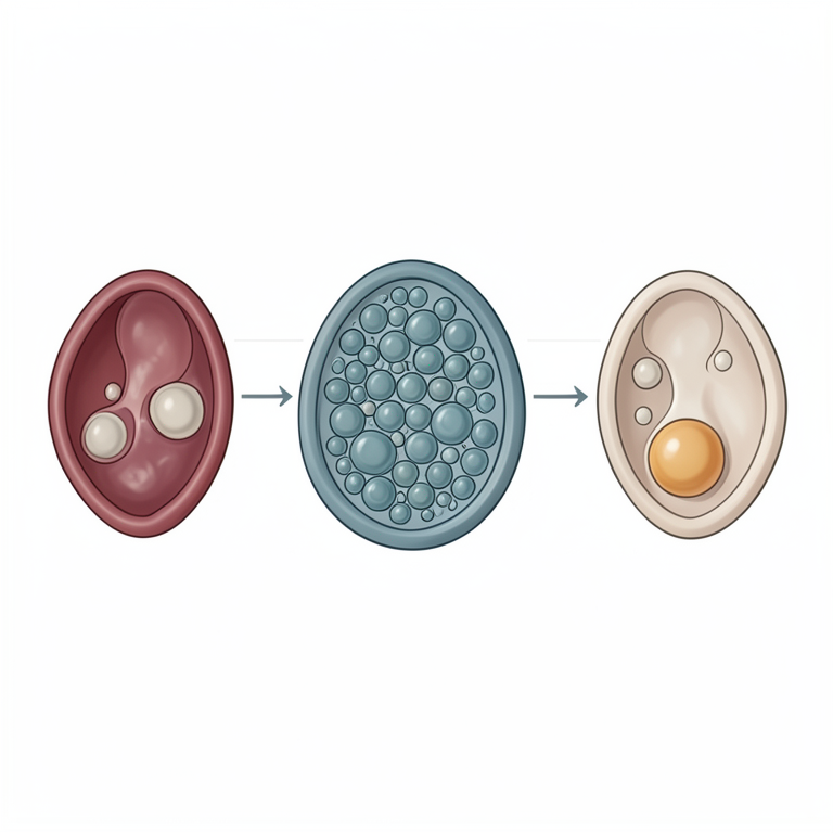
Factors Influencing Follicular Population
AI-generated conceptual illustration (Murrah buffalo ovaries). It explains the idea and does not show study data.
View source evidence ↗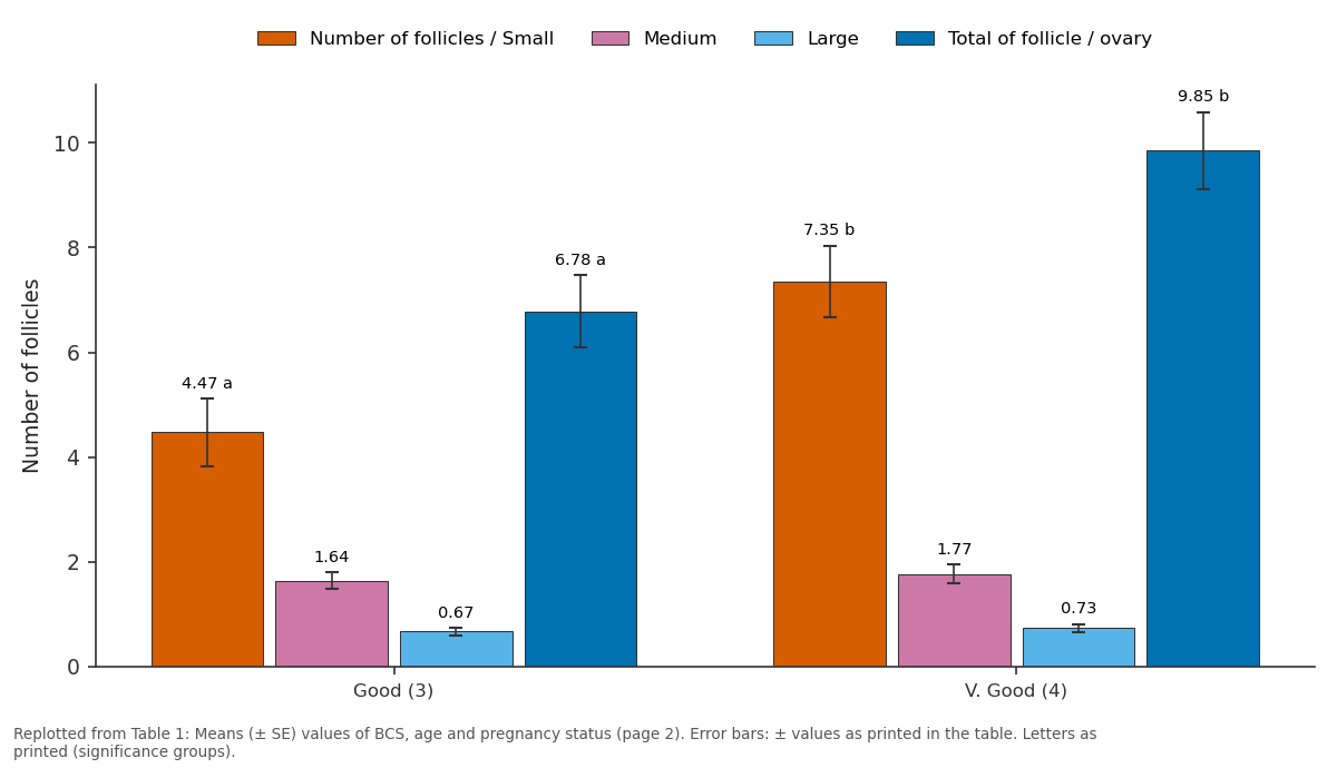
Number of follicles / Small and Medium and more: Good (3) vs V. Good (4)
Replotted from Table 1 (page 2) of the original paper. Every bar is the paper's own table value.
View source evidence ↗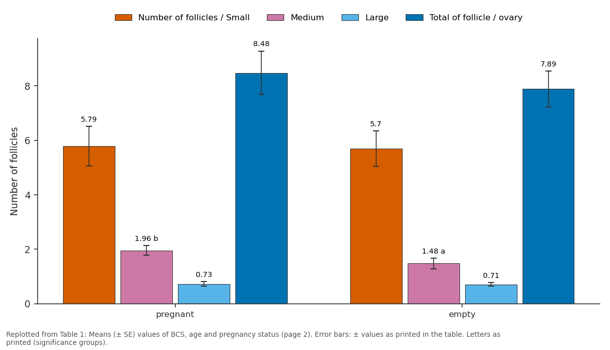
Number of follicles / Small and Medium and more: pregnant vs empty
Replotted from Table 1 (page 2) of the original paper. Every bar is the paper's own table value.
View source evidence ↗Figures & tables
From the original paper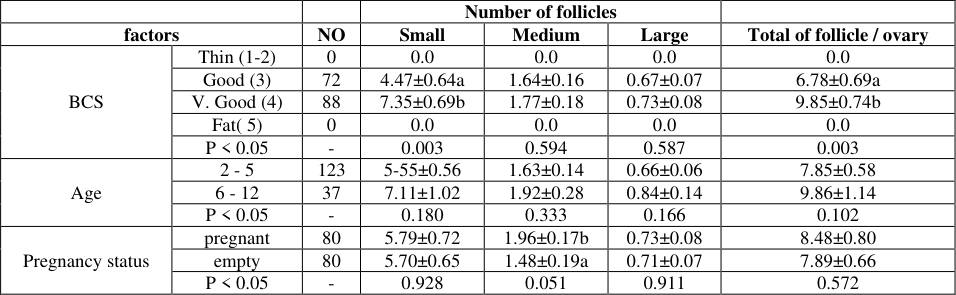 Table 1
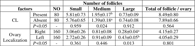 Table 2
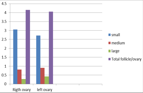 Figure 5
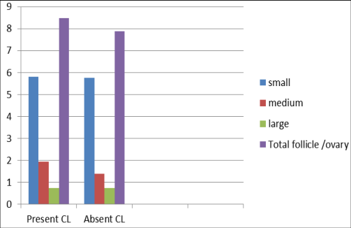 Figure 4
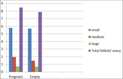 Figure 3
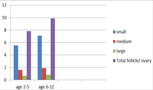 Figure 2
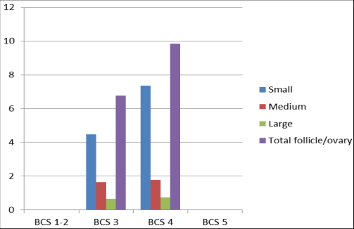 Figure 1
Introduction
Ultrasound graphy technique is simple, non invasive imaging technique without side effect and has been used for various functions such as a tool in buffalo reproductive management (Giuseppina, 2012) [4] . Gyan et al. (2017) [6] reported that this technique has helped in diagnosis of various ovary and uterus diseases such as ovarian cyst and tumors, in addition to helped in predicting estrus in dairy animals after prostaglandin administration. Presicce et al (2003) [12] registrated that the ovarian follicular dynamics and follicular growth in buffalo was similar to that observed in cattle and was characterized by waves of follicular growth and regression. Ultrasound graphy has proved to be a valuable tool in assessing the status of ovarian structures such as follicle and corpus luteum in cyclic and non-cyclic buffaloes. For this purpose the current study was designed to investigate the influence of related factors such as age, body condition score (BCS), CL, pregnancy status and ovary localization on follicular population in Murrah buffalos using Ultrasound graphy technique.
Materials and methods
Materials and methods
2.1Study area
The present research was conducted in year 2018 -2019 at Central Institute for Research on Buffalo, Hisar Haryana, India, located between Latitude: 29°09′14″ N Longitude: 75°43′22″ E and Elevation above sea level: 216 m.
Experimental animals
The study was conducted at buffalo's farm in Central Institute for Research on Buffaloes (CIRB) during period from September, 2018 to January, 2019. 160 female Murrah buffaloes with average age of 2-11 years were used. The animals were maintained in a semi intensive system of housing at CIRB animal farm and were fed balance ration consisting of green fodder, wheat straw and concentration with specific mineral mixture developed by the institute.
Determination of animal's Body Condition Scoring
Animal body condition scoring was determined according to that method described by (Anitha et al., 2011) [1] and classified animals from 1-5 score.
Transrectal Ultrasound graphy examination
Ultrasound graphy was performed by using a portable realtime B-mode transrectal ultrasound scanner (PVF -738F, TOSHIBA, SSA220 Japan) with an intra operative 7.0 MHz micro convex transducer. Each ovary was scanned several planes by maneuvering the transducer along it surface to ovary identity the ovarian structures. Positions and sizes of follicles (≥ 3mm) were measured after freezing the image using inbuilt calipers. However, follicles < 3mm were counted by seeing the image on the ultrasound screen appears as black circular structure surrounded by echogenic ovarian tissues. Ovary sizes, follicular population, localization of CL and uterus structure were examined and the diagnosis was made on basis of echogenicity of images. The pregnancy was confirmed by the presence of a visible fetus on viewing screen of the ultrasound machine which was irregularly shaped echogenic structure surrounded by non echogenic black color image in uterus (Sharma et al., 2012) [14] .
Statistical Analysis
Data were analyzed using SPSS (Statistical Package for Social Sciences) Version 18. The analysis of variance and Duncan's test statistics were used to analyze appropriate data sets. Differences were significant at P<0.05 (Kouamo et al., 2014) [8,9] .
Results
Results
Effect of non ovarian factors on follicular population
The Effect of non ovarian factors on follicular population was presented in Table 1. The number of small follicles in animals having BCS 4 (7.35±0.69) were significantly higher (P<0.05) as compared to animals having BCS 3 (4.47±0.64). However, no significant difference was observed between medium and large follicles in animals having different BCS. The total number of follicle per ovary was significantly greater (P<0.05) in animals having BCS 4 (9.85±0.74) as compared to BCS 3 animals 6.78±0.69. This effect was owing to more number of small follicles in the group. On the other hand, no significant different (P<0.05) was observed between the age of animal and the follicular population. Number of medium follicles was significantly higher (P<0.05) in pregnant animals when compared with non-pregnant cyclic animals. However, there was no significant different between the number of small follicles, large follicles and total number of follicle in pregnant and non-pregnant animals.
| — | — | — | Number of follicles | — | — | — |
|---|---|---|---|---|---|---|
| factors | NO | Small | Medium | Large | Total of follicle / ovary | |
| BCS | Thin (1-2) | 0 | 0.0 | 0.0 | 0.0 | 0.0 |
| Good (3) | 72 | 4.47±0.64a | 1.64±0.16 | 0.67±0.07 | 6.78±0.69a | |
| V. Good (4) | 88 | 7.35±0.69b | 1.77±0.18 | 0.73±0.08 | 9.85±0.74b | |
| Fat( 5) | 0 | 0.0 | 0.0 | 0.0 | 0.0 | |
| P < 0.05 | - | 0.003 | 0.594 | 0.587 | 0.003 | |
| Age | 2 - 5 | 123 | 5-55±0.56 | 1.63±0.14 | 0.66±0.06 | 7.85±0.58 |
| 6 - 12 | 37 | 7.11±1.02 | 1.92±0.28 | 0.84±0.14 | 9.86±1.14 | |
| P < 0.05 | - | 0.180 | 0.333 | 0.166 | 0.102 | |
| Pregnancy status | pregnant | 80 | 5.79±0.72 | 1.96±0.17b | 0.73±0.08 | 8.48±0.80 |
| empty | 80 | 5.70±0.65 | 1.48±0.19a | 0.71±0.07 | 7.89±0.66 | |
| P < 0.05 | - | 0.928 | 0.051 | 0.911 | 0.572 |
Table text extracted from the original PDF; superscript letters are shown inline. The original table image is below.
View original table image
Effect of ovarian factors on follicular population
The effect of ovarian factors on follicular population was presented in Table 2. The ovary having CL had more number of medium size follicles (1.95±0.17) as compared to ovary without CL (1.39±0.18)and difference was significant (P<0.05). There was no significant difference in the total number of follicles present in both ovaries. Right ovary was having significantly lower (P<0.05) number of large follicles than left ovary (0.28±0.04 vs.0.43±0.05). This decrease has no effect on the total number of follicle per ovary. While, no significant observed between numbers of small follicle, large follicle and ovary localization at the present study.
| — | — | — | Number of follicles | — | — | — |
|---|---|---|---|---|---|---|
| factors | NO | Small | Medium | Large | Total of follicle / ovary | |
| CL | Present | 80 | 5.81±0.73 | 1.95±0.17b | 0.73±0.09 | 8.49±0.80 |
| Absent | 80 | 5.76±0.65 | 1.39±0.18a | 0.74±0.08 | 7.89±0.66 | |
| P<0.05 | - | 0.959 | 0.024 | 0.912 | 0.564 | |
| Ovary Localization | Right | 160 | 3.06±0.26 | 0.81±0.08 | 0.28±0.04a | 4.15±0.27 |
| Left | 160 | 2.72±0.26 | 0.91±0.09 | 0.43±0.05b | 4.05±0.29 | |
| P<0.05 | - | 0.361 | 0.446 | 0.013 | 0.801 |
Table text extracted from the original PDF; superscript letters are shown inline. The original table image is below.
View original table image
Discussion
The present study demonstrated that the average number of small follicles were significantly higher (P<0.003)in animals that had BCS 4.0 as compared to those having BCS 3.0. However, there was no significant difference in average number of medium and large follicles between these two groups. The results were similar to that found in cow (Kouamo et al., 2014) [8,9] . This has been attributed to low Blood concentration of growth hormones such as Insulin like grow factor -1 (IGF-1). High plasma levels of IGF-1 resulting from improved nutrition, increases the sensitivity of granulosa cells to FSH stimulation (O'Callaghan and Boland, 1999) [10] . Ryan et al. (1994) [13] also found a relationship between the blood concentration of IGF-1 and fat or thin animals presented low concentrations of IGF-1. On the other hand the effect of age on follicular population showed no significant different between animals. May be this related to most of animals their ages between 2-5 years were heifers and have BCS grade 3. The results are similar to that reported in young steer and heifer's cow (Breier et al., 1988; Granger et al., 1989) [2,5] .The feed restriction causes a decrease in hepatic concentration of IGF-I and lead to deceleration of follicular growth. This study also demonstrated that the pregnancy status in buffaloes did not affect the follicular population. Further, the ovary bearing Corpus luteum has more number of medium size follicles as compared to ovary without CL. This might be due to more blood supply in the ovary and continuous follicular growth waves every 8 to 10 days without dominance during pregnancy despite the continuous production of progesterone (Ginther et al., 1989) [3] . The corpus luteum has a negative influence for the growth follicles larger than approximately 7mm after day 21 or 22 of pregnancy (Pierson and Ginther, 1989) [11] . The left ovary has significantly greater number of large follicles than right ovary. The presence corpus luteum in the ovary may contribute to unfavorable condition for follicular growth. As a result, follicle regressed and led to lower COCs recovered from ovaries with a presence of corpus luteum (Hafez and Hafez, 2000) [7] .
Conclusion
This study revealed that the ovaries of Murrah buffaloes with BCS 4 have higher number of follicles than those with BCS 3.The ovaries from pregnant animals have more number of medium size follicles as compared to those of non-pregnant buffaloes. The right ovaries in Murrah buffaloes have lower number of large follicles than left ovaries. Future studies are require to study any seasonal variation if any, between different BCS, age, pregnancy status, presence or absence of CL and ovary localization.
Figures and tables from the paper
References
14 in the original paper- Anitha A, Sarjan Rao K, Suresh J, Srinivasa Moorthy PR, Kotilinga Reddy Y. A body Condition Score (BCS) system in Murrah Buffaloes. Buffalo Bulletin. 2011; 1:79-99.
- Breier BH, Gluckman PD, Bass JJ. Influence of nutritional status and oestradiol-17b on plasma growth hormone, insulin-like growth factors-I and -II and the response to exogenous growth hormone in young steers. Journal Endocrinolology. 1988; 118:243-250.
- Ginther OJ, Knopf L, Kastelic JP. Ovarian follicular dynamics in heifers during early pregnancy. Biology. Reproduction. 1989; 41:247-254.
- Giuseppina MT. Ultrasonography and Reproduction in Buffalo. Journal of Buffalo Science. 2012; 1:163-173.
- Granger AL, Wyatt WE, Craig WM, Thompson DL, Hembry FG. Effects of breed and wintering diet on. growth, puberty and plasma concentrations of growth hormone and insulin like growth factor I in heifers. Domestic Animal Endocrinology. 1989; 6:253-262.
- Gyan S, Chandolia RK, Dutt R, Saini A, Dalal J, Malik RK. Comparative Study of Follicular Turn Over in Cyclic and Non-Cyclic Murrah Buffaloes through 2D TransRectal Ultrasonography. International Journal of Current Microbiology and Applied Sciences. 2017; 6:3590-3602.
- Hafez ES, Hafez EB. Folliculogenesis, egg maturation, and ovulation. Reproduction in Farm Animals, 7th Edition. 2000; pp. 68-81.
- Kouamo J, Dawaye SM, Zoli AP, Bah GS. Evaluation of bovine (Bos indicus) ovarian potential for in vitro embryo production in the Adamawa plateau (Cameroon), Open Veterinary Journal. 2014; 4(2):128-136.
- Kouamo J, Dawaye SM, Zoli AP, Bah GS. Evaluation of bovine (Bos indicus) ovarian potential for in vitro embryo production in the Adamawa plateau (Cameroon), Open Veterinary Journal. 2014; 4(2):128-136.
- O'callaghan D, Boland MP. Nutritional effects on ovulation, embryo development and establishment of pregnancy in ruminants. Animal. Science. 1999; 68:299314.
- Pierson RA, Ginther OJ. Intraovarian effect of the corpus luteum on ovarian follicles during early pregnancy in Heifers. Animal reproduction Science. 1989; 15:53-60.
- Presicce GA, Parmeggiani A, Sentore EM, Stecco R, Barille VL, De Maruo GJ, et al. Hormonal dynamics and follicular turnover in prepubertal Mediteranean Italian buffalo. Theriogenology. 2003; 8860:1-9.
- Ryan DP, Spoon RA, Griffith MK, Williams GL. Ovarian follicular recruitment, granulosa cell steroidogenic potential and growth hormone/ insulin-like growth factor-I relationships in suckled beef cows consuming high lipid diets: Effect of graded differences in body condition maintained during the puerperium. Domestic Animal Endocrinology. 1994; 11:161-174.
- Sharma RK, Singh JK, Khanna S, Phulia SK, Sarkar SK, Inderjeet S. Fetal age determination in Murrah buffaloes from days 22 through 60 with ultrasonography. Indian Journal of Animal Sciences. 2012; 82(4):374-376.