
Research article · 2018 · Full text
International Journal of Veterinary Sciences and Animal HusbandryManagement of femur fracture in bonnet macaque (Macaca radiata): A case report
Volume 3(2) · Pages 08–09
Research areasPrimate Behavior and Ecology
Abstract
Author-writtenA male Macaca radiata around 6 to 8 month old was rescued and brought to the Wild Animal Rescue and Rehabilitation Center, Katraz Pune. Clinical and radiological examination revealed that oblique displaced fracture of right femur. Treatment was carried out with internal fixation methods of fracture management under Ketamine anesthesia. After 2 months of surgery, the animal showed proper leg movement and walking.
Summary
Source-checkedPlain-language reading of the full paper, prepared with AI and checked against the text. Each point links to the authors' own words.
Why it matters
- The bonnet macaque (Macaca radiata) is a highly adaptive species found in both jungle and village environments in Maharashtra. View evidence ↗
- Long bone fractures are considered rare occurrences in non-human primates. View evidence ↗
The question
- The study aims to report the successful surgical management of a femur fracture in a bonnet macaque. View evidence ↗
What they did
- The subject was a male bonnet macaque, approximately 6 to 8 months old, treated at the Wild Animal Rescue and Rehabilitation Center in Katraz, Pune. View evidence ↗
- Radiological examination was used to confirm that the animal had an oblique displaced fracture of the right femur. View evidence ↗
- The surgical procedure was conducted under general inhalation anesthesia using ketamine hydrochloride at a dose of 10 mg/kg body weight. View evidence ↗
- The chosen surgical intervention for managing the fracture was the placement of intramedullary pins. View evidence ↗
- The intramedullary pin was inserted using a retrograde method, moving from the proximal end to the distal end of the fractured bone. View evidence ↗
- Post-operative care included meloxicam (1 mg/kg i/m) for pain and Cefotaxim (20 mg/kg body weight) as an antibiotic for 5 days. View evidence ↗
- The animal was kept in a confined cage to restrict movement of the affected limb and support recovery. View evidence ↗
What they found
- Two weeks after the operation, the fractured bone was stable and the animal began moving its legs. View evidence ↗
- Full recovery was achieved 2 months after surgery, with the animal demonstrating proper walking and leg movement. View evidence ↗
What it means
- The authors conclude that intramedullary pinning is an effective method for managing femur fractures in this species, especially since plaster bandaging is rarely indicated for wild animals. View evidence ↗
Visual summary
1 visualsIllustrations explain the paper; they do not show study data.
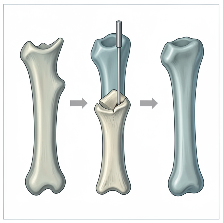
Retrograde Pinning Concept
AI-generated conceptual illustration (bonnet macaque femur bone). It explains the idea and does not show study data.
View source evidence ↗Figures & tables
From the original paper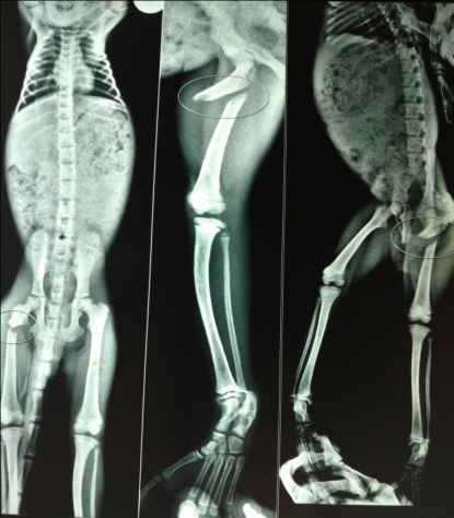 Figure 1
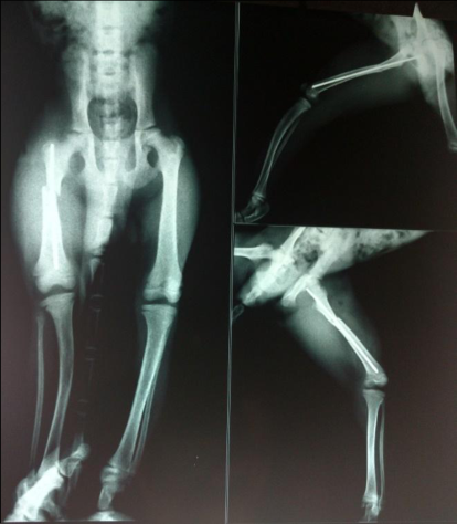 Figure 2
Introduction
Macaca radiata is distributed in both villages and jungles of Maharshtra and the species is highly adaptive. The incidence of long bone fracture in the non-human primates are rare (Bulstrode, 1990) [1]. The selected workers have been reported fracture of long bone like Femoral Fracture in a Rhesus Monkey (Singh et al. 2012) [7], humerus fracture in wild langur (Presbytis entellus) (Majie et al. 2014) [3], Femur fracture in Hanuman Langur (Semnopithecus entellus) (Ragunath et al. 2008) [6], Radial Fracture in a Capped Langur (Trachypithecus pileatus durga) (Debnath et al. 2016) [16]. This report present a case of successful surgical management of femur fracture in Bonnet macaque (Macaca radiata).
Case Details A male Macaca radiata around 6 to 8 month old was rescued and brought to the Wild Animal Rescue and Rehabilitation Center, Katraz Pune. According to history the primate came under an automobile accident and unable to bare weight by right hind limb. A through clinical examination was carried out. Clinical examination revealed crepitating sound at the right hind limb in femur region.
For confirmation of fracture and its type fracture radiological examination was carried out. Radiological examination revealed oblique displaced fracture of right femur (Fig. 1). For treatment of fracture decision was taken to do surgical intervention by placing intramedullary pins. Surgery was performed under general inhalation anesthesia with ketamine hydrochloride 10 mg/kg body weight.
Surgical site was prepared aseptically, lateral oblique incision was given on tancerfacia lata muscle which further dissected bluntly. The fractured fragment was exposed. Pin size was selected according to medullary diameter and depth of insertion. Insertition of intramedullary pin was done by retrograde method i.e first inserted proximally and then distally in fractured bone.
The pin was inserted in proximal fragment of bone up to trochanter fossa one half inch then reduction of distal fragment and alignment of both fragments was done. Then pin was inserted in distal fragment till it reaches up to epiphysis. (Fig 2) The fascia was sutured with Vicryl 2-0 in a simple interrupeted pattern, and the skin was sutured with nylon suture material in horizontal mattress pattern. Postoperative analgesia was provided with meloxicam @ 1 mg/kg i/m and antibiotic Cefotaxim @ 20 mg /kg bwt for 5 days. For additional support animal was kept in confined cage to prevent movement of affected part and post-operative care.
Skin sutures were removed after 2 weeks. Animal was kept in closed confiened area to restrict the movement of limb. Through periodical radiological examination healing and callus formation at fractured site was monitored. VET 2018; 3(2): 08-09 Ankush G Dubey Animal Rescue and Rehabilitation Centre, Katraj, Pune, Maharashtra, India GD Pardeshi Animal Rescue and Rehabilitation Centre, Katraj, Pune, Maharashtra, India NK Nighot Animal Rescue and Rehabilitation Centre, Katraj, Pune, Maharashtra, India RV Jadhav Animal Rescue and Rehabilitation Centre, Katraj, Pune, Maharashtra, India Correspondence Ankush G Dubey Animal Rescue and Rehabilitation Centre, Katraj, Pune, Maharashtra, India Reference 1.
Bulstrode C. What happens to wild animals with broken bones. Iowa Orthopedic Journal, 1990; 10:19-23. 2. Debnath K, Baishya MP, Gupta AK.
Intramedullary After two weeks postoperative fracture bone was stable and animal starts movement of legs. Complete recovery was achived after 2 months of surgery, the animal showed proper leg movement and walking. Pinning for Management of Radial Fracture in a Capped Langur, Intas Polivet. 2016; 17(I):13-15 3. Majie AK, Mondal P, Ghosh SK, Banerjee DN.
Invasive management of humerus fracture in wild langur (Presbytis entellus) with a view to rehabilitation back to the wild. J Wildlife Rehab. 2014; 34(1):7-10 4. Nolosco TP, Rao GD, William BJ, Prathaban S. Diazepam-ketamine anesthesia for the surgical management of electric burns in Bonnet macaque monkeys (Macaca radiata)-Report of 2 cases.
Tamil Nadu Veterinary and Animal Sciences University, Journal Veterinary & Animal Sciences. 2007; 3(2):113- 115 5. Ranganath LB, Ranganath N, Srinivas CL, Nagaraja R, Jayadevappa SM. Treatment of fracture of tibia and fibula in male panther (Panthera pardis). Indian Veterinary Journal, 1998; 74:783-784. 6.
Raghunath M, Singh N, Gopinathan A, Singh T, Sangwan V, Mohindroo J et al. Fracture Fixation of Femur in Hanuman Langur (Semnopithecus entellus) with Intramedullary Pinning, J Wildlife Rehab. 2008; 29(2-3):32-34. 7. Singh J, Aithal HP, Monsang SW. Intramedullary Fig 1: Oblique displaced fracture of right femur Pinning for the Management of Femoral Fracture in a Rhesus Monkey.
Intas Polivet. 2012; 13(II):429-431 Fig 2: Postoperative xray after intramedulart pin
Discussion
The femur fracture is very rare in bonnet macaque and normally occurs due to traumatic injuries. In the present clinical situation, the fracture was simple without any signs of commination or sharp ends, soft tissue swelling could have been due to present of haematoma around fracture bone. In the non-human primate for anesthesia generally xyalazine and ketamine combination was used (Nolosco et al. 2007) [4]. In the present case only ketamine was used for anesthesia due to docile nature of animal which is in general agreement with the report of Ragunath et al. (2008) [6].
Ranganath et al. 1998 [5] reported that plaster of paris bandaging is rarly indicated in management of fractures in wild animals. Hence, in the present case intramedullary pinning was selected for the management of femur fracture.
References
0 in the original paperCited by 1
From OpenAlex, OpenCitations and reference lists read by Scinovex
- Fibular and metatarsal osteosynthesis in a southern brown howler monkey (Alouatta guariba clamitans) ↗
Lívia Eichenberg Surita, Daniela Nicknich, Eduardo Almeida Ruivo dos Santos, Roberta Picoli, Eduardo Raposo Monteiro, Marcelo Meller Alievi · Journal of Medical Primatology · 2020
Here we describe a successful surgical management of a distal fibular fracture combined with a tarsocrural luxation and multiple metatarsal fractures in the left foot of a southern brown howler monkey (Alouatta guariba clamitans). We achieved satisfactory outcome by applying intramedullary pinning for each of the bone fractures and closed reduction of joint luxation-kept in place only by bone alignment,…
❝ 2