
Research article · 2018 · Full text
International Journal of Veterinary Sciences and Animal HusbandryMicrobial analysis of water system for veterinary vaccine manufacturing facility
Volume 3(4) · Pages 24–29
Research areasMineral Processing and Grinding
Abstract
Author-writtenWater is very widely used in the pharmaceutical, biopharmaceutical and biological industry for various purpose like as raw material, ingredient as a solvent in processing, formulation and manufacturing of the wide range of the finish products, as API and intermediates and analytical reagents. That’s why regulatory requirements over control of the quality of various types of the waters throughout the production, storage and distribution processes, including microbiological and chemical quality, are very stringent. Lack of monitoring over any of the parameters for the Potable, Purified or Water for Injection can leads to the loss of the costly drug product or drug substance. Veterinary vaccines are considered as the sterile parenteral dosage form and water used for the various purposes in the veterinary vaccine manufacturing facility must have to comply with the regulatory requirements. So the aim of the present work is to bioburden testing and check the suitability of the different types of the water used for the various applications in vaccine manufacturing facility using the various techniques like MPN method, Plate count method, Filtration method and based on the results of the bioburden testing and identification of pathogens and conclusion to be done for the compliance and suitability of the water used for the manufacturing of the vaccines as sterile parenteral dosage form to have the claimed safety, integrity, strength, purity and quality.
Summary
Source-checkedPlain-language reading of the full paper, prepared with AI and checked against the text. Each point links to the authors' own words.
Why it matters
- Water is a critical component in pharmaceutical and biological industries, serving as a raw material, solvent, and reagent for various products. View evidence ↗
- Failure to monitor the quality of potable, purified, or injection water can result in the loss of expensive drug substances or products. View evidence ↗
- Gram-negative bacteria are common water contaminants and pose a specific risk of endotoxin contamination in water used for parenteral production. View evidence ↗
The question
- The study aims to perform bioburden testing to determine if different types of water used in a veterinary vaccine facility are suitable for their intended applications. View evidence ↗
What they did
- The researchers tested four types of water: non-potable, potable, purified, and water for injection. View evidence ↗
- Bioburden was assessed using three primary techniques: the Most Probable Number (MPN) method, the plate count method, and the membrane filtration method. View evidence ↗
- Water samples of approximately 100 ml were collected in bottles and immediately covered with aluminum foil to prevent external contamination. View evidence ↗
- The membrane filtration technique utilized a sterile filter with a rating of 0.45μm. View evidence ↗
What they found
- No pathogenic organisms were detected in the purified water or water for injection (WFI) samples from either the generation or user points. View evidence ↗
- The common pathogenic microorganisms identified in the study's overall testing were E. coli, Salmonella abony, Pseudomonas aeruginosa, and Staphylococcus aureus. View evidence ↗
What it means
- The authors concluded that the bioburden levels for purified water and water for injection are well within acceptable regulatory limits. View evidence ↗
- The authors state that the potable, purified, and injection water used in the facility comply with microbiological limits and are suitable for manufacturing sterile parenteral vaccines. View evidence ↗
Visual summary
2 visualsIllustrations explain the paper; charts are replotted from the paper's own table values.
Water Testing Workflow
AI-generated conceptual illustration (Laboratory water samples and microbiological testing equipment). It explains the idea and does not show study data.
View source evidence ↗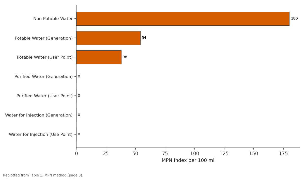
MPN Index per 100 ml: Non Potable Water vs Potable Water... vs Potable Water (User... vs Purified Water... vs Purified Water (User...
Replotted from Table 1 (page 3) of the original paper. Every bar is the paper's own table value.
View source evidence ↗Figures & tables
From the original paper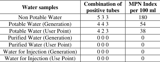 Table 1
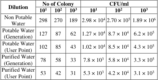 Table 2
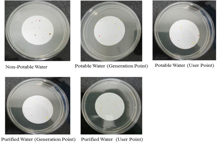 Figure 1
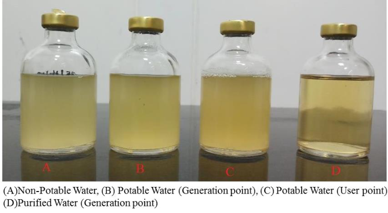 Figure 2
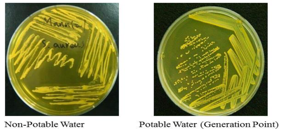 Figure 3
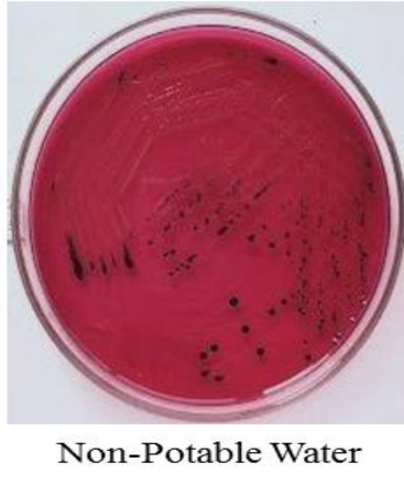 Figure 4
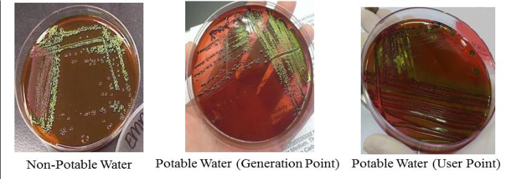 Figure 5
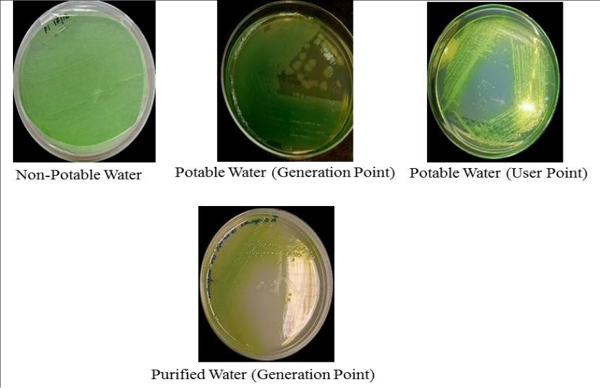 Figure 6
Introduction
Water must follow quality needs as stated in principles given by the United States Pharmacopeia (USP), European Pharmacopoeia (EP) before its usage in pharmaceutical manufacturing (Kolpin et al., 2002) [1] . Due to its criticality in pharmaceutical production, microbiological control of water is very important. Since water is ever present, each grade of water is a potential wellspring of microbiological contamination, particularly when not appropriately controlled (Sandle et al., 2004) [2] . The nature of protected water sources can be disintegrated because of poor site choice, lacking assurance and unhygienic management of facilities. Water may become contaminated at any point in between its collection and use. Despite the fact that most pharmaceutical water frameworks are controlled, microorganisms will once in a while be available, although in low numbers (Jiminez et al., 2004) [3] . A few of the microorganisms noted will be found in pharmaceutical water generation plants, starting from the supplied potable water. It is evaluated that there are 70 distinct kinds of bacteria in wastewater. A few unique kinds of microbes cross water-treatment barriers and are found in pharmaceutical waters. Most microbial contaminants are Gram-negative bacteria that represent the extra risk of Endotoxin contamination of waters utilized for parenteral production (Traeger et al., 2003) [4] . To check whether the water is contaminated with the microorganisms or not different types of microbiological test are performed to check the purity of water such as membrane filtration method, plate count method, most probable number etc. Microbial conditions are usually checked by experimental method that need as much as 48 to 72 hours for giving results. (Twinbrook 2012) [5] . Among the microorganisms generally found in water, these organisms are Mainly Gramnegative bacteria. Common species include: Pseudomonas aeruginosa, Salmonella spp, Staphylococcus aureus and Escherichia coli. Very less grampositive bacteria are isolated those that are identified include Bacillus spp. Validation and qualification of water purification, storage and distribution system are a fundamental part of GMP and form an integral part of the GMP inspection. Water in pharmaceutical industry is purified first by various processes before using it for any operation. The treatments for purification of water include using of softeners, ultrafiltration, reverse osmosis, distillation etc. Water which is obtained after passing through all these treatments is completely pure and sterile which does not contain any kind of microbial contamination (Murthy et al., 1984) [6] . If appropriate steps are not taken to lessen their numbers or eliminate them, these microorganisms may compromise subsequent water purification steps. Endogenous sources of microbial contamination can arise from unit operations if in a water purification system is not properly maintained and operated.
Materials and Methods
Sample collection: While collecting bulk water at the points of use first allow a forceful flow of water for about 1-3 min before sampling. Approx. 100 ml of sample is collected in the bottles form the quality control lab and they are covered by aluminium foil immediately to prevent any external contamination. Samples are processed for testing within one hour after collection to obtained most accurate result. Types of Water Samples to be tested are as below.
Membrane Filteration (MF) Technique
Filtration assembly having six filtration cup together is sterilised to avoid any unwanted contamination. Sterile filtration assembly is set up inside the laminar air flow. Using a sterile forceps, a sterile membrane filter with a rating of 0.45μm is placed under the filtration cup on the porous plate. The filter unit is connected to a suitable vacuum pump. The vacuum motor pump is switched on to draw the samples through the filter. A fixed volume of water is then added into the filtration cup. Upon completion of filtration microorganisms are retained on the surface of the membrane. After the filtration, the funnel is unlocked and removed. The membrane filter is removed with sterile forceps and placed on the SCDA plate in the centre carefully to avoid the entrapment of air. The SCDA plates are incubated at 37°C for 24 hours. On the next day observe the plates and count the colonies.
Most Probable Number (MPN) Method 2.2.1 Preparations for presumptive test
Single strength broth and double strength broth are prepared. Double strength broth contains double the concentration of ingredients except water. Prepare five tubes of MLBB having 10 ml (2x) medium with 10 ml of sample in each. Another set of five tubes of MLBB having 5 ml (x) medium with 1 ml of sample in each. Prepare third set of five tubes of MLBB having 5 ml (x) medium with 0.1 ml of sample in each.
Presumptive test
First of all shake the water sample to ensure uniform distribution of microorganisms. Prepare MacConkey media of single and double strength in sterile test tubes with Durham's tube and autoclave it. Prepare three sets of test tubes having five tubes in each set, one set with 10 ml of double strength and other two having five ml of single strength. One tube having five ml of medium is left uninoculated, and marked it as control tube. Using sterile pipettes and transfer 10 ml of water to each tubes of double strength broth. Transfer one ml of water sample to each of the five tubes of one set of single strength broth and transfer 0.1 ml water to five tubes of remaining last set of single strength broth tubes. Incubate the tubes at 37 °C for 24 hours. After incubation, observe the tubes for gas production which is collected in Durham's tube and acid production is indicated by colour change of media from pink to yellow. If none of the tube show acid and gas production then reincubate all tubes for additional 24 hours. On the next day, observe and record the number of positive tubes in each set and compare with standard MPN index to give presumptive coliform count per 100 ml water sample.
Confirmed test
From positive presumptive tubes spread 0.1ml of sample on EMB agar plate. Incubate one plate at 37 °C for 24 hours and another at 44.5 °C for 24 hours.
Completed test
Select and mark a well isolated typical or atypical colony from the EMB agar plate. With the help of sterile nicrome wire loop, pick half of the previously marked typical/atypical colony and transfer it to BGLB broth tube. From the remaining half of the same colony streak on the surface of a nutrient agar slant. Incubate slant and broth at 37 °C for 24 hours. Check lactose broth for presence of acid and gas. Prepare Gram's stain of the growth from the surface of agar slant and observe the slide. Look for the presence of gramnegative non-spore forming short rods. Record the result and interpreted.
Pathogen Identification Testing 2.3.1 Test for identification of Escherichia coli
Take a loopful of sample from each positive SCDA broth and spread it on a EMB agar plate. Incubate the plate at 37̊ C for 24-48 hours. If the colonies exhibit greenish metallic sheen under reflected light and blue black appearance, then it confirms the presence of E. coli. Upon examination, if none of the colonies exhibits both a characteristic metallic sheen under reflected light, the sample meets the requirements of the test for the absence of E. coli.
Test for identification of Salmonella spp.
Take a loopful of sample from positive SCDA broth and spread it on a xylose-lysine-deoxy cholate agar (XLDA) plate and brilliant green agar (BGA) plate. Simultaneously carry out the positive control by streaking a loop full growth of salmonella on a surface of one of the above media, which is used for testing. For negative control incubate the agar plate without streaking or inoculation. Invert and incubate all the plates at 37̊ C for 18-24 hours. If the colonies appear small, transparent, colourless or pink colonies with pink or red zone on BGA plate, then it confirms the presence of Salmonella. On XLDA plate, colonies appear Red with or without black centers.
Secondary Test
Subculture any colonies showing the above characteristics in triple sugar-iron agar by first inoculating the surface of the slope and then making a stab culture with the same inoculating needle. At the same time inoculate a tube of urea broth. Incubate at 36 o C to 38 o C for 18-24 hours. Upon examination, no evidence of tubes having alkaline (red) slant and acid (yellow) butt (with or without concomitant blackening of the butt from hydrogen sulphide production), the sample meets the requirements of the test for absence of genus salmonella.
Test for identification of Staphylococcus aureus
Streak a portion from Soyabean casein digest medium on the surface of mannitol salt agar medium. Simultaneously carry out the positive control by streaking a loop full growth of Staphylococcus aureus on the surface of agar medium. For negative control incubate the agar plate without inoculation. Invert and incubate all the plates at 37 o C for 18 to 24 hours. If upon examination, if the plate contains colonies having yellow colonies with yellow zones, then it indicate the presence of Staphylococcus aureus.
Test for identification of Pseudomonas spp.
Streak a portion from Soyabean casein digest medium on the surface of Citramide agar medium; simultaneously carry out the positive control by streaking a loop full growth of P.aeruginosa on the surface of Citramide agar. For negative control incubate the Citramide agar plate without inoculation. Invert and incubate all the plates at 35 o C to 37 o C for 18 to 24 hours. If upon examination, if the plate contains colonies having greenish yellow growth, the sample indicates the presence of Pseudomonas aeruginosa.
Confirmative Test
The suspected colonies from Citramide agar plate are smeared on the Oxidase disc. The test is positive, if purple colour is produced within 5-10 seconds.
Plate Count Method
Prepare a series of at least 3 test tubes containing 9 ml of sterile distilled water. Using a sterile pipette, add 1 ml of sample in the first tube of the set and label it as 10 -1. Mix the contents well by swirling the tube upside down few times. From the first tube, take 1 ml of the sample and transfer to second tube. Label it as 10 -2. Repeat the procedure with all the remaining tubes labelling them until 10 -3. Pipette out 0.1 ml from the appropriate desired dilution series onto the center of the surface of an agar plate. Spread the sample evenly over the surface of agar using the sterile glass spreader. Incubate the plate at 37 o C for 24 hours. Calculate the colony forming unit (CFU) value of the sample. Calculation of result:
(No. of colonies) Χ (Dilution factor) CFU/ml = Volume of culture plated
Results
Results
Membrane Filtration Method
Membrane Filtration Method
Discussion
The study result suggests that the common pathogenic microorganisms which get identified are commonly E. coli, Salmonella abony, Pseudomonas aeruginosa and Staphylococcus aureus. On EMB agar plate E.coli produce Greenish metallic sheen under reflected light while on Mannitol salt agar plate S. aureus produce yellowish colonies. Ps. aeruginosa produce greenish yellow pigment on citramide agar plate. When a well isolated colony from citramide agar plate is transferred to oxidase disc it gives purple colour within five seconds. This confirms the presence of pseudomonas. There is an absence of any pathogenic organisms in WFI and purified water in both the samples from generation and user point. On performing Membrane filtration method, each bacterium gets trapped on the filter which is then grows into a separate colony.
Conclusion
Bioburden testing of the Potable water, Purified water and Water for injection used for the manufacturing of the veterinary vaccines at the different stage of the process done for the generation points as well as the user points to be done using the MPN method, Plate count method and Filtration method. Identification of the pathogens was also done for the Potable water, Purified water and Water for injection used for the manufacturing of the veterinary vaccines. Results of Bioburden using all the methods shows that the bioburden limit for purified and water for injection are well within acceptable regulatory limits. So as far as the microorganisms are concerned based on the testing and its respective results reveals that Potable water, Purified water and Water for injection used for the manufacturing of the veterinary vaccines comply with the acceptance limit of the microbiology and states the control for the microorganism in water system as suitable for the manufacturing of the vaccines as sterile parenteral dosage form to have the claimed safety, integrity, strength, purity and quality.
Figures and tables from the paper
| Water samples | Combination of positive tubes | MPN Index per 100 ml |
|---|---|---|
| Non Potable Water | 5 3 3 | 180 |
| Potable Water (Generation) | 4 4 3 | 54 |
| Potable Water (User Point) | 4 2 3 | 38 |
| Purified Water (Generation) | 0 0 0 | 0 |
| Purified Water (User Point) | 0 0 0 | 0 |
| Water for Injection (Generation) | 0 0 0 | 0 |
| Water for Injection (Use Point) | 0 0 0 | 0 |
Table text extracted from the original PDF; superscript letters are shown inline. The original table image is below.
View original table image
| Dilution | No of Colony | — | — | CFU/ml | — | — |
|---|---|---|---|---|---|---|
| 101 | 102 | 103 | 101 | 102 | 103 | |
| Non Potable Water | 298 | 270 | 189 | 2.98 × 10⁴ | 2.70 × 10⁵ | 1.89 × 10⁶ |
| Potable Water (Generation) | 127 | 87 | 62 | 1.27 × 10⁴ | 8.7 × 10⁴ | 6.2 × 10⁵ |
| Potable Water (User Point) | 102 | 85 | 43 | 1.02 × 10⁴ | 8.5 × 10⁴ | 4.3 × 10⁵ |
| Purified Water (Generation) | 78 | 58 | 33 | 7.8 × 10³ | 5.8 × 10⁴ | 3.3 × 10⁵ |
| Purified Water (User Point) | 53 | 42 | 31 | 5.3 × 10³ | 4.2 × 10⁴ | 3.1 × 10³ |
Table text extracted from the original PDF; superscript letters are shown inline. The original table image is below.
View original table image
References
6 in the original paper- Kolpin DW, Furlong ET, Meyer MT, Thurman EM, Zaugg SD, Barber LB, et al. Pharmaceuticals, hormones and other organic wastewater contaminants in U.S. Environ. Sci. Technol. 2002; 36:1202-1211.
- Sandle T. An approach for the reporting of microbiological results from water systems. PDA J Pharm Sci. Technol. 2004; 58(4):231-7.
- Jiminez L. Microorganisms in the Environment and their relevance to Pharmaceutical Processes. In; Jiminez, L, editor. Microbial Contamination Control in the Pharmaceutical Industry. New York: Taylor & Francis; 2004, 8-9.
- Traeger H, Pharmaceuticals, The Presence of Bacteria, Endotoxins, and Biofilms in Pharmaceutical Water, Ultrapure Water, March 2003, 98-102.
- United States Pharmacopeia 35, General Chapter <1231>, United States Pharmacopeial Convention, Inc. 12601 Twinbrook Parkway, Rockville, MD, 2012.
- Murthy YS, Subbiah V, Rao DS, Reddy RC, Kumar LS, Elyas SI, et al. Deshmukh SB-Treatment and disposal of waste water from synthetic drugs plant (I.D.P.L.) 1984, 25-68.
Cited by 2
From OpenAlex, OpenCitations and reference lists read by Scinovex
- Isolation and Identification of Coliform Bacteria and Multidrug-Resistant Escherichia coli from Water Intended for Drug Compounding in Community Pharmacies in Jordan ↗
Mohammad Abu‐Sini, Rafeef A. Maharmah, Dina H. Abulebdah, Mohammad Nafi Solaiman Al-Sabi · Healthcare · 2023
(1) Background: Water is necessary for the preparation of some medicines found in pharmacies where the local water source does not meet the required purity. This study aimed to investigate the presence of coliform contamination in water used for drug reconstitution in community pharmacies in Jordan. (2) Methods: Two water samples from 50 randomly selected community pharmacies representing all Jordanian…
❝ 36PDF - Juncus rigidus high biomass and cellulose productivity under wastewater salinity stress - A paradigm shift to the valorization of RO reject water ↗
Krupali Dipakbhai Vyas, Aneesha Singh · The Science of The Total Environment · 2024
The use of water purifiers is intensively catching up and disposing of reverse osmosis reject water is of great concern. Reject water management using conventional methods is costly and harmful to the environment. To address this issue, the present study aims to utilize reverse osmosis reject wastewater using an eco-friendly approach. Juncus rigidus was treated with reject wastewater containing different…
❝ 7PDF