
Research article · 2019 · Full text
International Journal of Veterinary Sciences and Animal HusbandryGlasser’s disease in pig: A case report
Volume 4(4) · Pages 16–18 · DOI: 10.22271/veterinary.2019.v4.i4a.194 ↗
Research areasGenetic and phenotypic traits in livestockAnimal Behavior and Welfare StudiesAnimal health and immunology
Abstract
Author-writtenGlasser’s disease is an infectious and acute disease seen in pig after weaning, which is characterized by fibrinous polysterositis, arthritis and meningoencephalitis. A case of 3 month old piglet breed “Chwanche” (local) was presented to Veterinary Teaching Hospital (VTH), Institute of Agriculture and Animal Science (IAAS), Paklihawa Campus which chief complain of constipation since three day. Piglet was admitted on VTH and physical and clinical examination was done and heart rate, respiratory rate and temperature were 130/min, 60/min and 106°F respectively. Auscultations abdomen revealed burble sound. First phase treatment was done and further examination was started with hematology. PCV and WBC count were 38% and (28×103/µl) but after five hours the pig was died and postmortem examination was done. Necropsy findings reveals severe polyserositis, fibrinous covering around the lungs and heart, Swollen liver with fibrinous deposition and congested intestinal epithelium. Sever polyserositis indicates that this case is a glasser`s disease.
Summary
Source-checkedPlain-language reading of the full paper, prepared with AI and checked against the text. Each point links to the authors' own words.
Why it matters
- Glasser's disease, also known as porcine polyserositis, is an infectious swine disease that can cause inflammation of the serous membranes, joints, brain, and lungs. View evidence ↗
- The disease is caused by Haemophilus parasuis, which normally lives as a commensal organism in the upper respiratory tract of pigs. View evidence ↗
- The disease typically affects pigs between 4 and 8 weeks of age, often triggered by stress factors such as weaning or environmental changes. View evidence ↗
The question
- The authors report on a 3-month-old local 'Chwanche' piglet presented with a three-day history of constipation to determine the underlying cause of its illness. View evidence ↗
What they did
- Clinical assessment included physical examination, abdominal auscultation, and measurement of heart rate, respiratory rate, and body temperature. View evidence ↗
- Hematological analysis was performed to measure the Packed Cell Volume (PCV) and total White Blood Cell (WBC) count. View evidence ↗
- Following the animal's death, a necropsy was conducted to examine the abdominal cavity, lungs, heart, liver, and intestines. View evidence ↗
What they found
- The piglet exhibited tachycardia (130/min), tachypnea (60/min), and a fever of 106°F. View evidence ↗
- Blood tests showed a PCV of 38% and an elevated total WBC count of 28×103/µl. View evidence ↗
- Necropsy revealed severe peritonitis with fluid accumulation, fibrinous deposits on the lungs, heart, and pericardium, a swollen liver, and congested intestinal epithelium. View evidence ↗
- The presence of severe polyserositis led the authors to diagnose the case as Glasser's disease. View evidence ↗
What it means
- The authors conclude that porcine polyserositis is a new and underdiagnosed condition within the context of Nepal. View evidence ↗
- The authors suggest that the disease can be prevented through proper nutrition and care following the weaning process. View evidence ↗
Figures & tables
From the original paper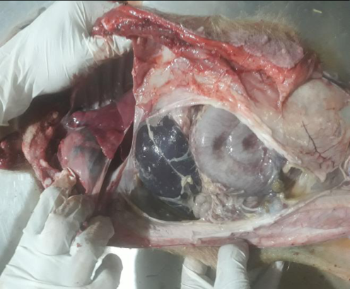 Figure 1
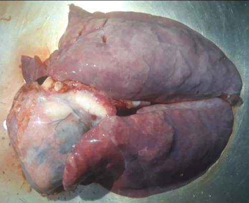 Figure 2
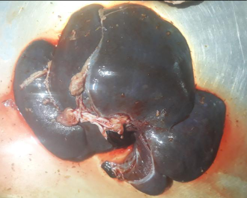 Figure 4
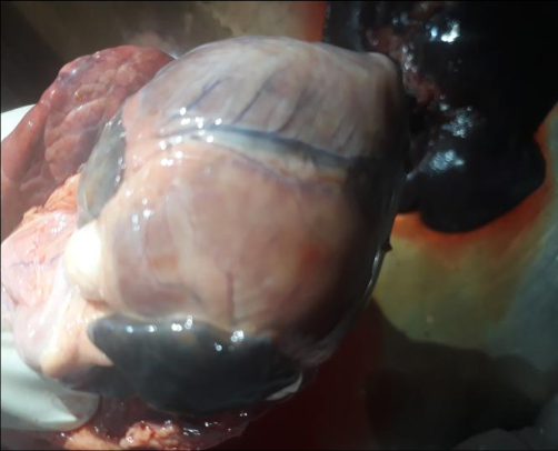 Figure 5
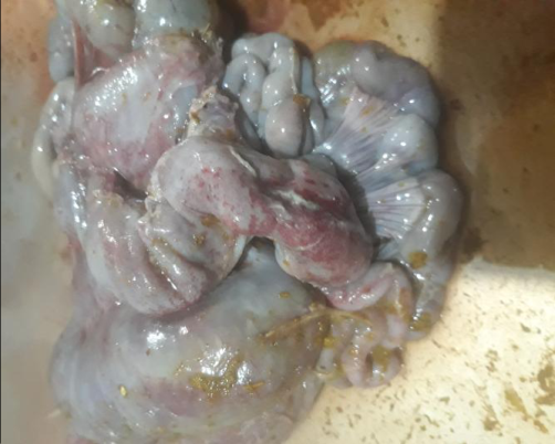 Figure 6
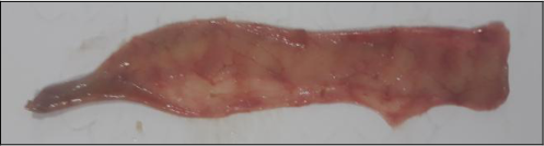 Figure 3
Introduction
Glasser's disease or porcine polyserositis is an infectious disease of swine characterized by polyserositis, arthritis, meningoencephalitis and pneumonia at various degree depending upon the severely. It is caused by Haemophilus parasuis, a commensal organism of upper respiratory tract of swine. Serotypes 3, 6, 7, 8, 9, and 11 are considered to be avirulent, 15 more pathogenic and 1, 4, 5, 10, 12, 13, and 14 are virulent. (Radostits, 2006) [5] . It mainly occurs between 4 weeks to 8 weeks in swine especially during stressful events like weaning, environmental stress or as a mixed infection with other microbial agents. (Co., 2016) [2] . Disease often has a sudden onset with high mortality and morbidity but chronic form of the disease is also prevalent. (Chakrabarti, 2012) [1] Case description A 3 month old piglet breed "Chwanche" (local) from Livestoct farm of Paklihawa Campus was presented to Veterinary Teaching Hospital, Institute of Agriculture and Animal science, Paklihawa with severe constipation, no fecal discharge since past 3 days, dry snout, large cyanotic patch on the lateral part of the left abdomen, slight dyspnea and swollen abdominal cavity, slight incoordination, mouth breathing, coughing and anorexia. Then the piglet was admitted to Veterinary Teaching Hospital and Physical examination and clinical examination was done. Auscultation of abdomen revealed burble sound. Heart rate and respiration rate were 130/min and 60/min (increased) respectively and temperature was 106°F. Based on history and Physical examination the case was tentatively diagnosed as constipation. Curative treatment in initial phase was done and further investigation was started with blood examination. Upon, blood examination PCV and Total WBC count was found to be 38% and 28×10 3 /µl respectively. Unfortunately, after 5 hours the pig was died and post mortem examination of the pig revealed Severe peritonitis with accumulation of fluid in abdominal cavity (Fig. 1), fibrinous covering around the lungs and heart (Fig. 2), ecchymotic patches on the outer surface of (Fig. 3), Swollen liver with fibrinous deposition (Fig. 4), fibrinous covering around the pericardium (Fig. 5), congested intestinal epithelium (Fig. 6).
Discussion
Pathophysiology: Glasser's disease is often very common in young pig as maternal antibody doesn't last long and may be gone by 2-4 weeks of age. (Radostits, 2006) [5] . Some case reveal, presence of antibody up to 8 weeks when sow's antibody titer is high enough. Animals become infected when maternal antibody are gone with low immune response.
Haemophilus parasuis is a commensal of upper respiratory tract of pig (Co., 2016) [2] . There are 15 serovars of H. parasuis. (Kielstein P., 1992) [3] Out of them serovar 15 is highly pathogenic (Radostits, 2006) [5] . The severity of the disease increases with the increase in microbial load. Upon infection it causes severe septicemia followed by fibrinous polyserositis, pericarditis, meningitis, polyarthritis and sometimes pneumonia. Experimental studies showed that not all field isolates are pathogenic and route of infection and dosage are most important in determining the outcome of the infection. Upon infection there is marked rise in neutrophils and reduction in CD25 + marker cells. There is variable tropism of this bacteria depending upon the serotypes. Some proliferate in the lungs and peritoneum while other in pericardium, joints, brain and kidney, which suggest that systemic sites are the best areas for the identification of the pathogenic H. parasuis. (Radostits, 2006) [5] . Some case showed mixed infection with Actinobacillus pleuropneumoniae, Actinobacillus suis, Streptococcus suis, Erysipelothrix rhusiopathiae, Mycoplasma hyorhinis, Influenzavirus which should be excluded during differential diagnosis. (Nicolet, 1992) [4] Polyserositis in this case could be due to high proliferation of the bacteria in the peritoneum. Peritonitis caused severe fibrin and fluid accumulation in the abdominal and thoracic cavity, leading to swollen abdomen and hydrothorax. Cyanosis at the lateral side of the body might be due to vasculitis of the blood vessel. Constipation which was very prominent in the pig was due to severe hemorrhage on outer part and congestion in inner surface of intestinal epithelium. Fever in this case was due to the presence of lipopolysaccharide layer in the cell wall of gram-negative causative bacteria which entered the blood and facilitated the release of prostaglandin E2 leading to fever. High WBC count in this case is due to increased immune response of body against the pathogenic bacteria. There were no other marked symptoms in this case, may be because of acute case.
Conclusion
Polyserositis in pig is an underdiagnosed and a new disease in the context of Nepal. As pigs are colonized with large no of commensal organisms, they are likely to encounter infection when the immune system is weak. Glasser's disease follows similar pattern and generally infects young ones between 4-8 weeks after weaning. Onset of this disease is generally acute and shows high morbidity and mortality. Survivors of the disease can develop severe fibrosis in the thoracic and abdominal cavity leading to reduced growth and meat condemnation at slaughter. As the disease mostly depends on individual's immune system it is less likely to spread over a herd at a short period of time. Although, proper care and nutrition after weaning can prevent the disease.
References
5 in the original paper- Chakrabarti A. A Textbook of Preventive Veterinary Medicine. New Delhi: Kalyani publishers, 2012.
- Co M. The Merck Veterinary Manual. New Jersey: Merck & Co, 2016.
- Kielstein P, RG V. Designation of 15 serovars of Haemophilus parasuis based immunodifusion using heatstable antigen extracts. Journal of Clinical Microbiology. 1992; 30:862-865.
- Nicolet J. Haemophilus parasuis. In Diseases of swine (7th ed., pp. 526-528). Iowa State University, 1992.
- Radostits MO, Gay CC, Hinchcliff KW, Constable PD. A textbook of the diseases of cattle, horses, sheep, pigs and goats. London: Saunders Ltd, 2006.
Cited by 1
From OpenAlex, OpenCitations and reference lists read by Scinovex
- Bacterial and fungal infections of pig and their management ↗
Vivek Joshi, Ajay Kumar Yadav, Mayur M. Jadav, Deepsikha Deepsikha, Sanjana, Umesh Dimri · Elsevier eBooks · 2025
❝ 0