
Research article · 2019 · Full text
International Journal of Veterinary Sciences and Animal HusbandryThe effect of tetracycline on the development and hatchability of Fasciola gigantica eggs following variable periods of incubation
Volume 4(3) · Pages 22–24
Research areasHelminth infection and controlCoccidia and coccidiosis researchAgricultural safety and regulations
Abstract
Author-writtenLiver fluke, Fasciola gigantica is a digenetic trematode belonging to the family fasciolidae. Found in the liver of ruminants and pigs, it causes the disease Fascioliasis. Adult flukes expel eggs in hosts' faeces. In this study, parasite eggs were extracted from gall bladders of Fascioliasis infested cattle, washed with distilled water, and viewed under microscope to confirm presence of eggs. Five milliliters of the egg sediment was dispensed into 14 labeled test tubes. Into three of the tubes was dispensed 1.5mls of pen-streptomycin, while 1.5mls of 2500ug/ml dilution of tetracycline was dispensed into the rest. All the tubes were incubated in dark cupboard at room temperatures (27-31˚C). Samples from different test tubes were observed at days 5,10,15,20 and 21. Samples incubated with tetracycline showed no evidence of zygote development while those incubated with pen-streptomycin did, with some eggs hatching and releasing miracidia.
Summary
Source-checkedPlain-language reading of the full paper, prepared with AI and checked against the text. Each point links to the authors' own words.
Why it matters
- Fasciola gigantica is a liver fluke that causes Fascioliasis in various animals, including cattle, sheep, goats, and pigs. View evidence ↗
- The parasite is economically significant because it negatively impacts the productivity and growth rates of ruminants. View evidence ↗
- In Nigerian research, penicillin-streptomycin is typically used as the antibiotic during the incubation of parasite eggs to allow them to hatch into miracidia. View evidence ↗
The question
- The study aimed to determine how adding tetracycline as an antibiotic affects the development of zygotes and the ability of F. gigantica eggs to hatch and release miracidia over different incubation periods. View evidence ↗
What they did
- The experimental setup involved dispensing 5ml of egg sediment into 14 labeled test tubes. View evidence ↗
- Test tubes 1-3 received 1.5ml of penicillin-streptomycin, while tubes 4-6 and 7A,B-10A,B received 1.5ml of tetracycline at the minimum inhibitory concentration. View evidence ↗
- All samples were incubated in dark cupboards at room temperatures ranging from 27-31°C (average 29°C). View evidence ↗
- Zygote development was monitored via stereomicroscope at 5, 10, 15, and 20 days for some tubes, and at 21 days for the remaining tubes. View evidence ↗
What they found
- Eggs incubated with tetracycline showed no zygote development at the 5, 10, 15, and 20-day observation points. View evidence ↗
- At 21 days of incubation, eggs treated with penicillin-streptomycin successfully hatched, with miracidia observed under the microscope. View evidence ↗
What it means
- The authors concluded that tetracyclines prevent the hatching of F. gigantica eggs regardless of the incubation period. View evidence ↗
- The failure to hatch is attributed to tetracycline's ability to enter the eggs and inhibit embryo development by interfering with protein synthesis. View evidence ↗
- The authors recommend further research to determine how this inhibitory effect can be used for therapeutic benefits. View evidence ↗
Visual summary
1 visualsIllustrations explain the paper; they do not show study data.
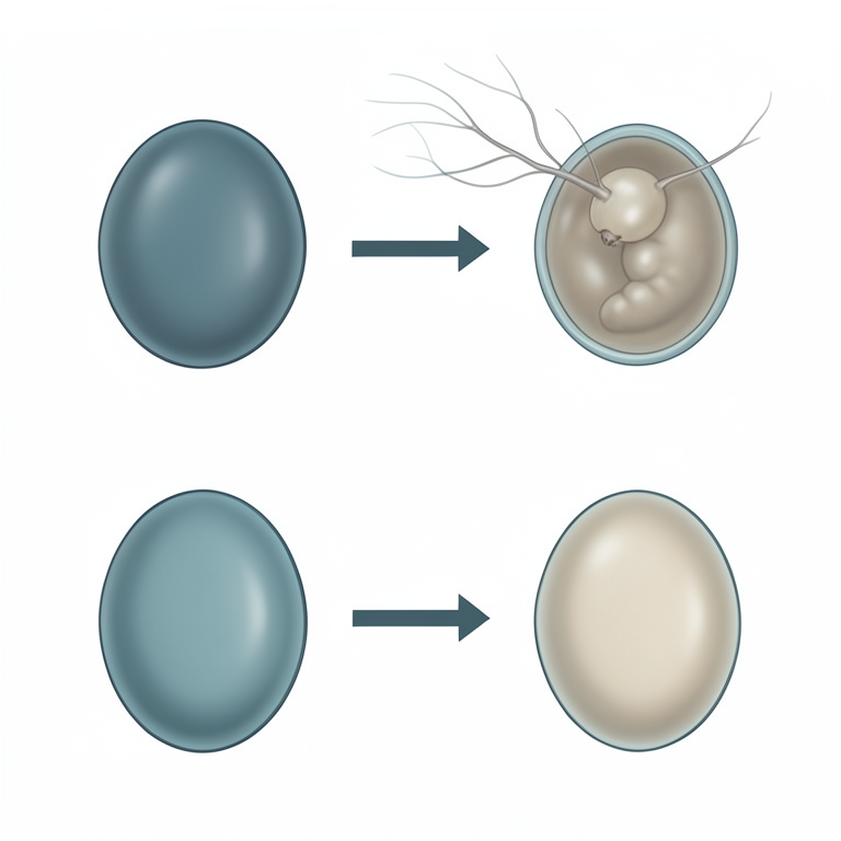
Comparison of Egg Hatching
AI-generated conceptual illustration (Fasciola gigantica eggs and miracidia). It explains the idea and does not show study data.
View source evidence ↗Figures & tables
From the original paper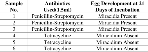 Table 2
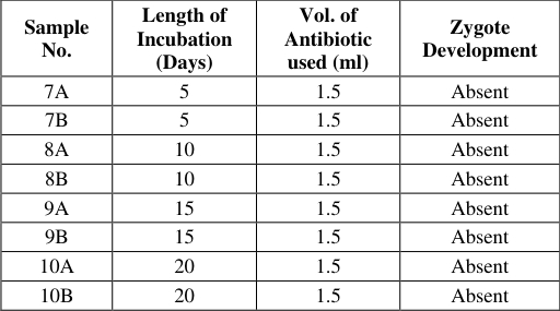 Table 1
Introduction
The liver fluke, Fasciola gigantica is a digenetic trematode that belongs to the family fasciolidae. It is found in the liver of cattle, sheep, goats, and pigs, where it causes the disease Fascioliasis. Infestation with the parasite has profound effect on growth rate and productivity of ruminants, making it economically significant [1] . The local breeds of cattle have been found to have lower prevalence rates and fluke burdens than exotic breeds and crosses of local and exotic breeds [2] . F. gigantica is predominant in the tropical regions of the world [3] . The snail intermediate host for the parasite in Nigeria is Lymnaea natalensis, an aquatic snail and the only lymnaeid snail encountered here [4] . Apart from this species, there are other lymnaeid snails (intermediate hosts), such as Lymnaea viridis, L. columella, L. cousin, L. ollula, and L. auricularia rubiginosa [5] . The adult flukes expel eggs through the feces of the definitive host. Miracidia are hatched from the eggs after 16-20 days [6] , from where they penetrate into the lymnaeid snails and then develop into the sporocysts, rediae, and cercariae. Cercariae separate from the snails and are encysted to become metacercariae on vegetation (infective stage). The metacercariae can then be found on vegetations [7] . These vegetations are important sources of fodder for the definitive hosts. The life cycle gets completed when the definitive host ingests vegetation containing the metacercariae. In studies involving the parasite in Nigeria, the eggs are mainly incubated with penicillinstreptomycin combination as antibiotic, they would hatch releasing miracidia at the end of the incubation period. The aim of the present study is to incubate F.gigantica eggs with tetracycline added as antibiotic for variable number of days and determine the effect of the antibiotic on zygote development and eggs hatchability to release miracidia.
Materials and Methods
Materials and Methods
Sample Collection and Preparation
The gall bladders used for this study (five in number) were obtained from the condemned liver of cattle infested with Fascioliasis after post mortem inspection at the Bodija abattoir, Ibadan, Oyo State Nigeria. The bladders were excised from the livers, wrapped in aluminum foil and transported in a cooler packed with ice to the laboratory. Eggs from the parasite were recovered from the bile after the bladders were incised with scalpel blade. They are washed several times with distilled water in a crucible to reduce the viscosity of bile and for easy sedimentation of eggs. Drops of sediment placed on a glass slide were examined under stereomicroscope, where elliptiform large eggs operculate at one pole and yellowish in color were observed. Into 14 test tubes labeled 1-6 and 7 A,B -10 A,B were dispensed 5mls of the sediment each.
Preparation of Tetracycline Dilutions
Two capsules of tetracycline (250mg) were used in the study. The 500mg of tetracycline was added to 10ml of distilled water and shaken to dissolve. One milliliter (1ml) of this solution was added to 9 ml of distilled water to give 5000ug/ml and then 5ml of this solution was added to 5ml of distilled water to give a concentration of 2500ug/ml regarded as the minimum inhibitory concentration.
Egg Incubation and Examination
One and half milliliter (1.5 ml) of penicillin-streptomycin antibiotic was added to test tubes labeled 1-3, while equivalent volume (1.5 ml) of the minimum inhibitory concentration of tetracycline was added to the ones labeled 4-6 and 7 A,B -10 A,B . All the test tubes were partially covered with cotton wool to allow for aeration. The 14 test tubes were placed on test tube rackets and incubated in dark cupboards at room temperature (27-31˚C; av. 29˚C). The first three test tubes (labeled 1-3) containing pen-streptomycin and the test tubes labeled 4-6 incubated with tetracycline were placed close to each other in the dark cupboard for 21 days. The remaining test tubes labeled 7 A,B -10 A,B were also placed in the same dark cupboard. Egg samples were taken from test tubes (7 A,B -10 A,B ) and observed at variable periods of 5, 10, 15 and 20 days respectively for zygote development under a stereomicroscope. Eggs from test tubes labeled 1-3 and 4-6 were examined at day 21 of incubation.
Results
The egg samples taken from test tubes 7A and 7B at day 5 of incubation, showed no zygote development when observed under the microscope after exposure to light. The same was observed for test tubes 8 (A and B) and 9 (A and B) at days 10 and 15 of incubation respectively. Eggs observed from test tubes 10 (A and B) also showed no development at day 20 (table.1). Egg samples taken from test tubes labeled 1-3 incubated with pen-streptomycin were examined under microscope at 21 day of incubation after exposure to light. Miracidia were observed, some still attached to the eggs. However, samples of egg taken from test tubes labeled 4-6 incubated with tetracycline showed no zygote development when examined under microscope (Table .2).
Discussion
The present study showed that eggs of F. gigantica incubated in test tubes in to which tetracycline was added as antibiotic in a dark cupboard at room temperatures, did not hatch to release miracidia on exposure to light after 21 days. When examined under a stereomicroscope no zygote development was observed. The eggs incubated in test tubes examined at days 5, 10, 15 and 20 also did not show any evidence of zygote development.
The phenomenon of failure of eggs to develop and hatch when incubated in the presence of tetracycline relates to its mode of action. This is consistent with the studies of [8.9] which showed that tetracyclines inhibit protein syntheses in the mitochondria due to their interactions with the ribosomal subunits in these organelles. Since protein synthesis is of key importance in the development of F. gigantica zygotes, these zygotes are susceptible to tetracycline inhibitory activities.
Reports from [10,11] showed that tetracycline is broadspectrum, bacteriostatic and effective against a wide range of microbes including bacteria, protozoa and helminthes. It has been reported that mitochondria are organelles that are derived from free-living proteobacteria acquired by eukaryotic cells via endosymbiosis [12] . Accordingly, the eukaryotic mitochondria possess ribosomes that are similar to those found in bacteria. Consequent to these ribosomal similarities, it is believed that the anti-protozoal activity of tetracycline is achieved through its interactions with the mitochondrial ribosomes of these parasites in manners comparable to the interactions with bacterial ribosomes [13] . Meanwhile, it has been established that two processes appears to be required for these antibiotics to gain access to the ribosomes; passive diffusion through hydrophilic pores and energy dependent active system [14] . The anti-eukaryotic activity of tetracyclines has been linked to their weak inhibition of protein synthesis at the 80S ribosomal sub units [15] . Despite the fact that different studies reported same interaction sites for tetracyclines, [16] showed that the apparent sites for drug interaction in the ribosome may not necessarily reflect the actual binding site. Following their binding, tetracyclines prevent accesses of amino-acryl tRNA (transfer ribonucleic acid) to the acceptor site on the mRNA (messenger ribonucleic acid). This prevents the addition of amino acid to the growing peptide chain providing an explanation for the bacteriostatic effects of these antibiotics as reported by [17] . Furthermore, tetracyclines are known to have acidic PH in aqueous solution, and may therefore have the ability to prevent the hatching of the eggs in vitro [18] . Conversely, the eggs incubated with penicillin streptomycin combination for 21 days hatched. Miracidia were observed on examination under microscope, the mechanisms of action of penicillin and streptomycin being different from those of tetracyclines.
Conclusion and recommendation
This study showed that tetracyclines prevent the hatching of F. gigantica eggs incubated with them for variable number of days. Eggs examined under microscope after 21 days of incubation did not show zygote development and did not hatch releasing miracidia. The inability of eggs to hatch is attributed to the ability of tetracyclines to gain access in to the eggs and inhibit embryo development by interfering with protein synthesis mechanism. It is recommended that further studies be carried out to ascertain ways of harnessing therapeutic benefits from this phenomenon.
Acknowledgement
AcknowledgmentsThe authors would like to thank the laboratory staff of the Department of Veterinary Parasitology, University of Ibadan who assisted and provided some of the facilities used for the study.
Figures and tables from the paper
References
18 in the original paper- Kuchai JA, Chishti MZ, Zaki MM. Some epidemiological aspects of fascioliasis among cattle of Ladakh. Global Vet. 2011; 7(4):342-346.
- Castelino JP, Preston JM. The Influence of breeds and age on the prevalence of Bovine Fascioliasis in Kenya. British Vet. J. 1979; 135(2):198-203.
- Mas-Coma S, Barques MD, Valero MA. Fascioliasis and other plant-borne trematode zoonoses. Int J Parasitol. 2005; 35(11-12):1255-1278.
- Ukoli FMA, Asumu DI. Fresh Water Snails of the Proposed Federal Capital Territory in Nigeria. J Nat. Sci. 1979; 1(1):49-58.
- Lee CG, Cho SH, Lee CY. Metacercarial production of Lymnaea viridis experimentally infected with Fasciola hepatica. Vet Parasitol. 1995; 58(4):313-318.
- Hussein AA, Hassan IM, Khalifa RMA. Development and hatching mechanism of Fasciola eggs, light and scanning electron microscopic studies. Saudi J Biol Sci. 2010; 17(3):247-251.
- Yoshihara S, Ueno H. Ingestion of Fasciola gigantica metacercariae by the intermediate host snail, Lymnaea ollula, and infectivity of discharged metacercariae. Southeast Asian J Trop Med Public Health. 2004; 35(3):535-539.
- Chopra I, Roberts M. Tetracycline antibiotics: mode of action, applications, molecular biology, and epidemiology of bacterial resistance. Microbiol Mol Biol Rev. 2001; 65(2):232-260.
- Gale EF, Cundliffe E, Reynolds PE, Richmond MH, Waring MJ. The molecular basis of antibiotic action, 2nd ed. John Wiley & Sons, Inc, New York, 1981.
- Chopra I, Hawkey PM, Hinton M. Tetracyclines, molecular and clinical aspects. J Antimicrob Chemother. 1992; 29(3):245-277.
- Hoerauf A. Filariasis: new drugs and new opportunities for lymphatic filariasis and onchocerciasis. Curr Opin Infect Dis. 2008; 21(6):673-681.
- Edlind TD. Protein synthesis as a target for antiprotozoal drugs. In Coombs G, North M, (eds), Biochemical protozoology, 1991, 569-586.
- Katiyar SK, Edlind TD. Enhanced antiparasitic activity of lipophilic tetracyclines: role of uptake. Antimicrob Agents Chemother. 1991; 35(11):2198-2202.
- Chopra I. Howe JGB. Bacterial resistance to tetracycline. Microbiol. Rev. 1978; 42(4):707-724.
- Van den Bogert C, Kroon A M. Tissue distribution and effects on mitochondrial protein synthesis of tetracyclines after prolonged continuous intravenous administration to rats. Biochem Pharmacol. 1981; 30:1706-1709.
- Schnappinger D, Hillen W. Tetracyclines: antibiotic action, uptake, and resistance mechanisms. Arch Microbiol. 1996; 165(6):359-369.
- Chopra I, Hawkey PM, Hinton M. Tetracyclines, molecular and clinical aspects. J Antimicrob Chemother. 1992; 29(3):245-277.
- Yakhchali M and Bahramnejad. Inhibition effect of pH on the hatchability of Fasciola Miracidia under Laboratory Conditions. Iran journal of Parasitology. 2016 11(1):30-34.