
Research article · 2019 · Full text
International Journal of Veterinary Sciences and Animal HusbandryManagement of Dystocia Due to Fetal Ascites in Boer Goat: A Case Report
Volume 4(2) · Pages 10–11
Research areasReproductive Physiology in LivestockAnimal health and immunology
Abstract
Author-writtenDystocia in goats is due to fetal causes is uncommon. However, there are reports suggesting dystocia due to fetal dropsy. Present case report show dystocia in Boer goat due to fetal ascites. This case report is about the successful management of dystocia due to fetal ascites in Boer goat by giving an incision on the hind limb and making an entry into the fetal abdomen to take out the fluid. Evisceration was done for completely removing the fetus.
Summary
Source-checkedPlain-language reading of the full paper, prepared with AI and checked against the text. Each point links to the authors' own words.
Why it matters
- Fetal ascites can cause dystocia across various species, although it is a rare occurrence in does. View evidence ↗
- In cows, dystocia may be caused by peritoneal dropsy, which often accompanies fetal infectious diseases. View evidence ↗
The question
- The authors aim to report a rare case of dystocia in a Boer goat caused by fetal ascites. View evidence ↗
What they did
- The subject was a 3-year-old Boer goat in her second parity who had completed full gestation but failed to deliver the fetus since 6 A.M. View evidence ↗
- Examination revealed the fetus was in a dorso-sacral position and posterior longitudinal presentation, with both hind limbs protruding from the vulva. View evidence ↗
- The diagnosis of fetal ascites was made after fetal ballottement revealed a fluid-filled abdomen. View evidence ↗
- Prior to the procedure, the doe was treated with antihistamines, analgesics, and dexamethasone. View evidence ↗
- The surgical approach involved making an incision lateral to the fetal hock joint and entering the abdominal cavity via the medial side of the thigh. View evidence ↗
What they found
- The procedure resulted in the sudden release of approximately 4 to 5 liters of straw-colored fluid from the fetal abdomen. View evidence ↗
- Because traction alone failed to deliver the fetus after fluid removal, evisceration was performed to further reduce size, leading to successful delivery. View evidence ↗
- The use of evisceration helped complete the delivery process in approximately 10 minutes. View evidence ↗
What it means
- The authors conclude that subcutaneous abdominal entry prevents injury to the female genitalia, while evisceration facilitates delivery by reducing fetal size. View evidence ↗
- The authors suggest that subcutaneous fetotomy may preserve future fertility better than a cesarean section. View evidence ↗
- This approach is recommended as a helpful alternative for farmers who cannot afford cesarean sections or are concerned about post-surgical fertility. View evidence ↗
Visual summary
1 visualsIllustrations explain the paper; they do not show study data.
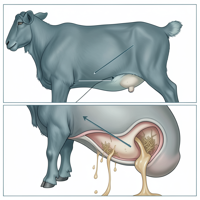
Subcutaneous Abdominal Entry
AI-generated conceptual illustration (Boer goat fetus). It explains the idea and does not show study data.
View source evidence ↗Introduction
Foetal ascites is seen as a cause of dystocia in many species but occurs rarely in does. Dropsy of the peritoneum is a common accompaniment of infectious disease of the fetus and may cause dystocia in cows (Arthur et al., 1986) [2]. A case of dystocia due to fetal ascites in a Boer goat is reported which is a very rare event. Case history and observation A 3 year Boer goat in second parity completed full gestation period was presented at Teaching Veterinary Clinical Complex, Nagpur Veterinary College Nagpur during noon hours for not delivering fetus since today morning 6 A.M.
The water bag has ruptured but second stage was not progressing further. All the vital signs were normal and straining continuously. Traction on both the hind limbs was not able to deliver the fetus by the owner itself. On external examination, swollen fetal hind limbs were presented in the hanging position from the vulva.
Before the per-vaginal examination, the external genitalia is cleaned with a weak potassium permanganate solution. Lubrication of sleeved hand is done with sweet oil and careful per- vaginal examination was done. The cervix was dilated completely and the obstetrician was able to pass his hand up to fetal abdomen. Slight traction was applied on hind limbs and the fluid ripples moved towards the anterior side, thus making a lock while traction.
The fetus was in posterior longitudinal presentation, dorso-sacral position with both hind limbs was hanging from the vulva. Upon fetal ballottement, the fluid feeling of the abdomen was noticed. Thus the diagnosis was made that dystocia was due to the fetal origin and was due to fetal ascites. Thus making fetus relatively oversized.
Treatment Before handling, the doe was administered with dexamethasone, analgesics and antihistamines. An incision was given on the hind limb of fetus lateral to the hock joint and subcutaneous tissue was separated. The obstetrician’s hand entered into the abdominal cavity from the medial side of the thigh. Suddenly almost 4 to 5 liters of straw-colored fluid was released out as a burst.
On applying traction, the fetus was still not coming out so evisceration was done for reducing the size further. After properly lubricating the fetus’s dorsal surface, the fetus was delivered successfully. Post obstetrical treatment was done with Inj. Ringer’s lactate 250 ml I/v, Inj.
Dextrose 25% 250 ml I/v, Inj. Calcium Borogluconate @ 1ml/kg BW I/v, Inj. Ceftriaxone @ 15 mg/kg I/v, Inj. Melonex @ 0.5 mg/kg I/m. the owner was advised to continue the same treatment for 3 days.
VET 2019; 4(2): 10-11 Abhishek Bhardwaj M.V.Sc. Scholar, Department of Animal Reproduction, Gynaecology and Obstetrics Nagpur Veterinary College Maharashtra Animal and Fishery Sciences University, Maharashtra, India Sheetal SK Assistant Professor, Department of Animal Reproduction, Gynaecology and Obstetrics Nagpur Veterinary College Maharashtra Animal and Fishery Sciences University, Maharashtra, India Sahatpure SK Professor and HOD, Department of Animal Reproduction, Gynaecology and Obstetrics Nagpur Veterinary College Maharashtra Animal and Fishery Sciences University, Maharashtra, India Bawaskar MS Assistant Professor, Department of Animal Reproduction, Gynaecology and Obstetrics Nagpur Veterinary College Maharashtra Animal and Fishery Sciences University, Maharashtra, India Correspondence Abhishek Bhardwaj M.V.Sc. Scholar, Department of Animal Reproduction, Gynaecology and Obstetrics Nagpur Veterinary College Maharashtra Animal and Fishery Sciences University, Maharashtra, India Ahuja 2017 [1] have found the same in cows and buffaloes. In this particular case, evisceration was also done which helped the process to be completed almost in 10 minutes.
The poor farmers cannot bear the cost of the cessarean section and post cessarean fertility is also questionable. Thus this approach is very helpful from the farmer’s point of view.
Discussion
It may be caused either by the overproduction or insufficient drainage of peritoneal fluid. Obstruction of the lymphatics, for various reasons, may prevent the disposal of peritoneal fluid (Sloss and Duffy, 1980) [6]. S. Prakash 2017 [4] noticed the ascites case in the posterior presentation in a non-descript doe.
M. Honparkhe 2003 [3], S. K. Sheetal 2017 [5], Ankit Kumar
Conclusion
The subcutaneous entry into abdomen save the female genitalia from the injuries and evisceration helps in reducing the size of the fetus for easy delivery. Subcutaneous fetotomy can save the future fertility which is sometimes affected in cesarean section.
References
6 in the original paper- Ankit Kumar Ahuja, Pooja Dogra, Shivkumar, Shahbaz Singh Dhindsa, Harpreet Singh. A case of Dystocia due to Fetal Ascites in Murrah Buffalo International Journal of Environment, Agriculture and Biotechnology. 2017; 2(4) ISSN: 2456-1878.
- Arthur GH, Noakes DE, Pearson H, Parkinson TJ. Veterinary Reproduction and obstetrics. 7th ed., W.B. Saunders Co, Ltd, Philadelphia, 1996, 302-307.
- Hoparkhe M, Kumar A, Gandotra VK. Dystocia due to accumulation of fluid in peritoneal cavity and intestines of fetus in a cross breed cow, Indian Journal of Animal Reproduction. 2003; 24(1):83-84.
- Prakash S, Selvaraju M, Ravikumar K, Palanisamy M, Manokaran S. Dystocia Due to Fetal Ascites with Brachynathism in a Doe Indian Vet J. 2017; 94(02):66-67
- Sheetal SK, Shiv Prasad, Gupta HP. Dystocia due to foetal ascites in a murrah buffalo Buffalo Bulletin 2017; 36(3):573-575
- Sloss V, Duffy JH. Handbook of Bovine Obstetrics. Williams and Wilkins, Baltimore, USA, 1980, 121.
Cited by 1
From OpenAlex, OpenCitations and reference lists read by Scinovex
- A rare case of fetal ascites in goat and it’s surgical management ↗
Abhinav Pratap Singh, Narayan Prasad, Avaneesh Kumar Singh, Jitendra Agrawal, Atul Saxena · Journal of Entomology and Zoology Studies · 2020