
Research article · 2018 · Full text
International Journal of Veterinary Sciences and Animal HusbandryAn In-Vitro antibacterial effect of Momordica foetida and Croton macrostachyus on Streptococcus agalactiae Isolated from bovine mastitis
Volume 3(6) · Pages 19–23
Research areasBiochemical and Structural CharacterizationTransgenic Plants and ApplicationsToxin Mechanisms and Immunotoxins
Abstract
Author-writtenTwo plants, Momordica foetida and Croton macrostachyus were collected from South Nation Nationality of People Region from Kembata Tembaro Zone specially Durame area. In-vitro antimicrobial sensitivity tests of both plants were conducted in Addis Ababa University College of Veterinary Medicine and Agriculture, Debre Zeit, from October 2016 to April 2017. The study was carried out with the objective of determining and comparing of the in-vitro antimicrobial effects of root of Momordica foetida and bark of Croton macrostachyus on three isolates of Streptococcus agalactiae coming from bovine mastitis cases. In this study, the 80% methanol crude extract preparation of root of Momordica foetida and bark of Croton macrostachyus had good antibacterial activity against the tested bacteria. The extracts of the plants inhibited the growth of Streptococcus agalactiae at all concentrations (0.625%, 1.25%, 2.5%, 5%, and 10%). In all cases, there was a dose dependent inhibition on the tested bacteria. Conventional antibiotic disc and DMSO (Dimethyl sulfoxide) impregnated discs were used as positive and negative controls, respectively and no inhibition was observed by DMSO which substantiates that the inhibition observed was exclusively by crude extracts. This finding suggests that there is a promise in combating drug resistant pathogens.
Summary
Source-checkedPlain-language reading of the full paper, prepared with AI and checked against the text. Each point links to the authors' own words.
Why it matters
- Bovine mastitis is a significant constraint in dairy cows, leading to reductions in both the quality and quantity of milk and milk products. View evidence ↗
- In developing countries like Ethiopia, the limited variety of conventional mastitis drugs and the development of antimicrobial resistance make the search for local, low-cost indigenous resources important. View evidence ↗
- Natural products are critical to pharmaceutical development, with over 50% of modern clinical drugs originating from natural sources. View evidence ↗
The question
- The study aimed to determine and compare the in-vitro antimicrobial effects of Momordica foetida root and Croton macrostachyus bark on three isolates of Streptococcus agalactiae from bovine mastitis. View evidence ↗
What they did
- Plants were collected from the Durame area in the Southern Nations Nationalities and Peoples Region of Ethiopia between October and November 2016. View evidence ↗
- Crude extracts were prepared by macerating 25 grams of each plant material in 80% methanol for seven days at room temperature. View evidence ↗
- The researchers prepared five serial dilutions of the extracts (10%, 5%, 2.5%, 1.25%, and 0.625%) using dimethyl sulfoxide (DMSO). View evidence ↗
- The agar disc diffusion method was used to test the extracts, with conventional antibiotic discs as references and DMSO-impregnated discs as negative controls. View evidence ↗
What they found
- Both plant extracts inhibited the growth of Streptococcus agalactiae at all tested concentrations (0.625% to 10%), showing a dose-dependent effect. View evidence ↗
- Momordica foetida produced larger mean inhibition zones at 10% and 5% concentrations, whereas Croton macrostachyus was more effective at 2.5%, 1.25%, and 0.625%. View evidence ↗
- At a 10% concentration, the inhibition zones of the plant extracts (20 mm for M. foetida and 18.33 mm for C. macrostachyus) were comparable to Gentamycin (20 mm) and superior to Tetracycline, which showed no inhibition. View evidence ↗
- The negative control (DMSO) showed no inhibition, confirming that the antibacterial activity was caused by the plant extracts themselves. View evidence ↗
What it means
- The authors conclude that both Momordica foetida root and Croton macrostachyus bark extracts have very good antimicrobial effects against S. agalactiae across all tested concentrations. View evidence ↗
- The results suggest these plants have potential for the synthesis of new phyto-remedies to treat microbial infections and combat drug resistance. View evidence ↗
- The authors state that future work should evaluate the toxicity of these plants to determine their safety margins. View evidence ↗
Figures & tables
From the original paper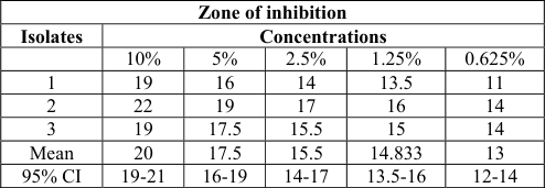 Table 2
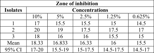 Table 2
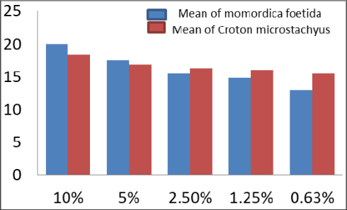 Figure 1
 Table 3
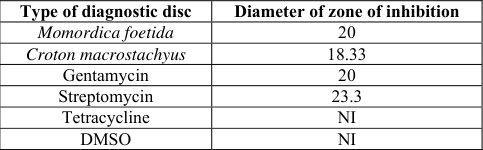 Table 4
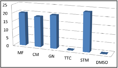 Figure 2
Introduction
People generally depend on nearby forest areas to supply their needs such as medicine, timber, fuel wood, wild vegetables and many more [1] . Nature has a source of medicinal plants for thousands of years and an impressive number of modern drugs have been isolated from natural sources, mainly based on their use in traditional medicine. It is reported that over 50% of all modern clinical drugs are natural product origin and natural plant play an important role in drug development in the pharmaceutical industry [2] . The plants that possess therapeutic properties or exert beneficial pharmacological effect on the animal body are generally designated as medicinal plants [3] . The use of higher plants and their extracts to treat infections is an ancient practice in traditional medicine. Humans have been using natural products of animals, plants and microbial source for thousands of years either in pure form or crude extracts [4] . Now days, the indigenous knowledge on medicinal plants is gaining worldwide recognition. The world health organization has estimated that more than 80% the world's population in developing countries depend primarily on herbal medicine for basic health care needs [5] . Africa is abundantly rich in herbal medicinal practice. For example, in Ethiopia, Madagascar and Tanzania practical steps are already begin to take to ascertain efficacy and determine optimum dosage for several herbal drugs. As a matter of fact, herbal medicinal care continues to remain the only type of health care for nearly 80% of people and animals in Ethiopia while the remaining 20% swing between the modern and the traditional system of health care [6] . Bovine mastitis is one of animal diseases that have been treated by using traditional medicinal plants. It is incriminated as important disease constraints in dairy cow and is responsible for reduction in quality and quantity milk and milk products [7] . Majority of organisms that are responsible for mastitis and spoilage of milk could be Staphylococcus aureus, Streptococcus agalactiae, coli forms (Eshershia coli, klebisella and Enterobacter aerogeneos), Serratia, Pseudomonas and Proteus species [8] . In Ethiopia, traditional healers use a number of plants or herbs for the treatment of bovine mastitis and efficacy of some these plants have been tested on a range of causative agents of mastitis.
In-vitro study conducted by Sahle [9] indicated that Persicaria senegalis, Cyphostemma adenocaule and Cucmis ficifolius, have shown some degree of growth inhibitory effect. Markos [10] has screened six herbal preparations against mastitis causing pathogen. Mengistu [11] has screend some herbal preparations; namely Brucea antidysenterica, combertum molle, Cyphostemma adencuale, Periscara senegales, plantogalnceoleta and zhneriscabraon major isolate of bovie mastitis. Tadesse [12] has conducted in-vitro test of combertum molle on staphylococcus aureus isolate and also Mohammedamin [13] has conducted in-vitro test of laggera alata and xanthium stumariumon staphylococcus aureus isolate and observed encouraging result. The conventional drugs used for treatment of mastitis are limited in types in developing countries in general and in Ethiopia in particular. Due to this and other factors the causative agents were also reported to have developed variable degrees of resistance to the commonly used antimicrobial agents. It is therefore important to design and follow strategies that could help in the utilization and development of locally available low cost indigenous resource [14] . Therefore, the main objectives of the present study were: To determine and compare the in-vitro antimicrobial effect of two phytopreparations: namely Momoredica foetida and Croton macrostachyus on streptococcus agalactia isolated from bovine mastitis.
Materials and Methods
Materials and Methods
Study area
Plant collection was carried out in Kedida Gamela Woreda, Durame town, Kembata Tembaro zone in Southern Nations Nationalities and Peoples Region of Ethiopia from October to November 2016. Durame is located 370 km South to Addis Ababa with 07 o N' North latitude and 38 o 00' East longitude. The minimum and maximum average temperature of the area is 18-24 0 C; and the average rain fall being 1200-1300mm. The climate of the area seems favorable to crop, vegetable and livestock production [10] .
Study Design
An experimental study was conducted on in-vitro antimicrobial efficiencies on selected plants from November 2016 to May 2017 in AAU, College of Veterinary Medicine and Agriculture, Bishoftu.
2.2.1
Plant materials or herbal material used for the study 1. Momordica foetida: is a perennial climbing native of tropical Africa, closely related to the bitter melon (M. charantia) and balsam apple (M. balsamina). Its species name refers to unpleasant smell. The plant grows in forest edges and similar habitats (including disturbed and cultivated land), woodland, and wooded grassland. Its leaves are wrinkled, heart-shaped with irregular edges, up to 18 cm wide. The flowers are yellow to yellow-orange. The fruit is a spheroid, 3.5-7.5 cm long and 2.5-5 cm wide, bright orange and covered with soft spines. When fully ripe it splits from the bottom into three valves, exposing a cluster of black seeds individually covered by a bright red, sticky, sweet pulp. The plant has perennial tuberous roots.
Croton macrostachyus
Is commonly known as rush foil or broad-leaved Croton. It belongs to the Euphorbiaceae, a very large family with 300 genera and 8,000 to 10,000 species, and is the most numerous in the tropics. The genus contains over 1,200 species, which are distributed throughout the world. The most common Croton species is Croton macrostachyus which is a deciduous tree 3-25 m high, although more commonly 6-12 m high. Many parts of this tree have various medicinal value. Bark from the stems and roots are boiled in water and newly born babies are bathed in the mixture as a remedy for skin rash. Roots are used as an anthelminthic for tapeworm, for malaria, venereal diseases, as antidiabetic, and the seeds are widely used for constipation and as antihelminthic. It has active ingredient used as facial-rejuvenating chemical peels when used in a phenol-based solution since it has a caustic exfoliating effect on the dermal layer of the skin [15] .
Bacterial organism used for the test
One bacterial species, Streptococcus agalactiae isolated from bovine mastitis cases from Veterinary Teaching Hospital of Addis Ababa University, College of Veterinary Medicine and Agriculture; Bishoftu used in testing.
Study methodology 2.3.1 Plant collection
Both plants were collected from Durame area and after collection the plants were washed with tape water to remove unnecessary particles, dried under shade and grounded by grinding machine in National Veterinary Institute (NVI). The grounded material was then sieved and weighed before maceration.
Preparation of crude extract
Preparation of crude extraction of 25 gm of each of the 2 plant materials was macerated separately with 80% of methanol in large bottle flask for about 7 days at room temperature. Each extract filtered by watt man's filter paper. Then methanol was evaporated from the filter paper by evaporation using rota vapour at 60 0 C (heating bath), 150 torr (vacuum pump) and 100rpm leaving the plant residue. the plant residues were then taken out and put in evaporating dishes and kept in dry oven at 40 0 C. The resulting concentrated extracts were weighed and stored at 4 0 C until tested.
Preparation of antimicrobial discs from herb extracts
Five serial dilutions with different concentrations (10%, 5%, 2.5%, 1.25% and 0.625%) of each plant extract were prepared using dimethyl sulfoxide (DMSO) as described by [16] . In the first test tube, 2ml of DMSO was added and each of the remaining four was filled with 1ml DMSO. 1ml of 10% solution from the first tube was transferred to a second test tube to prepare 5% solution. 1ml of solution from the 5% preparation was then transferred to the third test tube to get a 2.5% concentration; and the procedure continued in this similar manner until 0.625% concentration is reached. Discs of 12mm diameter were impregnated by adding three drops from each reconstitution solution and allowed to dry at 37 0 C overnight. Dried discs were used to determine antimicrobial effect of the respective plant types.
Preparation of test bacteria a. Sample collection procedures:
Before specimen collection sterile test tube of 10ml capacity that had appropriate tight fitting caps were prepared. Milk specimen was collected from clinical cases of mastitis at AAU, CVMA Veterinary Teaching Hospital. The specimen was taken from untreated cows. During the specimen collection, the udder was cleaned by brushing off any dirt which was clinging to the skin and washing with tap water. It was washed again with clean gauze soaked in savlon, then; mammary gland was dried thoroughly with a single use swab.
Tubes were labeled to quarters of udder in such a way that front left (FL), front right (FR), hind left (HL) and hind right (HR). The caps of the tubes were carefully removed and held between the fingers in such a way that the inside of the capes were facing downward. The tubes were at stilt to prevent contamination of milk by falling particles. The test tubes contain the milk sample with appropriate tight fitting capes were transported from the clinic to the laboratory quickly. b. Microbiological culture was performed from collected milk samples: In streaking of milk on the blood agar which was supplemented with 7% sheep blood; a wire loop was sterilized on open flame of Bunsen burner and allowed to cool. A loop full of well mixed milk was streaked on the plat agar. The plates were incubated at 37 0 c for 24 hours. Presumptive identification of bacteria primary culture was made on the bases of morphological features of the colony, gram staining reaction and hemolytic characteristics. The pure colonies were again transferred to Edwards's medium to detect asculin hydrolysis. Finally, the positive bacteria were considered as Streptococcus agalactiae and this bacterium was maintained by sub culturing in every 10 days to prevent it from death.
In-vitro antibacterial activity tests of the alcoholic extracts
Agar disc diffusion method was used to test the plant extracts and antibiotic discs, the latter were used as reference and DMSO impregnated discs as a negative control. The well isolated colonies of the same morphological type taken from an agar plate culture and touched with a wire loop then transferred to the growth tube contains 4-5ml of tryptic soya broth.
This was incubated at 37 0 C until it archives 0.5 McFarland turbidity standard usually within 2-3hrs.McFarland turbidity standard was prepared by mixing 0.5 ml of 1.175% aqueous solution of barium chloride (0.048ml Bacl2. 2H20) with 9.95ml of 1% H2So4 (0.036NH2S04) [17] . Within 15 minutes after adjusting the turbidity of inoculums suspension, a sterile swab was dipped and rotated several times, pressing firmly on the inside wall of the tube above the fluid level to remove excess inoculums from the swab. Finally, the surface of Muller Hinton agar plate was inoculated by swabbing over the entire agar surface. The appropriate crude extract and DMSO impregnated discs and antibiotic discs were placed on the surface of inoculums agar plates using sterile forceps. Each disc was gently pressed down to ensure complete contact with the agar surface with 15 minutes after the discs were applied, the plates were inverted and incubated at 37 0 C. After 24 hrs of inoculation, each plate was then taken out of incubator and the diameter (mm) of zone of inhibition exhibited by each disc was measured using a millimeter caliper. The test was repeated three times for each concentration and taken as the diameter zone of inhibition.
Data analysis
Data was transferred into Microsoft excel spread sheet, and converted to SPSS version 20.0 Software. Then, descriptive statistical methods were used for data analysis and results were presented as tables and graphs.
Results
Results
Effects of crude extracts
Effects of crude extracts
Effects of 10% herbal extracts in comparison with commonly used conventional antibiotic discs
In general, the size of diameter of inhibition zones exhibited by 10% concentration of both the plant extracts was found almost comparable to those of noble antibiotics discs (figure 2). The DMSO impregnated disc hasn't showed any inhibition against the organism which implies that the inhibition observed was exclusively by the crude extracts. The diameter of both crude extract and impregnated disc are equal.
Discussion
In this study, an in-vitro antimicrobial efficacy test of bark extract and root extract from two medicinal plants, Croton macrostachyus and Momordica foetida, respectively was performed on three isolates of Streptococcus agalactiae coming from bovine mastitis cases. The results of this study indicated that methanol bark extract of Croton macrostachyus and root extract of Momordica foetida plants were effective strongly against the test bacteria.
The absolute methanol extract of Croton macrostachyus showed a good inhibitory effect on test organism. There was no work done on this plant previously, especially on Streptococcus agalactiae, the causal agent of mastitis. But on different study conducted by Karunamoorthi and Ilango [18] , it was effective larvicidal agent against Anopheles arabiensis which is the causative agent of malaria in humans. Its effectiveness was also reported on different organisms like causative agent of gonorrhea and syphilis by Mesfin [19] ; typhoid and measles by Wagate et al [20] ; promastigote and amastigote form of Leishmania aethiopica by Habtamu et al [21] ; and E. coli and Pastuerella aeuregenosa by Johani [22] . It inhibited the growth of Streptococcus agalactiae isolates at all concentrations. When compared to Momordica foetida, it showed a wider zone of inhibition especially at the concentrations of 2.5%, 1.25%, and 0.625%.In this study, a direct relationship between concentration and zone of inhibition was observed. Therefore, in all cases of the test plants with antimicrobial activity, there was a dose dependent inhibition on the tested bacteria showing greatest activity at highest concentrations of the crude extracts.
An increasing zone of inhibition with increasing concentrations of methanol extracts of root of Momerdica foetida on Streptococcus agalactiae agrees with the previous worker on this plant [10] even if there was a variation in zones of inhibition at each concentration levels. When compared to each mean inhibition zone to previous [10] , current MIZ is better. Thus, the difference and similarities of this study with previous may be due to the method of antimicrobial sensitivity test adopted, the solvent used to prepare the bacteria, extractions method, storage condition and the geographical location of the plant. The shoot of the same plant, Momerdica foetida, was applied on fungus, C. albicanus, in Kenya and it was effective with the inhibition activity of 77.78% [23] . The current result was also in agreement with the report of Boily and Van, [24] who found out that methanolic extracts of leaf of this herb were active against C. albicans. This plant has been reported in other studies for the treatment of malaria [25,26,27,28,29] .
According to the report of Wasswa and Olila from Uganda [30] , Momordica foetida has been used as a treatment of ascaris and the plant killed the organism after 36 hours with 5% concentration.
In this study, the effect observed by the root extract of Momordica foetida and Croton macrostachyus on Streptococcus agalactiae was compared. When both plants are compared, Momordica foetida was found as a better medicinal plant in the first two concentrations 10% and 5% with 19mm and 16mm diameters, respectively; but Croton macrostachyus was found to be better the last three concentrations 2.5%, 1.25% and 0.625% with diameter of 16.33mm, 16mm and 15.5mm, respectively (Table 2). Besides, the diameter of the inhibition zone of Momordica foetida in the above concentrations was 15.5mm, 14.83mm and 13mm, respectively (Table 1). This shows that there was direct relationship observed between the concentration and zone of inhibition in both of the tested plants, exhibiting the greatest activity at the highest concentration of crude extracts. Finally, 10% Phyto-preparations from both plant parts were compared with conventional antimicrobial discs. The efficacy of those preparations at the mentioned concentration was satisfactory. The mean inhibition zone of both root extract of Momordica foetida and bark of Croton macrostachyus were greater than the inhibition zone obtained from Tetracycline. But Gentamycin inhibition zone was comparable with inhibition zone of the two methanol extracts of plants.
| Zone of inhibition | — | — | — | — | — |
|---|---|---|---|---|---|
| Isolates | Concentrations | ||||
| 10% | 5% | 2.5% | 1.25% | 0.625% | |
| 1 | 17 | 15.5 | 15.5 | 15 | 14.5 |
| 2 | 20 | 19 | 17.5 | 17.5 | 17 |
| 3 | 18 | 16 | 16 | 15.5 | 15 |
| Mean | 18.33 | 16.833 | 16.33 | 16 | 15.5 |
| 95% CI | 17-20 | 15.5-19 | 15-17.5 | 14.5-17.5 | 14.5-17 |
Table text extracted from the original PDF; superscript letters are shown inline. The original table image is below.
View original table image
| Zone of inhibition | — | — | — | — | — |
|---|---|---|---|---|---|
| Isolates | Concentrations | ||||
| 10% | 5% | 2.5% | 1.25% | 0.625% | |
| 1 | 19 | 16 | 14 | 13.5 | 11 |
| 2 | 22 | 19 | 17 | 16 | 14 |
| 3 | 19 | 17.5 | 15.5 | 15 | 14 |
| Mean | 20 | 17.5 | 15.5 | 14.833 | 13 |
| 95% CI | 19-21 | 16-19 | 14-17 | 13.5-16 | 12-14 |
Table text extracted from the original PDF; superscript letters are shown inline. The original table image is below.
View original table image
Conclusion and Recommendations
In this study, the root extract of Momordica foetida and Croton macrostachyus had demonstrated very good antimicrobial effect against Streptococcus agalactiae at concentrations of 10%, 5%, 2.5%, 1.25% and 0.625%. The plant extracts would seem to justify their future potential in different synthesis of new Phyto-remedies and their use in treatment of microbial infections. One way of control of drug resistance problem is through the development of alternative drugs by screening and testing medicinal plants for their susceptible antimicrobial effects. Therefore, the following points are recommended:
These medicinal plants may have an effect against range of pathogenic microorganisms that have veterinary and public health importance, therefore, further researches should be undertaken to reveal such and other effects. In parallel to testing the efficacy of these plants for antimicrobial activities, their toxicity should also be evaluated to determine the safety margin they possess.
Acknowledgement
We forward great thanks Professor Fekadu Regassa, Head of Research and Technology transfer in Addis Ababa University, Ethiopia, for his technical and material support throughout the work.
Conflict of Interest
There is no any conflict of interest in the publication of this manuscript.
Figures and tables from the paper
| Control | Zone of inhibition (mm) |
|---|---|
| GN | 20 |
| TTC | NI |
| STM | 23.3 |
Table text extracted from the original PDF; superscript letters are shown inline. The original table image is below.
View original table image
| Type of diagnostic disc | Diameter of zone of inhibition |
|---|---|
| Momordica foetida | 20 |
| Croton macrostachyus | 18.33 |
| Gentamycin | 20 |
| Streptomycin | 23.3 |
| Tetracycline | NI |
| DMSO | NI |
Table text extracted from the original PDF; superscript letters are shown inline. The original table image is below.
View original table image
References
29 in the original paper- Joshi AR, Shrestha SL, Joshi K. Environmental management and sustainable development at the crossroad. Ankus Kathmandu. 2003; 181:10-11.
- Latha PS, Kannabiran K. Antimicrobial activity and Phytochemicals of Solanum trilobatum Linn. African Journal of Biotechnology. 2006; 5:2402-2404.
- Edward R. No remedy insights for herbal ransack. New Science. 2004; 18:10-11.
- Parek J, Chanda S. In vitro antimicrobial activity of trapanatanslinn. Fruit rind extracted in different solvents. Afr. J biothecnol. 2007; 6:766-72.
- Vines G. Herbal harvest with a future: towards sustainable sources for medical plants life international. Internet, 2004. https://www.plantlife.org.uk, [June. 29, 2017].
- Abebe D. Plants in the health care delivery system of Africa in leeuwenberg. A.J.M. Medicinal and poisonous plants of the tropics, proceeding symposium of the international botanical congress, Berlin, 1987, 73-87.
- Rhadostis OM, Gay CC, Blood DC, Hinchliff KW. Mastitis, in Veterinary Medicine, 9 th ed., Harcourt ltd., London, 2007, 174-754.
- Erskine RJ. Mastitis control in dairy farm, in Rhadostis, O.M. (Eds), herd health food animal production medicine, 3 rd edition, W.B Sauders Company, Philadelphia penny Sylvania, 2001, 397-432.
- Sahle S. A study on medicinal plants used in the traditional veterinary practice for treatment of bovine mastitis in selected sites of central Ethiopia, DVM Thesis, Faculty of Veterinary Medicine, AAU, Ethiopia, 2002.
- Markos T. Survey and screening of selected traditionally used medicinal plants for treatment of bovine mastitis and skin disease in Kembata, Southern Ethiopia, DVM Thesis, Faculty of Veterinary Medicine, Addis Ababa University, Ethiopia, 2003.
- Mengistu AK. The effect of herbal preparation on Staphylococcus aureus and Streptoccocus agalactia isolated from clinical bovine mastitis, MSc Thesis, Faculty of Veterinary Medicine, AAU, Ethiopia, 2004.
- Tadesse T. In-vitro antimicrobial effects of Combertum Molle on staphylococcus aureus isolates, DVM thesis, Faculty of Veterinary Medicine, AAU, Ethiopia, 2007.
- Mohammedamin K. An in vitro antimicrobial effect of Lagger Alata and Xanthium on Staphylococcus aureus isolated from bovine mastitis, DVM Thesis, Faculty of Veterinary Medicine, AAU, Ethiopia, 2011.
- Tolosa T, Wagaye H, Regessa F. A study on in-vitro antimicrobial effect of some selected plants on staphylococcus aureus isolated from bovine clinical mastitis, DVM thesis, Faculty of Veterinary Medicine, AAU, Ethiopia, 2010.
- Gidey M, Teklehaymanot T, Animut A, Mekonnen Y. Medicinal plants of the Shinasha, Agew and Amhara Peoples in the Northwest Ethiopia. J Ethnopharm. 2007; 110:20-22.
- Olila D, Opuda Asibo J, Odyek O. Antimicrobial and antifungal activities of extracts of Zanthoxylum chalybeum and Wubergia Urgandensis, Ugandan Medicinal plants. Afri. Hlth. scie. 2001; 1:66-72.
- Quinn PJ, Carter ME, Markey BK, Carter GR. Clinical Veterinary Microbiology. London, UK: Wolfe, 1999, 96117.
- Karunamoorthi K, Ilango K. Larvicidal activity of Cymbopogon citratus (DC) Stapf. and Croton macrostachyus Del. against Anopheles arabiensis Patton, a potent malaria vector. E. R. Med. Pharmaco. Sci. 2010; 14:57-62.
- Mesfin T, Abera G, Asfaw D. In vitro anti-Neisseria gonorrhoeae activity of Albizia gummifera and Croton macrostachyus. Ciencias biológicas. 2010; 41:1-11.
- Wagate C, Mbaria J, Gakuya D, Nanyngi M, Kareru P, Njuguna A, et al. Screening of some Kenyan medicinal plants for antibacterial activity. Phytother Res. 2010; 24: 150-153.
- Habtamu G, Legesse A, Yinebeb T. Isolation of Crotepoxide from berries of Croton macrostachyus and Evaluation of its anti Leishmanial activity. J Pharmacogn. Phytochem. 2012; 1:15-24.
- Johani. Antibacterial properties of Croton species. M.Sc. Thesis, University of Eastern Finland, Faculty of Health Sciences, Institute of public health and clinical nutrition, Finland, 2012.
- Odhiambo A, Siboe M, Lukhoba W, Dossaji F. Antifungal Activity of Crude extracts of Selected Medicinal Plants used in Combinations in Lake Victoria Basin, Kenya. Pl. Prod. Res. J. 2009; 13:35-43.
- Boily Y, Van Puyvelde L. Screening of medicinal plants of Rwanda, Central Africa, for Antimicrobial activity. Journal of Ethnopharmacology. 1986; 16:1-13.
- Marquis VO, Adanlawo TA, Oaniyi AA. The effect of foetidin from Momerdica foetida on blood glucose level of albino rats. Planta Med. 1977; 31:367-374.
- Assase A, Oteng-Yeboaha A, Odamttena G, Simmonds M. Ethnobotanical study of some Ghanaian anti-malarial plants. Journal of Ethnopharmacology. 2005; 99:273-279.
- Waako P, Gumede B, Smith P, Folb P. The in vitro and in vivo antimalarial activity of Cardiospermum halicacabum and Momordica foetida. J. Ethinopharma. 2005; 99:137143.
- John R. Herbal medicines used in the treatment of malaria in Uganda: A case study of Budiope County, Mekerere University, Uganda, 2006.
- Wasswa P, Olila D. The in vitro ascaricidal activity of selected Indigeneous medicinal plants used in ethno veterinary practices in Uganda. Afr. J Trad. CAM. 2006; 3:94-103.
Cited by 1
From OpenAlex, OpenCitations and reference lists read by Scinovex
- Global evidence on the potential of some Ugandan herbal medicines to mitigate antibiotic resistance: a systematic review and meta-analysis from 1996 to 2021 ↗
Abdul Walusansa, Jamilu Edirisa Ssenku, Arthur K. Tugume, Savina Asiimwe, Hussein Mukasa Kafeero, Dickson Aruhomukama et al. · Research Square · 2022
❝ 0PDF