
Research article · 2018 · Full text
International Journal of Veterinary Sciences and Animal HusbandryReview: overview on bovine dermatophytosis
Volume 3(2) · Pages 16–19
Research areasNail Diseases and TreatmentsPlant Pathogens and Fungal DiseasesFungal Biology and Applications
Abstract
Author-writtenDermatophytosis or known as (Ring worm) is a major contagious fungal disease affecting cattle of worldwide distribution. It invade and digested the keratinized tissues of skin and hair of host causing severe skin damage. Sever economic losses including damage or low grade type of skin besides the reduction of meat and milk production. Moreover, contagiousness of this disease due to its zoontic and public health concern to human throw direct or indirect contact with infected animals or materials. T. verrucosum considered a common fungi causing ringworm in cattle which characterized by rapidly spreading among susceptible animals. Longevity of Spores of T. verrucosum in different environmental conditions appear to be a big obstacle that prevent its eradication from the environment and difficulty of treatment. Mycological examination as well as Molecular techniques as PCR were used effectively for diagnosis the disease in suspected cases. Recently, new modern alternative methods for treatment were applied as use of some plant extract or essential oils to avoid problems and side effects of traditional antifungal therapy. This review provide update information about some important points in risk factors and new trend in control of bovine dermatophytosis.
Summary
Source-checkedPlain-language reading of the full paper, prepared with AI and checked against the text. Each point links to the authors' own words.
Why it matters
- Bovine dermatophytosis, commonly known as ringworm, is a highly contagious fungal infection found in cattle globally. View evidence ↗
- The disease causes significant economic losses by damaging skin quality and reducing the production of milk and meat. View evidence ↗
- Ringworm is a zoonotic disease, meaning it can be transmitted to humans through direct or indirect contact with infected animals or materials. View evidence ↗
The question
- The aim of this review is to provide updated information on the risk factors and current trends in the control of bovine dermatophytosis. View evidence ↗
What they did
- Dermatophytes are categorized into three main genera, Epidermophyton, Microsporum, and Trichophyton, based on the shape of their microconidia. View evidence ↗
- A standard laboratory method for diagnosing suspected lesions involves examining skin scrapings using 20% KOH. View evidence ↗
- Definitive identification is achieved by isolating the fungus on Sabauroud dextrose agar (SDA) with chloramphenicol and actidion, incubated at 37°C for 3 to 4 weeks. View evidence ↗
- Molecular techniques, specifically PCR, are used for species identification and are considered more accurate and stable than traditional phenotypic methods. View evidence ↗
What they found
- Trichophyton verrucosum is identified as the most common fungus responsible for ringworm in cattle. View evidence ↗
- T. verrucosum spores are highly resilient, remaining infective in the environment for 5 to 7 years. View evidence ↗
- Calves under one year of age are more susceptible to infection due to their underdeveloped immune systems. View evidence ↗
- Male calves are reported to have a higher prevalence of the disease than females, potentially due to close contact in fattening programs. View evidence ↗
- Typical clinical lesions appear as circular or circumscribed plaques of hair loss (alopecia) measuring 1 to 5 inches. View evidence ↗
- In vitro studies have shown that clove oil has highly effective antifungal activity and can be used as a topical treatment. View evidence ↗
- Immunizing young calves with a live strain of T. verrucosum has been shown to provide good protection against the disease. View evidence ↗
What it means
- The authors conclude that the review successfully highlights the etiology, clinical signs, and both traditional and advanced diagnostic tools for bovine dermatophytosis. View evidence ↗
- The authors state that further research is needed to evaluate the pharmacological properties and interactions of plant-based compounds compared to traditional drugs. View evidence ↗
Visual summary
1 visualsIllustrations explain the paper; they do not show study data.
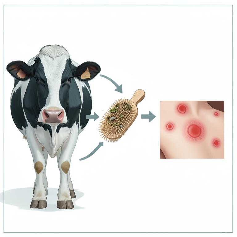
Zoonotic Transmission Cycle
AI-generated conceptual illustration (Holstein cattle and human skin). It explains the idea and does not show study data.
View source evidence ↗Introduction
Dermatophytes are a group of fungi that classified according to microconidia shape into three main genera (Epidermophyton, Microsporum and Trichophyton) that affecting superficial layers of skin and hair in animals and man. Almost 40 dermatophyte species were identified (Smith and McGinnis, 2011) [35] . Only, three genera (Trichophyton, Microsporum and Epidermophyton) are known to be pathogenic for human and animals (Weitzman and, Summerbell, (1995) [37] . The high prevalence of dermatophytosis recently may be attributed to intensive use of corticosteroids and poorly hygienic management for animals (Woodfolk, 2005) [38] . Dermatophytes have the ability to invade as well as borrow the skin layers of host, particularly the superficial layers that rich in keratin-content (Fernandes et al., 2001) [20] , although deep layers may be occasionally involved in some sever complicated cases. The diseased animals, infected materials and contaminated environment act as the main source and reservoir for infection among susceptible hosts throw direct or indirect contact (Murray et al., 2005) [28] . The disease appear to be more predominant in poorly closed housed animals (Al-Ani et al., 2002) [3] , particularly, in worm humid climate countries that allowed and favored spore formation and consequently highly infection (Radostits et al., 2007) [31] . Dermatophytosis mainly affected cattle especially young calves, which have high skin PH and immune system is remain less developed (Radostits et al., 1997) [30] , infection to other animals as small ruminants and human has been reported (Ming et al., 2006) [26] .
Etiology
Dermatophytes are divided into three main types according to habitat, geophilic (soil), anthrophilic (man) and zoophilic (animal). T. verrucosum belong to zoophilic type that considered the main frequently fungi responsible for ringworm in cattle (Abou-Eisha, 2008; El-Diasty et al. 2013) [1] , although T. mentagrophytes may also causing such disease in cattle and to less extend M. canis. There are difference between these species in diagnostic culturing and examination as shown in the blow table (1). Moreover, T. verrucosum can remain infective in environment for long periods (5-7) years (Mahmoudabadi and Zarrin, 2008; Singh and Kushwaha, 2010) [24,34] , thus, prevent complete eradication and control of the disease in farm animals. The pathogensis of dermatophytes begins firstly from contact between skin abrasion of host and contaminated environment or infected animal lesion containing arthrocondia, which germination occur in hair follicles then the invasion the cell wall of skin (stratum corneum) with digestion of keratin content and highly formation of hyphea and conidia (ecto or endothrix). The growing hair carrying fungal elements (ecto or endothrix) leading to rapid spreading of infection in form of ring shape lesion covered by scales and crusts that may coalescence together forming large area of alopecia. Moreover skin hyperkertosis may developed (Radostits et al. 2007) [30] . Table ( [13] and in other countries (Cam et al. 2007; Akbarmehr, 2011) [8,2] , all described the skin lesion of dermatophytosis as a circular or circumscribed plaques of alopecia (1-5 inches), which may gathered to each other forming large irregular area of gryish-whitish colour, thick scabs that usually firmly attached to the skin, particularly distributed in head, neck, face and around eyes as showed in photo (1&2). The presence of lesion on face and around eyes may act as source of new infection throw animal habit of licking and grooming. Other frequent studies supposed the higher incidence of ringworm lesions on the flank and back areas as these sites are more exposed to contamination, injuries and wetness than other body parts (Swai and Sanka, 2012) [36] . The frequency and distribution of infection influenced by host factors (age, stress, transportation and management), geographic area and climate condition (Al-Rubiay et al. 2006) [4] . The factors as species, numbers and age of animal besides environmental aspects will serves a significant role in dermatophytes infection and incidence (Nassif and Osman, 2003) [29] . Furthermore, calves of less than one year was more affected age among animals which explained that ill developed immune system not exposed to previous infection (Nassif and Osman, 2003 and Cafarchia et al. 2004) [29,7] . Regarding to the sex factor on dermatophyosis prevalence, males reported to be more affected higher than females, this could be Attributed to, program of farm fatting mainly acting only on males calves of closely contact to each other (Cafarchia et al. 2004) [7] . Adult animals of nonsymptomatic lesions appear to carry and spread the infection to young calves during early stage of their life (Singh and Kushwaha, 2010) [34] .
Photo (1): showing cow suffering from ringworm in face and around eyes
Photo 2: showed ring worm I neck region (gryish-whitish circumscribed lesion, above the skin) [11,36,8] . Examination of T. verrucosum by 20% KOH was a routine laboratory procedure was classically applied in dermatophytes suspected lesions, briefly 20% KOH (one drop ) put on a microscopic slide with 1cm of lesion(skin scraped) was added with gently heating and applied a cover slip. Then, after 2 h, examine the slide for the presence of arthrospores and hyphae (ecto and endothrix) under a light microscope according to (Ellis et al. 2007) [14] . Then, laboratory identification through isolation on Sabauroud dextrose agar (SDA) containing chloramphenicol and actidion incubated at 37°C for 3-4 weeks, followed by examination of culture characters including (texture, shape and color), pigment production and microscopical features of spores as described by (Ellis et al. 2007; Cowen, 1990) [14,10] . The main culture characters of T. verrucosum showed, very slow growing white, cottony, heaped up, button like with folded and nonpigmented reversed side colonies (Eman-abdeen, 2011; Dalis et al. 2014) [16,11] . In contrast to microscopical picture, T. verrucosum apper as septated hyphae and microconidia with presence of chlamydospores which arranged in chain as described by (Forbes et al. 2002; Shams-Gahfarokhi et al.
2009) [21,32] with characteristic shape of microcondia which give calvate to pyriform shape (Eman-abdeen, 2011; Swai and Sanka, 2012) [16,36] , or thin wall of cigar shape (Mucoma, 2000) [27] . The phenotypic characteristics of dermatophyte are influenced by environmental, nutritional and chemical factors besides time consuming in traditional method. So, molecular methods to identify dermatophytes become more likely used (Liu et al. 2000b) [23] . These techniques as PCR have been efficiently used for identification of dermatophyte species which proved to be more specific, accurate and stable than phenotypic characterization (Gräser et al. 2000) [22] . PCR able to detected species-specific DNA polymorphisms in dermatophyte species through characteristic patterns of each species (Faggi et al. 2001) [18] . As well as random amplified polymorphic DNA (RAPD), multiplex PCR and Specific nucleotide sequence are used as helpful advanced methods for detection of dermatophytes species and sub-species as well as in epidemiological parameters control (Faggi et al. 2001; Dhib et al. 2014) [18,12] .
Photo 3: T. verrcusum showing very slow growing, with heaped up, button like appearance folded white colored colony
Treatment and Control
The disease is self-limiting and spontaneous recovery usually occur in mild cases, although, some topical iodine and sulpher preparation are applied for treatment of severely affected lesions. Remove the scales and crusts before applying of ointment preparation is recommended. The systemic antifungal therapy that used for dermatophytes infection may left some residues problems or toxic effect on animals or human body (Araujo et al. 2009) [6] . The high cost of treatment, economic losses, long course of treatment and its public health importance activated more concern for development of natural plant extract of antifungal activity that effective and less toxic. Such natural plants include garlic, lemon grass, datura, acacia, a triplex, ginger, black seed, neem, basil, eucalyptus, alfalfa and basil (Aly and Bafiel, 2008) [5] . The properties of these plants should be safe, economic and easily applied under field conditions (Shelef, 1983) [33] . Extraction of a new compounds from plants have been recently used for treatment of some infectious diseases (Fardos, 2009) [19] . For example, Clove oil proved highly effective antifungal activity against Dermatophytes species in vitro and can used as a topical spray and ointment for treatment of dermatophytes in cattle and guinea-pigs as reported by (Eman-abdeen and El-Diasty, 2015 and chee and Lee et al, 2007) [17,9] respectively. A new trend for control of animal ring worm through immunization of young calves by live T. verrucosum strain which gave a good protection against ringworm as early reported by (Eman-abdeen, 2011; Mikaili et al. 2012) [16,25] .
Conclusion
This review spot high lights on bovine dermatophytosis and the main etiology as well as the descriptive clinical lesion. Also provide a considerable knowledge about the laboratory methods and advanced tools for rapid diagnosis of dermatophytosis. As well as it discuss the new trend in control of this disease by compounds from medical plants.
But, more research should be investigated the interaction of these compounds and the evaluated its activity and pharmacological properties with comparing with traditional drugs.
References
38 in the original paper- Abou-Eisha AM, Sobih MA, Hanaa MF, Heba SE. Dermatophytes in animals and their zoonotic importance in Suez Canal area. SCVMJ, 2008, XIII(2).
- Akbarmehr J. The prevalence of cattle ringworm in native dairy farms of Sarab city (East Azarbayjan Province) in Iran. Afr. J Microbiol. Res. 2011;
- Al-Ani FK, Younes FA, Al-Rawashdeh OF. Ringworm Infection in Cattle and Horses in Jordan. Acta. Vet. Brno. 2002; 71:55-60.
- Al-Rubiay KK. Dermatoepidemiology: A household survey among two urban areas in Basra city, Iraq. Int J Dermatol. 2006; 4:1-4.
- Aly MM, Bafiel S. Screening for antimicrobial activity of some medicinal plants in Saudi Arabia. World conference on medical and aromatic, 2008.
- Araujo CR, Miranda KC, Fernandes OFL, Soares AJ, Silva MR. In vitro susceptibility testing of dermatophytes isolated in Goiania, Brazil, against five antifungal agents by broth microdilution method. Rev. Inst. Med. trop. S. Paulo. 2009; 51:9-12.
- Cafarchia C, Romito D, Sasanelli M, Lia R, Capelli G, Otranto D. The epidemiology of canine and feline dermatophytoses in southern Italy. Mycoses. 2004; 47(11-12):508-13.
- Cam Y, Gümüssoy KS, Kibar M, Apaydin N, an Atalay. Efficacy of ethylenediamine dihydriodise for the treatment of ringworm in young cattle. Vet. Rec. 2007; 160:408-410.
- Chee HY, Min Hee Lee MH. Antifungal Activity of Clove Essential Oil and its Volatile Vapour against Dermatophytic Fungi. Mycobiology. 2007; 35(4):241243.
- Cowen P. Microscopy of skin scrapings for dermatophyte diagnosis. Aust. Fam. Physician. 1990; 19(5):685-690.
- Dalis JS, Kazeem HM, Kwaga JKP, Kwanashie CN. An outbreak of ringworm caused by Trichophyton verrucosum in a group of calves in Vom, Nigeria. Afr. J Microbiol. Res. 2014; 8(8):783-787.
- Dhib I, Fathallah A, Yaacoub A, Hadj Slama F, Said MB, Zemni R. Multiplex PCR assay for the detection of common dermatophyte nail infections. Mycoses. 2014; 57(1):19-26.
- El-Attar AA. Characters of zoophilic dermatophytes commonly attacking man in Suez Canal and Sinai districts. Ph.D. Thesis (Microbiology), Fac. Vet. Med. Suez Canal Univ, 1992.
- Ellis D, Davis S, Alexiou H, Handke R, Bartley R. Description of medical Fungi. Mycology Unit, Women's and Children's Hospital, North Adelaide, 2007.
- Eman ME, Ahmed MA, Nagwa O, Salwa FM, Samaa IE, Hanaa MA et al. Antifungal activity of zinc oxide nanoparticles against dermatophytic lesions of cattle. Romanian j biophys, Bucharest. 2013; 23(3):191-202.
- Eman-abdeen E. Studies on the evaluation of the Immune response to Some Fungal Vaccines. Ph.D. Thesis (Microbiology), Fac. Vet. Med. Minufiya University, 2011.
- Eman-abdeen E, El-Diasty EM. Antifungal Activity of Clove Oil on Dermatophytes and Other Fungi. International Journal of Advanced Research. 2015; 3(12):1299-1305.
- Faggi E, Pini G, Campisi E, Bertellini C, Difonzo E, Mancianti F. Application of PCR to distinguish common species of dermatophytes. J Clin. Microbiol. 2001; 39:3382-3385.
- Fardos MB. Antifungal activity of some medicinal plants used in Jeddah, Saudi Arabia. Mycopath. 2009; 7(1):5157.
- Fernandes NC, Akiti T, Barreiros MG. Dermatophytoses in children: Study of 137 cases. Revista do Instituto de Medicina Tropical de Sao Paulo. 2001; 43:83-85.
- Forbes BA, Sahm DF, Weissfeld AS. Laboratory Methods in Basic Mycology. In Bailey and Scott's Diagnostic Microbiology 11th edition, Mosby Inc, 11830 west line Industrial Drive St. Louis, Missouri 63146, US a, 2002.
- Gräser Y, Kuijpers AF, Presber W, de Hoog GS. Molecular taxonomy of the Trichophyton rubrum complex. J Clin. Microbiol. 2000; 38:3329-3336.
- Liu D, Coloe S, Baird R, Pederson J. Rapid minipreparation of fungal DNA for PCR. J Clin. Microbiol. 2000; 38:471.
- Mahmoudabadi AA, Zarrin M. Isolation of dermatophytes and related keratinophilic fungi from the two public parks in Ahvaz. Jundishapur J Microbiol. 2008; 1(20): 20-23.
- Mikaili A, Chalabi M, Ghashghaie A, Mostafaie A. Immunization against bovine dermatophytosis with live Trichophyton verrucosum. Afr. J Microbiol. Res. 2012; 6(23):4950-4953.
- Ming PX, Ti YLXm Bulmer GS. Outbreak of Trichophyton verrucosum in China transmitted from cows to humans. Mycopatho-logia. 2006; 161:225-228.
- Mucoma FS. Dermatophytes: Their Taxonomy, Ecology and Pathogenicity. Department of Biological Sciences, University of Botsuana, Gaborone, 2000, 1-10.
- Murray PR, Rosenthal KS, Pfaller MA. Superficial and cutaneous mycosis. In: Medical Microbiology, 5th edition. Philadelphia, USA, 2005, 745-751.
- Nassif MM, Osman SA. An outbreak of dermatophytosis in goat kids. Assiut Vet. Med. J, 2003; 49(99):142-155.
- Radostits OM, Blood DC, Gay CC. Veterinary Medicine, 10th Ed, Bailliere Tindall, London, 1997.
- Radostits OM, Done SH. Veterinary medicine: a textbook of the diseases of cattle, sheep, pigs, goats, and horses. Elsevier Saunders, Oxford, UK, 2007.
- Shams-Gahfarokhi M, Mosleh TF, Ranjbar BS, Razzaghi AM. An epidemiological survey on cattle ringworm in major dairy farms of Mashad city, Eastern Iran. Iran. J Microbiol. 2009; 1(3):31-36.
- Shelef LA. Antimicrobial effects of spices. J. Food Safety. 1983; 6:29-44
- Singh I, Kushwaha RKS. Dermatophytes and related fungi in soils of parks and agricultural fields of Uttar Pradesh. Indian Journal of Dermatology. 2010; 53:306308.
- Smith MB, McGinnis MR. Dermatophytosis in Tropical Infectious Diseases (Third Edition), 2011.
- Swai ES, Sanka PN. Bovine dermatophytosis caused by trichophyton verrucousum: A case report. Vet. Wod. 2012; 5(5):297-3.
- Weitzman I, Summerbell RC. The dermatophytes. Clin Microbiol Rev. 1995; 8:240-25.
- Woodfolk JA. Allergy and dermatophytes. Clin. Microbiol. 2005; Rev. 18(1):30-43.
Cited by 3
From OpenAlex, OpenCitations and reference lists read by Scinovex
- Prophylaxis and Therapeutic Ability of Inactivated Dermatophytic Vaccine Against Dermatophytosis in the Rabbits as an Animal Model ↗
Ali Abdul Hussein S. AL‐Janabi, Falah Hasan Obayes Al-Khikani · Turkish Journal of Pharmaceutical Sciences · 2020
Rabbits as an Animal Model ABSTRACT Objectives: Dermatophytosis is a group of cutaneous diseases widely distributed in human and animals.It causes serious infection in some human cases and economic losses in farm animals.The primary aim of this study is to conduct an investigation of prophylaxis and a potential therapeutic vaccine against dermatophytosis. Materials and Methods:The rabbit was chosen as an…
❝ 19PDF - Histopathologic Diagnosis of Fungal Infections of Lab Animals ↗
Abhilasha Shourie, Rajeev Kumar Singh, Anamika Singh · Fungal biology · 2021
❝ 3 - In-vitro evaluation of different commercial antimycotics and disinfectants against Trichophyton verrucosum isolated from beef farm in Beni-Suef, Egypt ↗
Manar Mohamed, Sherin Reda Rouby, Sahar Abd El Aziz · Journal of Veterinary Medical Research · 2022
Dermatophytosis is a fungal disease that affects both animals and humans. The improper use of antimycotics and disinfectants led to an acquired resistance of dermatophytes to most of the commonly used antimycotic agents. The aim of the present study was to estimate the prevalence of dermatophytosis in a calf-beef farm and to evaluate the in-vitro efficacy of six antifungal agents…
❝ 0PDF