
Research article · 2018 · Full text
International Journal of Veterinary Sciences and Animal HusbandryFunctional anatomy of the masticatory mechanism: A comparative study of physical characteristics of jaw- closing and jaw- opening muscles in sheep
Volume 3(1) · Pages 03–09
Research areasTemporomandibular Joint DisordersOropharyngeal Anatomy and Pathologies
Abstract
Author-writtenThe functional anatomy study of the mammalian muscles is very important for understanding the mastication mechanism during eating. It is also important for their health and breeding performance. Fifteen heads of sheep (Mean ± SD age: 18 ± 4 month) and of both sexes were included in this study. A comparative gross anatomical study and linear measurements of the two types of masticatory muscles were conducted on slaughtered sheep heads. The physiological cross- sectional area (PCSA), maximal isometric force, torque and kinetic energy were calculated. were calculated. The study showed that the sheep jaw-closing muscles were the masseter and temporal muscles whereas the digastricus was a jaw-opening muscle. The physiological cross- sectional area (PCSA) was 3.1, 2, 3.1 cm² for the masseter, temporal and the digastric muscle, respectively. The maximal isometric force for those muscles was 6.2, 4, 6.2 cm², respectively. The force of those muscles was 0.49, 0.098, 0.196 N, respectively. The torque of those muscles was 0.49, 0.098, 0.196 Nm, respectively. While the kinetic energy of the jaw-closing group was 90 Joule, that of the jaw-opening group was 30 Joule. The values of the PCSA, force, maximum isometric force, torque and kinetic energy of the jaw-closing group were higher than those of the jaw- opening. This is suggested to be due to the increased activity of the jaw-closing muscles.
Summary
Source-checkedPlain-language reading of the full paper, prepared with AI and checked against the text. Each point links to the authors' own words.
Why it matters
- Studying the functional anatomy of mammalian muscles is essential for understanding how animals eat, which in turn impacts their overall health and breeding performance. View evidence ↗
- There is a lack of existing literature detailing the physical properties of masticatory muscles specifically in sheep. View evidence ↗
The question
- The study aimed to compare the functional biomechanics of jaw-closing and jaw-opening muscles in adult sheep to understand their dynamics. View evidence ↗
What they did
- The researchers used fifteen sheep heads of both sexes, aged between 12 and 24 months, sourced from a slaughterhouse in the Qassim Region, KSA. View evidence ↗
- Ten heads were dissected for anatomical measurements and weight, while five live sheep were used to observe the timing of jaw movements during mastication. View evidence ↗
- Muscle volume was measured via water displacement, and the physiological cross-sectional area (PCSA) was calculated using mass, density, and fiber length. View evidence ↗
- The study calculated maximum isometric force as twice the PCSA, and used standard physics formulas to determine force, torque, and kinetic energy. View evidence ↗
What they found
- The masseter and temporal muscles were identified as the jaw-closing muscles, while the digastricus muscle served as the jaw-opener. View evidence ↗
- The jaw-closing muscle group had a total kinetic energy of 90 J, which was three times higher than the 30 J of the jaw-opening group. View evidence ↗
- Across all measured physical characteristics, PCSA, force, maximum isometric force, torque, and kinetic energy, the jaw-closing group outperformed the jaw-opening group. View evidence ↗
What it means
- The authors suggest that the higher physical values of the jaw-closing muscles are due to their increased activity during mastication. View evidence ↗
- The study concludes that the functional differences between jaw-closing and opening muscles are explained by their distinct physical characteristics and movement patterns. View evidence ↗
Visual summary
1 visualsIllustrations explain the paper; they do not show study data.
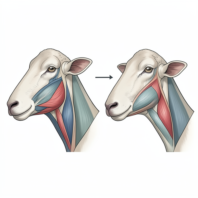
Masticatory Muscle Grouping
AI-generated conceptual illustration (sheep head and jaw muscles). It explains the idea and does not show study data.
View source evidence ↗Figures & tables
From the original paper Table 1
 Table 2
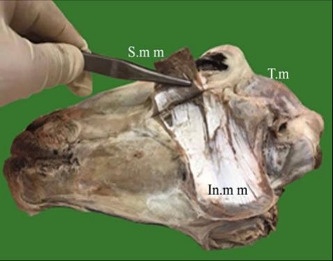 Figure 2
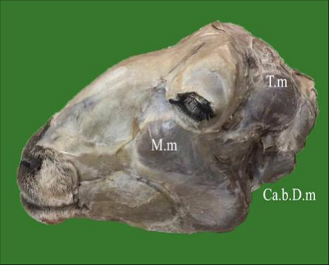 Figure 1
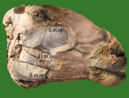 Figure 3
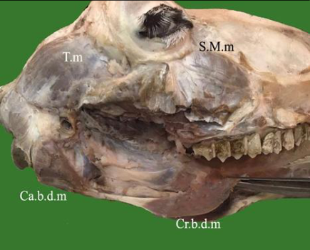 Figure 4
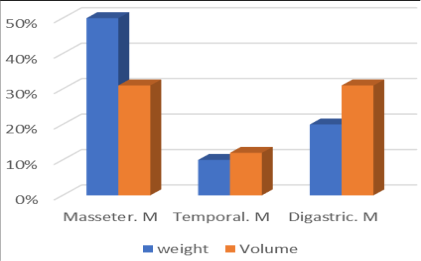 Figure 5
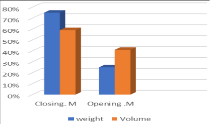 Figure 6
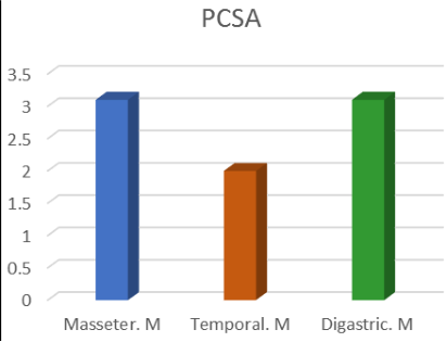 Figure 7
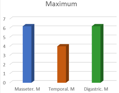 Figure 8
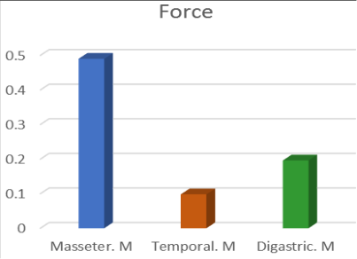 Figure 9
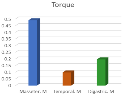 Figure 10
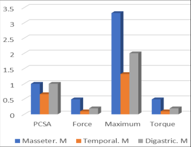 Figure 13
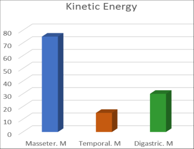 Figure 12
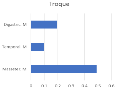 Figure 11
Introduction
The functional anatomical study of the masticatory muscles in sheep is very important to understand the mastication mechanism during eating. They are also important for the animal health and breeding success. There is very rare literature about the physical properties of the masticatory muscles that explain masticatory mechanism in sheep. The anatomy of the masticatory muscles was previously studied in ruminants and described carefully in details (Getty, 1976 and Dyce et al., 2010). Moreover; the function of the masticatory muscles correlates with resemblance generally skull shape and food natural of the deer reported by (Janis, 1990) [14] . However, no detailed description of masticatory mechanics is available in sheep. The oro-facial system including stomatognathic system, maxillamandibular apparatus and masticatory system was a functional unit (Patial and Bindra, 2012), while the masticatory system was completed by including a jaw opener digastric muscle using the origin and insertion sites (Weijs et al., 1987) [24] . The involvement actions of masticatory muscles in a variety of mammals in which feeding behavior and the configuration of the masticatory apparatus were different was reported by (Gans, 1978; Gorniak, 1985) [10] . The mechanism of jaw opening and closing is complex (Herring, 1992; Hiiemae, 2000) [12] . The mechanical advantage of the superficial layer of the masseter muscle was promoted by elevation of the jaw joint and expansion of the mandibular angle (Popowics and Herring, 2006) [17] . The present work which aimed to investigate the functional anatomy of the different masticatory muscles in the sheep could be considered as supporting The present study was aimed to present the research concerning the comparison of the masticatory functional biomechanics of the closing and opening movements of the masticatory muscles in sheep in addition to understand the dynamics of masticatory muscles of adult's sheep.
Materials and Methods
Materials and Methods
Materials
Fifteen heads of sheep of both sexes and different ages (12-24 months) were collected from Buraidah slaughterhouse, Qassim Region, KSA were used in this study. Ten heads were dissected using standard instruments to investigate the anatomical features including weight and measurements of masticatory muscles. In addition, five heads of a live sheep were used for taking the time expended by the jaw opening and jaw closing muscle during mastication mechanism.
Methods
An incision was made in the masticatory muscles along its length bell to reveal the muscle fibers. Linear measurements (cm) concerning the muscles longitudinal axis were taken. The muscle volume was determined using water displacement technique.
Taking the density of muscle by Mass/Volume. The physiological cross-sectional area (PCSA) was determined using the following equations: PCSA= (mass /density) / fiber length (Anapol and Barry 1996) [1] , maximum isometric force generation capacity of muscle =2 x PCSA (Woledge et al., 1985) [26] . Force =Mass x Gravity (g x 9.8). The muscle torque (T) = F. d = F. r. Sin θ: Where F (force) r (The vertical distance of the center of force for the axis of rotation. Sin θ a numerical value for the rotation angle of each masticatory muscle. The muscular Kinetic energy =KE= ½. m. v²: where M (mass of muscle) V (speed of muscle). The muscle weight was estimated by (g), volume (cm³), Density (g /cm³), force (N), PCSA (cm³), the torque (Nm), Kinetic energy (Joule).
Results
The sheep masticatory system is a complete functional unit including muscles, mandible bone and other tissues comprise ligaments, tendons, arteries and nerves. The masticatory muscle was divided into three groups: the closing, the opening and the unilateral (medially and laterally).
In the present study we focus on the anatomical, physiological and physical characteristics of masticatory muscles closing, and opening muscles was performed. They were presented as follow as the masseter, temporal and digastricus muscles. The closing muscles include both masseter and temporal muscles.
The masseter muscle (Figs. 1, 2, 3, 4) is the largest masticatory muscle. It occupies most of the lateral surface of the cheeks, representing about 83% of weight of closing muscle mass, and represents 10% of the volume of this muscle and 41% of the total volume of these muscles. The masseter mass weight is about 40-60 g. This muscle subdivides by two tendinous laminae forming three distinguished separate layers namely; superficial, middle and deep masseter muscle. The average length of the masseter mass is 12±2 cm cranio-caudally, 6±8 upper-lower and about 1 cm lateromedially. Length of the muscle fiber is about 8±2 cm which are oriented in different directions according to the layer of masseter muscle. The superficial masseter muscle fibers are oriented cranioventrally in the superior part in an angle about 30° degree to the horizontal line. The fibers of the superficial muscle are oriented almost cranio-ventrally pulling the mandible anteriorly, when is elevated the mandible to close the mouth as well as this part of the muscle aid to rotate to the medial side of the mandible. While the fibers of the intermediate layer of the masseter muscle are oriented ventrally according to the vertical line and due to these fibers orientation, this part is responsible for elevation the mandible to close the mouth. The fibers of the deep masseter muscle are oriented ventrally in a vertical direction and overlapped with the fibers of the temporal muscle. Because of this ventral orientation, it contributes to elevate the mandible and retracts the mandible posteriorly. The physical characteristics of the masseter muscle indicates the volume of the masseter muscle was 50 cm³, density of the muscle is 1.612 g/ cm³. Moreover, the physiological crosssectional area (PCSA) is the area of the cross section of the muscle perpendicular to its fibers. It is typically used to describe the contraction properties of pennate muscles: PCSA = 50/1.612) / 10 = 3.1cm². Furthermore, the maximum isometric force generation capacity of muscle is representing PCSA. 3.1 x 2= 6.2 Ncm². Regarding the estimated force that of the masseter muscle, it is 0.49 N. The torque is the measurement of the turning force on muscle. The torque is estimated about 0, 49 Nm due to the fibers direction which increases the torque of the movement by closing the lower jaw. The kinetic energy moves the masseter muscle of the mouth either it is vertically or horizontally, is about 75 Joule. It depends on the movement speed of the muscle. It represents the work needed to moves muscle mass from rest state to limited distance to the new position. The same used kinetic energy of work is done by the muscle to return from its new position to a rest state. The main function of the masseter muscle is to stabilize the tempomandibular joint elevating the mandible and aides in the simple rotating movement.
The temporal muscle (Figs. 5, 6, 7) is smaller than the masseter muscle. It locates in the temporal fossa of the temporal bone, presenting about 17% of weight of closing muscle mass, and represents 12% of the volume of this muscle. The temporal mass weight is about 8-14 g. This muscle has thick spherical shape and have a broader aponeurotic extension almost the length of the muscle. The average length of the temporal mass is 8±2 cm craniocaudally, 2±4 upper-lower and about 1-1.5 cm lateromedially. The length of the muscle fiber is about 8±2 cm which oriented caudo-ventrally to the horizontal line. The fibers of the temporal muscle oriented caudo-ventrally to pull the lower jaw caudally, they are considered responsible for elevation of the lower jaw to close the mouth and pulling it caudally. The indication of the physical characteristics of the temporal muscle are the volume of the temporal muscle was 12 cm³. The density of muscle is 0.83 g/cm³. Moreover, the physiological cross-sectional area (PCSA) is typically used to describe the contraction properties of pennate muscles: PCSA = 10/0.83)/6 = 2 cm. Furthermore, the maximum isometric force generation capacity of muscle is representing PCSA. 2 x 2= 4 Ncm². According to the estimated force of the temporal muscle. It is 0,098 N. The torque is a measure of the turning force on muscle. The estimated torque is about 0, 49 Nm due to the direction which to increment the torque of the movement by closing the mandible The kinetic energy of the temporal muscle which causes its moving whether it is vertical or horizontal is about 15 Joule. It represents the work needed to move a muscle from opining movement to closing movement. The same used kinetic energy of work is done by the muscle to return from the opening state to a closing state of the lower jaw.
The main function of the temporal is to stabilize the temporomandibular joint, elevation the mandible. The vertical movements of the lower jaw apparently are produced by action of the three parts of masseter and temporal muscles. The opening muscles include one muscle which is digastric muscle (Fig. 8). It occupies most of the medial surface of the mandible. The digastric muscle has a different shape from the other masticatory muscles. The digastricus muscle mass weight is about 18-22 g, representing 100% of weight of opening muscle mass, and represents 41% of the volume of this muscle. This muscle has two bellies between them a short tendinous junction namely; cranial, caudal belly. The average length of the digastric mass is 14±2 cm cranio-caudally, 3±1upper-lower and about 1 cm lateromedially. The length of the muscle fiber of each belly is about 8±2 cm which are oriented in two directions. The fibers are oriented cranioventrally in the caudal belly and oriented cranially in the cranial belly due to these fibers orientation the muscle is responsible for pulling down the mandible and acts reduction the mandible. The physical characteristics of theof digastric muscle indicates the volume of the of digastric muscle was 10 cm³, density of the muscle is 0.64 g/ cm³. Moreover, the physiological cross-sectional area (PCSA) = (mass/density)/fiber length, PCSA = 20/0.64)/ 10 = 3.1 cm².
Furthermore, the maximum isometric force generation capacity of muscle is representing PCSA. 3.1 x 2= 6.2 Ncm². Regarding the estimated force that of the digastric muscle. It is 0.196 N due to the fibers orientation and arrangement had a role on a muscle's ability to produce movement and to generate force. The torque is the force applied to the distance between the center of the axis of force and the axis of rotation. It is rotational force as a linear force is a push or a pull and it is a measure of the turning force on muscle. The estimated torque is about 0.196 Nm due to the fibers direction which increases the torque of the movement by closing the lower jaw. The estimated the kinetic energy involved in moving the digastricus muscle of the mouth whether it is vertical or horizontal movement is about 30 Joule. The kinetic energy is needed to move a muscle mass from closing to opening position. The main function of the digastricus muscle is to open the mouth. Finally, the closing and opening movements apparently are produced by action of the masseter, temporal and digastric muscles. Finally, the closing and opening pattern for moving the lower jaw vertically or horizontal depends a force both masseter, temporal and digastric muscles on both sides of the mandible, generally.
Table 1:
The physical functional characterizes of the masticatory muscles (jaw closing -opening) comparative the total masticatory mass. [4] in horse. On the other hand, the current work demonstrated three layers of masseter muscle, superficial, intermediate and deep. The current results were similar to those of Parker (1983) [16] in contrast to two layers, superficial and deep in cattle (Budras and Habel 2009), camel (Smut et al., 1987) [20] and horse (Budras et al., 2013) [4] . On the other hand, the camel has a large temporalis muscle is not unexpected (Maynard Smith & Savage, 1959). The digastric muscle or jaw-opening muscle had two bellies of spindle shape separated by an intermediate tendon which penetrates the tendon of the stylohyoideus. Our finding is compatible with (Kushkhov, 1991) [15] in sheep and (Budras and Habel, 2009) in bovine, (Smut et al., 1980) in camel and (Budras et al., 2013) [4] in horse. The dominance of the masseter musculature is in contrast to the emphasis of the temporalis musculature in carnivores (Turnbull, 1970) [22] . The relative mass of the closed and opening muscles group was functionally consistent. The closed and opening muscles group proportions were 60%. They were presented of masseter 50%, temporal 10%, the opening group were 20%; presented by digastric 2%.They were similar to white-tailed deer; masseter 46.1%, temporalis 29.3% among the mammals investigated by (Turnbull, 1970) [22] and that is agree with (Janis, 1990) [14] who recorded that the function of the masticatory muscles correlates with similarities generally skull shape and food natural of the deer. The present study revealed that the function of muscles was estimated through measuring the length and the orientation of muscle fibers. The length of muscular fibers of masticatory closed group was about 10 cm of masseter muscle, 6 cm of the temporal. However; the superficial masseter fibers directed cranioventrally, the intermediate masseter layer was ventrally direction while the deep masseter layer was ventrally and caudoventrally direction. While the length muscular fiber of opening group was about 5 cm of each belly and cranioventrally directed. These results were in accordance with those of Gorniak (1985) [10] who recorded that the horizontal movements are generated by muscles having fibers arranged in marked anteroposterior direction, whereas vertical movements are generated by muscles having more or less vertically arranged fibers. In ruminants, Suzuki (1977) [21] recorded that the masseter fibers of the ruminant differed from those of the other species in histochemical properties. Budras and Habel (2009) mentioned that a superficial layer with almost horizontal muscle fibers, and a deep layer with caudoventral fiber direction in bovine and added the digastric muscle the two parts have different fiber directions. On the contrary, Budraset et al., (2013) [4] recorded that the superficial fibers in horse run obliquely caudoventrally but deeper ones run nearly vertically. These fiber direction are responsible for the lateral and rotational chewing movement. Furthermore, the results showed that the physiological crosssectional area (PCSA) of the closed masticatory group in sheep was 5.1 cm². They were represented by 3.1 cm² of the masseter, 2 cm² of the temporal muscle. While (PCSA) was 3.1 cm² of the opening masticatory group. These results explain the PCSA of the closed mastication is larger than the PCSA of the opening mastication muscle about double value due to mass, density, length of fiber of the muscle and function of the muscle. (Watson et al., 2017) [23] recorded that in the rabbits, the PCSA was 5.3 cm² of the closing group represented by 4.7 cm² of the masseter, 1.5 cm² of the temporal muscles. They don't remember PCSA of digastric muscle. This result was in harmony with those of Watson et al., (2017) [23] as value of PCSA to closed and opening mastication group. On the other hand; our result Emphasizes Eijden et al (1995) [6] who recorded that the Muscle force is proportional to the physiological cross-sectional area (PCSA).
Muscle
The differences in PCSA measurements can be attributed to variations in the muscle mass and the type of the comparative studied animals (Fukunaga et al., 1990). Also, the present study revealed the force of the closed muscle group presented about 6 N collected by 4.9 from the masseter muscle and 0.9 from the temporal muscle. This force was necessary for closing operation of the mouth. It was produced due a large mass of the masseter muscle, whereas the increasing of masseter muscles size serves both the moving the mandible vertically. The force of the opening muscle presented was about 2 N from the digastric muscle. This force was necessary for the opening of the mouth. Also, this force was produced due to the mass, the extending of the digastric muscle serves both the moving the mandible vertically but distally. This explains why the force of the closed was equal to threefold of the opening force and force of the opening equal 1/3 of the closed force its due to the gravity which need more force to occlusion. This agree with (Watson et al., 2017) [23] mentioned that the closed force was completely attributed to a vertical component (97.4% of resultant), with only minor contributions from the anterior and medial components (20.6% and 9.8% of resultant, respectively. In the present study, it was clear that the strength of the chewing was limited by the maximum force of muscle, Maximum isometric force generation capacity of muscle was estimated by multiplying muscle PCSA A maximum muscle force was assigned to each mandible closed and opening muscle. They were 10.2 of the closed muscles, they were represented by 6.2 of within masseter muscle and 4 within the temporal muscle as well as it was 6.2 of within opening muscle represented by the digastric muscle. It had maximal effect on the closed force due to the large size of the muscle, while a maximum force within the temporal. It had minimal effect on the closed force due to the small size of the muscle. On the other hand; A maximum force within digastric muscle for the opening group. It had like maximal effect on the masseter muscle of the closed force due to large size and due to extension of the digastric muscle on the distance in the mandible. Watson et al., (2017) [23] reported that the rabbit has the highest maximum force of all the masticatory muscles (60.9 N) and the muscle vary significantly in their orientation due to its pennate structure. The current study revealed that the torque was estimated by 0.49 Nm of masseter muscle and 0.098 Nm of the temporal muscle due to the direction, which to increment the torque of the movement by rotation the mandible. While the torque of the digastric muscle had 0.196 Nm. The torque value of the closed group is higher than the torque value of the opening group due to area increased attached the muscle on the bone and the activity of the masseter and temporal muscle the in comparing with the activity of the digastric muscle. We find that when the area of muscular attachment to the bones was large, the torque values decreased as well as the torque was rotational force as a axial force to push or a pull and it was a measurement of the turning force on muscle. These results agree with Serway and Jewett (2003) [19] . Herring (2007) [11] recorded that the torques asymmetrical muscle usage sets up torques on the skull and combines with occlusal loads to produce bony deformations not only in the tooth-bearing jaw bones. On the other hand; Other biomechanical forces generated during mastication, such as joint torques (Widmer et al., 2002) [25] . Regarding the kinetic energy, it was about 90 Joule of the closed muscles group. They were represented by 75 Joule of masseter muscle and 15 Joule of the temporal muscle. In the closed group; the kinetic energy value of the masseter muscle higher than the value of the temporal muscle due to the size and the activity increasing of the masseter in comparing of the temporal muscle. While the kinetic energy of the opening muscle group represented by the digastric muscle was 30 Joule. So the kinetic energy value of the closed group about threefold (90: 30) the opening group because the gravity needed more KG to end the closing operation. Thus, these muscles need kinetic energy to move the muscle from its condition to another condition. These results were in parallel line with those of Sasaki and Neptune (2006) [18] who mentioned that the kinetic energy have the potential to be stored as elastic energy in compliant connective tissue and tendinous structures, and subsequently released to do positive work at a later point.
Conclusion
Finally, this study explained the cause of the functional differences which are responsible for the closing and opening muscles of the mandible. It explained these differences by representing the physical characteristics such as the PCSA, force Maximum isometric force, torque and kinetic energy as well as the differences in the pattern of mandible movements.
Competing Interest: The research was not founded officially.
Figures and tables from the paper
References
26 in the original paper- Anapol F, Barry K. Fiber architecture of the extensors of the handlimb in semterrestrial and arboreal guenons. Am. J. Phs. Anthropol. 1996; 99:429-447.
- BECHT C. Comparative biologic-anatomical researches on mastication in some mammals. I and 11. Proceedings Koninklijke Nederlandse Akademie van Welenschappen Amsterdam, Series C. 1953; 56:508-5 I 7, 5:18-527.
- Budras KD, Habel RE. Bovine Anatomy, An Illustrated Text, First Edition, Schlütersche Verlagsgesellschaft. Hans-Böckler-Allee 7, 30173 Hannover, Germany, 2011.
- Budras KD, Sack WO, Röck S. Anatomy of the Horse. Fifth, Revised, Edition. Schlütersche Verlagsges ellschaft. Hans-Böckler-Alle. 2013; 7:30173 Hannover.
- Dyce KM, Sack WO, Wensing CJG. Textbook of Veterinary Anatomy, 3rd edition (WB Saunders Company) Philadelphia, 2002.
- Eijden TM, Koolstra JH, Brugman P. Architecture of the human pterygoid muscles. J Dent Res. 1995; 74(8):148995.
- Fukunaga T, Roy TRR, Shellock SFG, Hodgson TJA, Day MK, Lee IPL, et al. Cross-Sectional Area of Human Leg Muscles Based on Magnetic Resonance Imaging, J Thopaedic Res, Raven Press, Ltd, New York, 1992.
- Gans C, Vree FD, Gorniak GC. Analysis of mammalian masticatory mechanisms: Progress and problems. Anat. Histol. Embryol. 1978; 7:226-244.
- Getty R. Sisson and Grossman's The Anatomy of the Domestic Animals, 5th edition (Saunders) Philadelphia 1975; ISBN-13:978-0721641027.
- Gorniak GC. Trends in the Actions of Mammalian Masticatory Muscles1, AMER. ZOOL. 1985; 25:331337.
- Herring SW. Masticatory Muscles and The Skull: A Comparative Perspective, 2007.
- Herring SW. Muscles of mastication: architecture and functional organization. In: Davidovitch, Z. (ed.) The Biological Mechanisms of Tooth Movement and Craniofacial Adaptation. The Ohio State University College of Dentistry, Columbus, Ohio, 1992, 541-548.
- Hiiemae KM. Feeding in mammals. In: Schwenk, K. (ed.) Feeding: Form, Function and Evolution in Tetrapod Vertebrates. Academic Press, San Diego, California, 2000, 411-448.
- Janis CM. Correlation of cranial and dental variables with dietary preferences in mammals: a comparison of macropodoids and ungulates. Mem Qld Mus. 1990; 28:349-366.
- Kushkhov KhT. Masticatory muscles of domestic sheep and swine in ontogenesis. Arkh Anat Gistol Embriol Jan. 1991; 100(1):88-93.
- Parker WS. Comparative Anatomy of the Internal and External Pterygoid Muscles, Functional Variations Among Species. The angle Orthodontist. 1983; 53(1):918.
- Popowics TE, Herring SW. Teeth, jaws and muscles in mammalian mastication, Bels-Ch05.qxd. 2006; 6:61-83, https://www.researchgate.net/publication/282881060 rsif.royalsocietypublishing.org J. R. Soc. Interface 11: 20140564
- Sasaki K, Neptune RR. Muscle mechanical work and elastic energy utilization during walking and running near the preferred gait transition speed/Gait & Posture. 2006; 23:383-3902005 Elsevier B.V. All rights reserved
- Serway RA, Jewett Jr JW. Physics for Scientists and Engineers. 6th Ed. Brooks Cole. 2003, ISBN 0-53440842-7.
- Smut M, Bezuidenhout A. Anatomy of the Dromedary. Clarendon: Oxford University Press, 1987.
- Suzuki A. A Comparative Histochemical Study of the Masseter Muscle of the Cattle, Sheep, Swine, Dog, Guinea Pig, and Rat. Histochemistry. Histochemistry 9 by Springer-Verlag 1977. 1977; 51:121-131
- Turnbull WD. Mammalian masticatory apparatus. Fieldiana Geolgy. 1970; 18:147-356.
- Watson JP, Groning F, Curtis N, Fitton CL, Herrel A, McCormack WS, et al. Masticatory biomechanics in the rabbit:a multi-body dynamics analysis, 2017.
- Weijs WA, Brugnan P, Klok EM. The growth of the skull and jaw muscles and its functional consequences in the New Zealand rabbit (Oryctolagus cuniculus). J.Morpho. 1987; 194:143-161.
- Widmer CG, English AW, Carrasco DI, Malick CL. Modeling rabbit temporomandibular joint torques during a power stroke. Angle Orthod. 2002; 72:331-337.
- Woledge RC, Curtin NA, Homsher E. Energetic aspects of muscle contraction. Monogr. Physiol. Soc. 1985; 41:1357.