
Research article · 2018 · Full text
International Journal of Statistics and Applied MathematicsA study on cerebrospinal fluid analysis
Volume 3(1) · Pages 107–109
Research areasCerebrospinal fluid and hydrocephalus
Abstract
Author-writtenCerebrospinal fluid (CSF) is a biological fluid mainly formed in the Ventricular choroid plexus distributed within the ventricular system. Analysis of the CSF provides invaluable diagnostic information because diseases take place either within its bounding membranes. To treat the CSF some test should include protein and glucose levels, cell counts are included with table data. Additional details with test such as opening pressure, supernatant color, latex agglutination and polymerase chain reaction are performed in primary care and secondary care mentioned with some advanced treatment like shunt valve.
Summary
Source-checkedPlain-language reading of the full paper, prepared with AI and checked against the text. Each point links to the authors' own words.
Why it matters
- Analyzing cerebrospinal fluid (CSF) is critical for diagnosis because diseases often occur within the membranes that bound the fluid. View evidence ↗
- CSF provides buoyancy to the brain, reducing its effective weight from 1400–1500 grams to 25-50 grams to prevent the brain's own weight from cutting off blood supply. View evidence ↗
- CSF acts as a shock absorber, protecting brain tissue from mechanical injury during jolts or hits. View evidence ↗
The question
- The paper aims to describe the necessary tests for CSF analysis, specifically focusing on protein levels, glucose levels, and cell counts. View evidence ↗
What they did
- Lumbar puncture is the primary method used in primary care to obtain CSF for analysis. View evidence ↗
- To measure CSF opening pressure, patients are placed in the lateral decubitus position with neutral neck and leg positions. View evidence ↗
What they found
- The normal range for CSF opening pressure is 50-175 mm H2O. View evidence ↗
- Normal CSF glucose levels typically range from 40–80 mg/dL. View evidence ↗
- The normal total protein concentration in CSF is 15–45 mg/dL. View evidence ↗
- Normal leukocyte counts are 0–5/microL for adults and children, and up to 30/microL for newborns. View evidence ↗
- In patients with bacterial meningitis, 87% exhibit a white blood cell (WBC) count exceeding 100 per mm3. View evidence ↗
- Bacterial meningitis is associated with an average protein level of 418 mg/dL, with a range from 21 to 2220 mg/dL. View evidence ↗
- Epilepsy is associated with an average protein level of 31 mg/dL, with a range from 7 to 200 mg/dL. View evidence ↗
- CSF appears turbid if it contains as few as 200 white blood cells per mm3 or 400 red blood cells per mm3. View evidence ↗
- The placement of a cerebral spinal shunt system is a life-saving and successful treatment for hydrocephalus. View evidence ↗
What it means
- The authors conclude that patients should be advised to maintain protein levels within the specified table values to prevent serious conditions. View evidence ↗
Visual summary
2 visualsIllustrations explain the paper; charts are replotted from the paper's own table values.
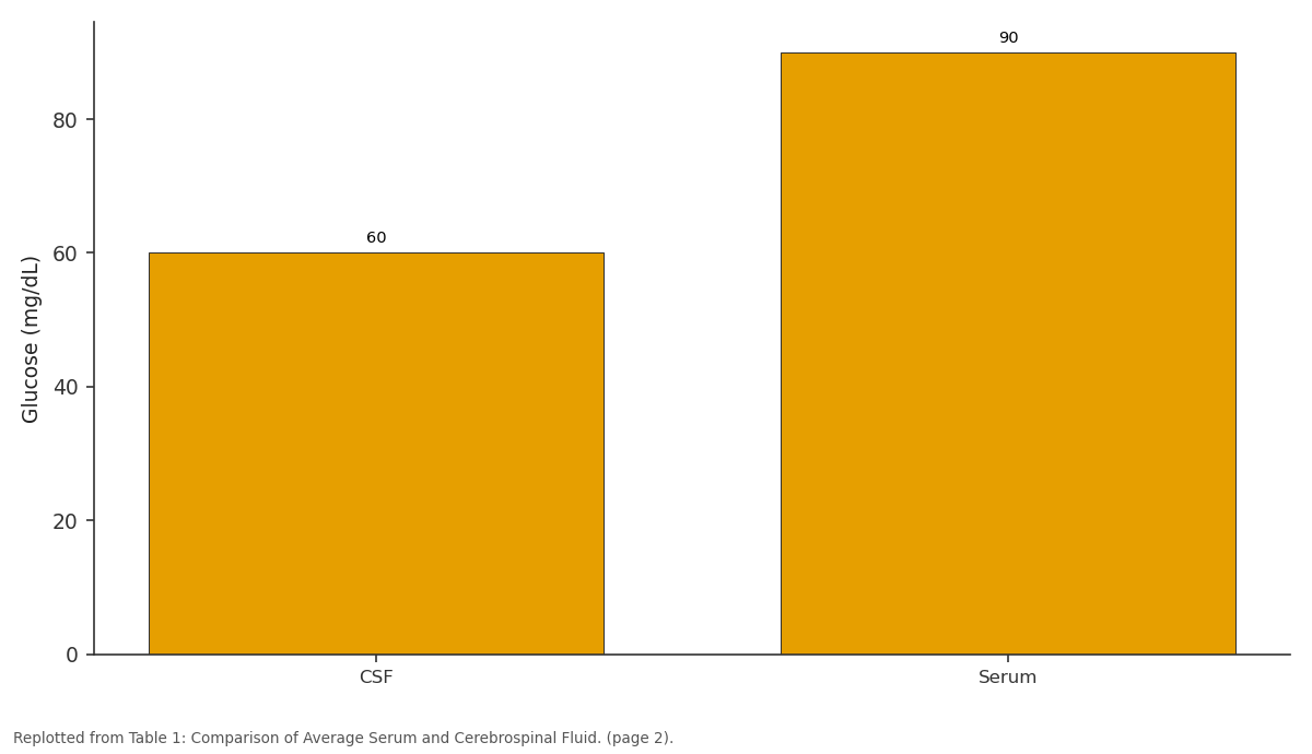
Glucose (mg/dL): CSF vs Serum
Replotted from Table 1 (page 2) of the original paper. Every bar is the paper's own table value.
View source evidence ↗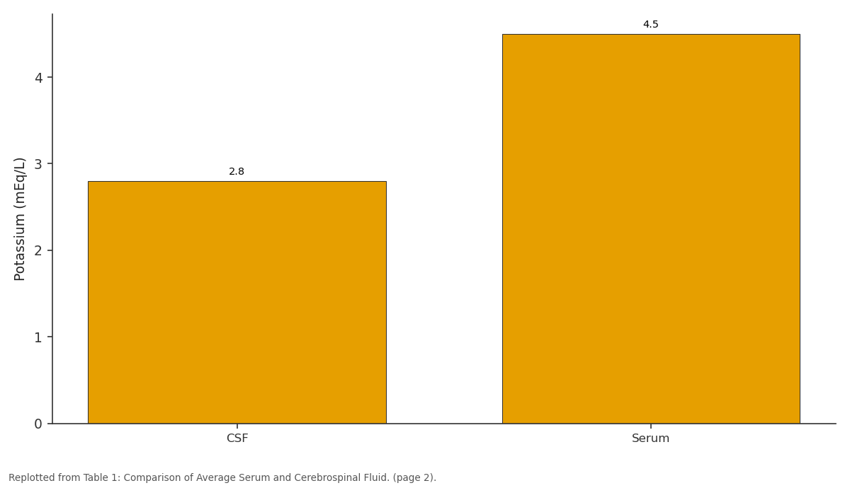
Potassium (mEq/L): CSF vs Serum
Replotted from Table 1 (page 2) of the original paper. Every bar is the paper's own table value.
View source evidence ↗Figures & tables
From the original paper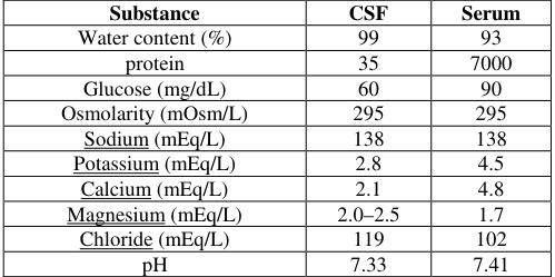 Table 1
 Table 2
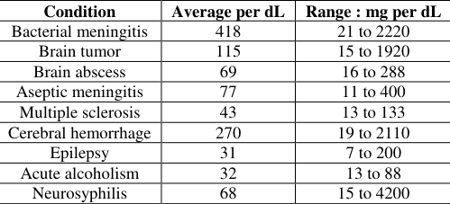 Table 3
Introduction
The diagnostic chain for various central nervous system pathologies is an important element of the cerebrospinal fluid (CSF). Primary care frequently performed using lumbar puncture. It deals with many serious complications are included with extreme caution and its benefits are overcome risks situation in that diseases. If pressure increased as much as it occur to brain tumor then it also leading with irreversible brain damage or brain death. Lumbar puncture usually are a dull head pain initially it shows as simple headache some people reported as throbbing sensation. Normally stiff neck and nausea may accompany the headache. Lumbar puncture Headaches typically begin within two days and it extend to several weeks or months.
Normal results
1. Gross appearance: Normal CSF is clear and colorless. 2. CSF opening pressure: 50-175 mm H2O. 3. Specific gravity: 1.006-1.009. 4. Glucose: 40-80 mg/dL. 5. Total protein: 15-45 mg/dL. 6. LD: 1/10 of serum level. 7. Lactate: less than 35 mg/dL. 8. Leukocytes (white blood cells): 0-5/microL (adults and children); up to 30/microL (newborns). 9. Differential: 60-80% lymphocytes; up to 30% monocytes and macrophages; other cells 2% or less. Monocytes and macrophages are somewhat higher in neonates. 10. Gram stain: Negative. 11. Culture: Sterile. 12. Syphilis serology: Negative. 13. Red blood cell count: There are no red blood cells in the CSF unless the needle passes through a blood vessel on route to the CSF.
Primary Care
Primary care is the main terms to prevent CSF. Knowing which tests to order and how to interpret them to allow for using CSF as a key diagnostic tool and it is necessary to identify variety of diseases. CSF is an invaluable diagonistic window to the central nervous system with the performance of Lumbar puncture using primary care physicians frequently.
Opening pressure
The identification of opening pressure and to measure CSF opening pressure, the patient must be in the lateral decubitus position with the legs and neck in a neutral position. Then the meniscus will fluctuate between 2 and 5 mm with the patients pulse and between 4 and 10 mm with respirations. Because of straining the patient can increase its opening pressure.
Supernatant color
Normal CSF is transparent clear as few as with 200 white blood cells per mm 3 or 400 red blood cells per mm 3 will cause CSF to appear turbid.
Cell Count
Normal CSF may contain up to 5 WBCs per mm 3 in adults and 20 WBCs per mm 3 in new borns. 87% of patient with bacterial meningitis will have a WBC count higher than 100 per mm 3 .99% will have less than 100 per mm 3 . Having less than 100 WBCs per mm# is more common in patients with viral meningitis.
Protein level
The most sensitive indicators of pathology within the CNS is CSF protein concentration. The new born patients have up to 150 mg per dL of protein and the adults range of 18 to 58 mg per dL is reach between 6 to 12 months of age.
Glucose level
The level of CSF Glucose measured during the preceding two to four hours in normal adults. A true normal range cannot be given because 50% of patients who have bacterial meningitis will have normal CSF glucose level. The only having the elevated level of glucose in the blood cause some problems and there is no pathologic process.
Secondary Care
Allergy tests with the rising incidence of atopic diseases, the important of identifying allergens is increasing. The methods available are the radio allergen serum test and skin tests. Radiological investigations such as sinus X-rays and X-rays of nasal bones, are of little value. Computerized tomography of sinuses there are extremely valuable in the following situations suspected chronic sinusitis suspected nasal or sinus tumor, facial fractures. The scans will demonstrate even minor degree of mucosal disease and anatomical abnormalities within the nose and sinuses. MRI scans of the sinuses: the appropriate sequence will highlight any sinus disease. Although it is more sensitive than CT and capable of greater soft-tissue differentiation, it has the disadvantage of being unable to demonstrate directly the bony anatomy of the sinuses and still cannot be used to make a definite pathological diagnosis. Many Primary and secondary treatments are developed but it cannot able to save the future of the patients. It also shows some side effects and difficulties to survive in the society. The concept of spinal fluid shunt system developed many levels with various treatment increased the life span. Mainly beneficiary are new borns and adults.
Cerebral spinal fluid shunt systems
The treatment of hydrocephalus is a common medical procedure of the placement of cerebral spinal shunt system and its purely life saving treatment and successful for many patients. In this treatment shunt system drain excess fluid from the brain to all the parts of the body and absorbed as part of the circulatory process. CSF mainly contain three components as follows as in flow tube and a value that regulated the fluid flow. It catheter in outflow that carries the fluid to the abdomen or the heart the fluid can be absorbed.
Types of CSF shunt valves
There are two types of valves: fixed and adjustable. Fixed shunt valves allow CSF to drain when its pressure exceeds a certain fixed threshold. But adjustable valves allow to the amount of fluid to flow through the valve. In adjustable shunt valve differentiate into magnetic and non magnetic. Normally it is used to adjust externally from outside of the body.
Magnetic Externally Adjustable Shunt Valves
The process os setting valve to drain more or less fluid from the brain is called as programming throught the process involves no electronics. Once values fixed in treatement no other additional surgical procedures are required and no need to change the setting of a magnetic externally adjustable shunt valve. It only deals with mechanical parts that can be moved non-invasively from outside of the body. If any need of adjustment in or near valve a clinical places a magnectic tools on the skin manually rotates it. The magnetic field of the programming tool passes thorough the skin to adjust the position of the valve.
Non-Magnetic Externally Adjustable Valves
These types of shunts do not use an external magnetic field for valve adjustments. Usage of minimally invasive surgical procedures adjustable are basically applicable but some valves have self-adjusting flow-regulating mechanisms. CSF serves several purposes: 1. Buoyancy: The actual mass of the human brain is about 1400-1500 grams; however, the net weight of the brain suspended in the CSF is equivalent to a mass of 25-50 grams. The brain therefore exists in neutral buoyancy, which allows the brain to maintain its density without being impaired by its own weight, which would cut off blood supply and kill neurons in the lower sections without CSF. 2. Protection: CSF protects the brain tissue from injury when jolted or hit, by providing a fluid buffer that acts as a shock absorber from some forms of mechanical injury. 3. Prevention of brain ischemia: The prevention of brain ischemia is made by decreasing the amount of CSF in the limited space inside the skull. This decreases total intracranial pressure and facilitates blood perfusion. 4. Homeostasis: CSF allows for regulation of the distribution of substances between cells of the brain and neuroendocrine factors, to which slight changes can cause problems or damage to the nervous system. For example, high glycine concentration disrupts temperature and blood pressure control, and high CSF pH causes dizziness and syncope. 5. Clearing waste: CSF allows for the removal of waste products from the brain and is critical in the brain's lymphatic system. Metabolic waste products spread rapidly into the CSF and are removed into the bloodstream as CSF is absorbed.
Conclusion
In this paper the importance of Cerebrospinal fluid are exposed with its primary and secondary care. To prevent the serious case of this fluid patients should advised to maintain the protein level are mentioned in the table value. The concept of CSF fluid is mainly problem in damaging brain tissues to be death and tends to critical situation are explained with recent treatments. Various treatments are implemented for CSF like shunt valve process maintain the patient to move confidently are included.
Figures and tables from the paper
| Substance | CSF | Serum |
|---|---|---|
| Water content (%) | 99 | 93 |
| protein | 35 | 7000 |
| Glucose (mg/dL) | 60 | 90 |
| Osmolarity (mOsm/L) | 295 | 295 |
| Sodium (mEq/L) | 138 | 138 |
| Potassium (mEq/L) | 2.8 | 4.5 |
| Calcium (mEq/L) | 2.1 | 4.8 |
| Magnesium (mEq/L) | 2.0–2.5 | 1.7 |
| Chloride (mEq/L) | 119 | 102 |
| pH | 7.33 | 7.41 |
Table text extracted from the original PDF; superscript letters are shown inline. The original table image is below.
View original table image
| Color of CSF Supernatant | Condition or Causes |
|---|---|
| Yellow | Blood breakdown products hyperbilirubinemia |
| Orange | Blood breakdown products High carotenoid ingestion |
| Pink | Blood breakdown products |
| Green | Hyperbilirubinemia Purulent CSF |
| Brown | Meningeal melanomatosis |
Table text extracted from the original PDF; superscript letters are shown inline. The original table image is below.
View original table image
| Condition | Average per dL | Range : mg per dL |
|---|---|---|
| Bacterial meningitis | 418 | 21 to 2220 |
| Brain tumor | 115 | 15 to 1920 |
| Brain abscess | 69 | 16 to 288 |
| Aseptic meningitis | 77 | 11 to 400 |
| Multiple sclerosis | 43 | 13 to 133 |
| Cerebral hemorrhage | 270 | 19 to 2110 |
| Epilepsy | 31 | 7 to 200 |
| Acute alcoholism | 32 | 13 to 88 |
| Neurosyphilis | 68 | 15 to 4200 |
Table text extracted from the original PDF; superscript letters are shown inline. The original table image is below.
View original table image
References
7 in the original paper- Daneman R. The Blood-Brain Barrier in Health and Disease, 2012.
- Hajdu SI. A note from history: discovery of the cerebrospinal fluid. Annals of Clinical and Laboratory Science. 2003; 33(3):334-6. PMID 12956452.
- Klarica M, Orešković D. A new look at cerebrospinal fluid movement. Fluids Barriers CNS, 2014.
- Mahdi Asgari, Diane De Zelicourt A, Vartan kurtcuoglu. Barrier dysfunction or drainage reduction: Differentiating causes of CSF protein increase, 2017.
- Nirmala J. Structure of Liquid and Comparison of Adhesive Force with Cohesion Force. International Journal of Engineering, Science and Mathematics, 2017.
- Seehusen DA, Reeves MM, Fomin DA. CSF analysis. Am Fam Physician, 2003.
- Shellock FG. MR safety and Cerebral Spinal Fluid Shunt (CSF) Valves, 2004.