
Research article · 2017 · Full text
International Journal of Veterinary Sciences and Animal HusbandryToxic effect of silver nanoparticles on bull and cattle spermatozoa
Volume 2(3) · Pages 04–09
Research areasNanoparticles: synthesis and applications
Abstract
Author-writtenNow-a-days, most of researchers are focusing upon synthesis and application of nanoparticles as therapeutic and diagnostic agents, medical products and drug delivery systems. The increased use of silver nanoparticles enhanced products will almost certainly increase environmental silver levels, resulting in increased exposures and the potential for increased adverse reactions. The anti-microbial and anti-inflammatory features of silver nanoparticles (Ag-NPs) make them one of the fastest growing product categories in the nanotechnology industry. Excessive exposure to silver nanoparticles may have adverse toxic effects on the animal body, however, their harmful characteristics to bovine sperm and the mechanisms were not well documented. In view of this, preliminary study was undertaken to evaluate the sperm toxicity of silver nano particles. Silver nanoparticles of 2 various plants leaves were synthesized. One plant was medicinal (Acorus calamus) and another plant was a weed (Lantana Camara). The silver nanoparticles of both the plants proved to be toxic to the semen samples. The semen sample showed immediate precipitation of sperm cells and the seminal fluid formed as the supernatant. Deheaded sperms, abnormality of sperms and coagulation of semen was observed immediately after the addition of the silver nanoparticles solution to the semen sample. Similarly the studies were carried out to check whether the same result is observed when the semen sample is treated with plant extracts and silver nitrate separately. The plant extracts did not lead to any coagulation of sperm cells but it reduced the progressive motility of sperms to zero no morphological changes in the sperms were observed. Whereas when the semen sample was treated with Silver nitrate solution even at the lowest concentration of 10µl it lead to immediate coagulation of sperms at the bottom of the tube and separation of seminal plasma fluid and the sperm cells, morphological defects in the sperms and zero motility of sperms was observed.
Summary
Source-checkedPlain-language reading of the full paper, prepared with AI and checked against the text. Each point links to the authors' own words.
Why it matters
- Silver nanoparticles (Ag-NPs) are widely used in the nanotechnology industry due to their anti-inflammatory and anti-microbial properties. View evidence ↗
- The growing use of Ag-NP products is expected to raise environmental silver levels, potentially increasing adverse reactions through exposure. View evidence ↗
- While Ag-NPs may have toxic effects on animals, their specific impact and mechanisms regarding bovine sperm have not been well documented. View evidence ↗
The question
- The study aimed to conduct a preliminary evaluation of the toxicity of silver nanoparticles on sperm. View evidence ↗
What they did
- Fresh semen samples were collected from healthy cattle and bulls at the BAIF-CRS Semen Laboratory using an artificial vagina. View evidence ↗
- Silver nanoparticles were biologically synthesized by incubating 50 ml of aqueous leaf extracts from two plants (Acorus calamus and Lantana camara) with an equal volume of silver nitrate solution (10-3) at 37 °C overnight. View evidence ↗
- The synthesized nanoparticles had an average size of 20.5 nm for Acorus calamus and 40 nm for Lantana camara. View evidence ↗
- Semen samples from eight different breeds were analyzed using Computer Assisted Semen Analysis (CASA). View evidence ↗
- Semen samples were treated with silver nanoparticle solutions at concentrations of 100µl, 200µl, 300µl, 400µl, and 500µl. View evidence ↗
What they found
- Treatment with silver nanoparticles caused immediate coagulation and precipitation of sperm cells at the bottom of the tube. View evidence ↗
- CASA analysis revealed zero motility, damaged acrosomes, and deheaded sperm following interaction with silver nanoparticles from both plant sources. View evidence ↗
- Plant extracts alone reduced progressive sperm motility to zero but did not cause coagulation or morphological changes. View evidence ↗
- Silver nitrate solution caused immediate coagulation, morphological defects, and zero motility even at a low concentration of 10µl. View evidence ↗
- Sperm monitored on CASA after nanoparticle treatment showed zero motility and acrosomal integrity of less than 15%. View evidence ↗
- Immediate precipitation and coagulation occurred at the lowest concentration of 100µl, with the effect increasing as the concentration of silver nanoparticles rose. View evidence ↗
What it means
- The authors conclude that silver nanoparticles are toxic to bull semen, causing increased coagulation and damage to the acrosome and tail as concentration increases. View evidence ↗
Visual summary
2 visualsIllustrations explain the paper; charts are replotted from the paper's own table values.
Dose-Response Effect of Ag-NPs on Sperm
AI-generated conceptual illustration (bull and cattle spermatozoa). It explains the idea and does not show study data.
View source evidence ↗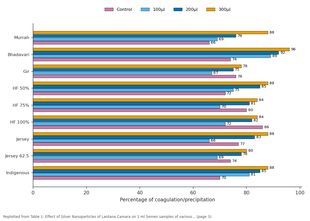
Control and 100µl and more: Murrah vs Bhadavari vs Gir vs HF 50% vs HF 75%
Replotted from Table 1 (page 3) of the original paper. Every bar is the paper's own table value.
View source evidence ↗Figures & tables
From the original paper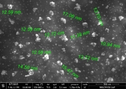 Figure 1
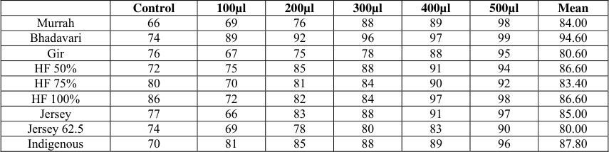 Table 1
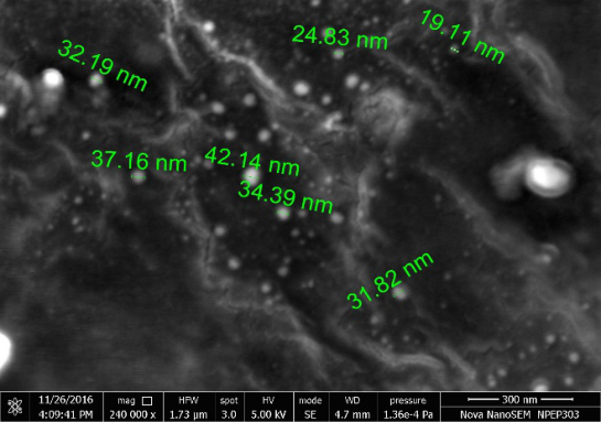 Figure 2
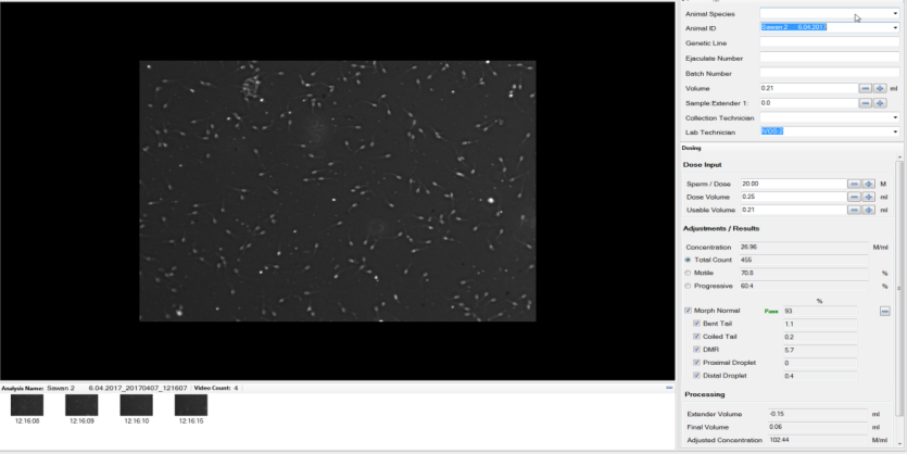 Figure 3
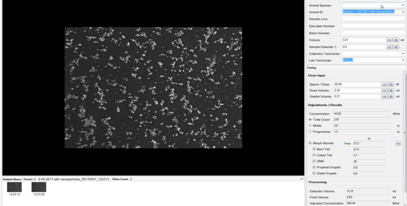 Figure 4
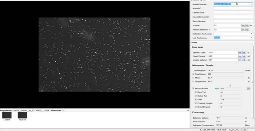 Figure 5
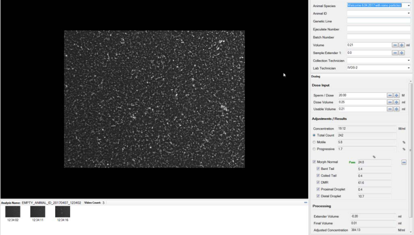 Figure 6
Introduction
The multidisciplinary field of nanotoxicology focuses on determining the extent to which nanomaterials (materials with at least one dimension <100 nm) pose a hazard to human health and the environment. The small size, large surface area-to-volume ratio, and quantum size effects of nanoscale materials may lead to biological effects that differ from those induced by their larger counterparts [1] . Although human beings have been exposed to airborne nanosized particles throughout their evolutionary stages, such exposures have increased dramatically over the last century [2] . The rapidly developing field of nanotechnology will result in new sources of this exposure, through inhalation, ingestion, skin uptake, and injection of engineered nanomaterials. Air pollution research has suggested that particles may be more toxic to cells at the nanoscale. Despite the wide use of nanomaterials, there is a lack of information about the impact of the nanoparticles (NPs) on health and environment. There is only limited knowledge about the toxicity of nanoparticles, including in reproductive medicine for example, regarding effects on spermatozoa. Spermatogenesis is a complex process that is very sensitive to environmental toxicants [3] , among these NPs may exert negative effects in many organs including testis. However, in the field of reproduction biology, studies on the effects of NPs on germ cells and ejaculated spermatozoa are scanty. Silver Nanoparticles exert a significant dose-dependent effect on motility and viability of sperms. Ag-NPs seem to show a slightly elevated toxicity as compared to gold nanoparticles [4] .
In a study on donor sperm stated that the gold nano-particles show dose-dependent effects on sperm motility [5] . Nanoparticles can reduce the sperm parameters such as motility and normal morphology and secondly affect sperm chromatin remodelling and cause the increase instability of chromatin and also increase the rate of sperm DNA damage in mice. These deleterious effects were more obvious in maximum dose and chronic phase [6] . Nuclear sperm DNA damage has a relationship with male infertility. Assessment of sperm DNA damage appears to be a potential tool for evaluating semen samples before the use in as reproduction, helping to select spermatozoa with intact DNA or with the least amount of DNA damage for use in assisted conception [7] . Studies also suggest that Titanium Oxide Nanoparticles may have cytotoxic effect on buffalo spermatozoa by affecting sperm functionality and causing high amount of DNA fragmentations [8] . A molecular examination revealed a reduction of free thiol residues on the cell membrane after nanoparticle exposure, which could explain the decrease in sperm motility [9] . Evaluation of sperm cell motility and morphology is an important parameter in the examination of sperm quality and in the establishment of correlations between sperm quality and fertility [10] . Computer-assisted sperm analysis (CASA) allows an objective assessment of different cell characteristics: motion, velocity, and morphology for quality assurance of semen and for the understanding of the diversity of sperm responses to changes in the environment CASA systems are important [11] . Moreover, filters can be applied to optimize sperm recognition/tail detection, debris rejection and advanced analysis for semen samples with "noisy" backgrounds (full of debris). Together these features greatly assist in the more accurate determination of percentage sperm motility [12] . In view of above, present study was undertaken to assess the sperm toxicity of silver nanoparticles by using Computer Assisted Semen Analysis (CASA). The trial of effect of the nanoparticles on semen sample was first carried out with the weed Lantana Camara but it proved to be toxic to the semen sample, so a considerable thought about the medicinal plant might prove to be beneficial was taken into trials. Hence later the experiment was carried out with the medicinal plant Acorus calamus which also proved to be toxic when treated with semen sample in the form of its nanoparticles. Lantana camara, a noxious weed, has been expanding and now established in many regions of the world, including India. As it poses major threats to ecosystem, it has been in the focus of control attempts [13] . Lantana camara is a small perennial shrub in the form of dense thickets in a variety of environments. It has been nominated as among 100 of the "World's Worst" invaders by the IUCN Invasive Species Specialist Group and it has been listed as a noxious weed in many countries. Phytochemical tests reveal the presence of alkaloids, tannins, saponins, flavanoids, carbohydrates, steroids and triterpinoids [14] . Acorus calamus (Sweet flag) is a monocot plant. Its scented leaves and rhizomes have been traditionally used medicinally against different ailments [15] . The results of preliminary phytochemical screening from Extracts of leaves of Acorus calamus revealed the presence of Carbohydrates, Tannins, Amino acids, Alkaloids, Sterols, Terpinoids, Glycosides, Saponins and Flavanoids [16] . The presence of triterpenoids and alkaloids of both the plants interferes with the metabolism and proves to be toxic to a number organisms like bacteria and fungi [17] .
Materials and Methods
Materials and Methods
Materials 2.1.1 Semen Sample
Fresh semen samples were collected from healthy bulls and cattle housed at Semen Laboratory, BAIF-CRS (Bhartiya Agro Industrial Foundation -Central Research Station) by using Artificial Vagina and monitored for progressive motility, acrosomal integrity and sperm shape on CASA. It was collected and transferred according to the published standard procedure in reproductive medicine (Semen analysis. US patent 20040146848).
Plants
The plants Acorus calamus and Lantana camara were obtained from More Garden Nursery campus, Manjri, Pune. These plants were authenticated by from BSI (Botanical Survey of India), Pune (Voucher No. BSI/WRC/100-1/Tech./ 2017/1).
2.1.3
Silver Nitrate (Fischer Chemicals) (mol wt 169.87 g/mol) was used.
Preparation of Silver Nanoparticles
Silver Nanoparticles were biologically synthesized by using 2 aqueous plant extracts.20 g of finely cut leaves were thoroughly washed with Distilled water and crushed to prepare aqueous extract and stored at 4 °C for further use. 50 ml of leaf extract of both the plants was added to equal volume of Silver Nitrate Solution (10 -3 ) and was incubated at 37 0 c overnight till the colour change (Bio reduction) was observed. All standard aseptic techniques were maintained.
Characterization of Silver Nanoparticles
The nano particles were confirmed, by the appearance of brown colour with naked eyes and characterized by UV-Visible double beam spectroscopy (UV-1800 Shimadzu UV spectrophotometer, Japan) at a wavelength of 400-600 nm. The crystalline nature of nanoparticles was checked by XRAY Diffraction at the Central Instrumentation Facility (CIF) Department, Savitribai Phule University of Pune. The topography of nanoparticles was studied by Field Emission Scanning Electron Microscope (FESEM) (FEI Nova Nano, Lincoln, SEM 450) and Energy Dispersive Analysis of X-rays (EDAX) (Bruker 9430) at the Central Instrumentation Facility (CIF), Savitribai Phule University. The average size of nanoparticles was found to be 20.
Methodology
The fresh semen sample was monitored for 8 various Breeds on CASA (Computer Assisted Semen Analysis) (HT CASA II, 2014, Beverly). Single sample of the following breeds was analysed:- Bhadavari Murrah Gir HF50% HF75% HF 100% Jersey Jersey 62.5% Indigenous These samples were analysed for motility, acrosomal integrity on CASA, and all of the samples monitored showed motility above 65%. Later these samples were treated with silver nanoparticles solution at various concentrationsi.e 100µl, 200µl, 300µl, 400µl and 500µl and immediate coagulation of the sperms at the bottom of the tube (as a precipitate) was observed through the naked eyes. In the next step these samples were analysed on CASA and they showed Zero Motility, the acrosomes were damaged, deheaded sperms were also observed as a result interaction of the Silver nano particles of both the plants i.e. Acorus calamus and Lantana camara.
Results and Discussion
Results and Discussion
Cattle Semen Sample
Cattle Semen Sample
Conclusions
We demonstrated in a preliminary, small study that the motility of spermatozoa was affected by the presence of silver nanoparticles. The Silver nanoparticles prove to be toxic to the Semen sample of all the breeds of bulls and cattle.
When the semen samples were treated with plant extracts it lead to zero progressive motility. When the semen sample was treated with Silver nanoparticles of both the plants it lead to immediate coagulation of the sperms and immediate precipitate of sperm cells was seen at the bottom of the tube separating the seminal fluid as the supernatant. Similarly when the semen sample was treated with silver nitrate solution it leads to strong precipitation and coagulation of the semen sample. Now with the plant extracts there was no coagulation of sperms seen. But the progressive motility reduced to zero. The motility of the control sperm was 75%. This states that silver in the nanoparticles leads to the damage of the sperms and motility reduction. Whereas the plant extract leads to destroying of the motility of sperms. The sperms were monitored on CASA and it leads to zero motility, less than 15% acrosomal integrity, and also Deheaded sperms were observed (Fig No 3 -Fig No 6). When the Silver Nanoparticles solution was added to the Semen sample immediate precipitation / coagulation of sperms was observed even at lowest concentrations of 100µl and as the concentration of silver nanoparticles increased the precipitation. In the mixture, penetration of silvernano particles into the sperm head and tails could be observed. With increasing concentration of the silver nanoparticle solution in the semen sample it lead to increased coagulation and damaged spermatozoa. We also noted fragmentation of sperm in the study sample that included the nanoparticle solution. Previously studies to test the effect of magnetite nanoparticles show that penetration of magnetite nanoparticles into sperm cells can be visualized (18) . Hence we can conclude that Silver nanoparticles are toxic to the bull semen. With increasing concentration the acrosomal damage, tail damage, Coagulation of sperms increased.
Acknowledgement
The authors gratefully acknowledge the Program Director Dr J R Khadse, BAIF Development Research Foundation for providing us with all the facilities to conduct present research work at BAIF CRS campus, Uruli kanchan, Pune. We express gratitude towards the Central Instrumentation Facility (CIF), Savitribai Phule University of Pune for providing us with all the facilities required for Characterization of Silver Nano particles At last but not the least, the authors are highly grateful to Miss Tejashree Shirsath for supporting us to improve the quality of this research article.
Figures and tables from the paper
| — | Control | 100µl | 200µl | 300µl | 400µl | 500µl | Mean |
|---|---|---|---|---|---|---|---|
| Murrah | 66 | 69 | 76 | 88 | 89 | 98 | 84.00 |
| Bhadavari | 74 | 89 | 92 | 96 | 97 | 99 | 94.60 |
| Gir | 76 | 67 | 75 | 78 | 88 | 95 | 80.60 |
| HF 50% | 72 | 75 | 85 | 88 | 91 | 94 | 86.60 |
| HF 75% | 80 | 70 | 81 | 84 | 90 | 92 | 83.40 |
| HF 100% | 86 | 72 | 82 | 84 | 97 | 98 | 86.60 |
| Jersey | 77 | 66 | 83 | 88 | 91 | 97 | 85.00 |
| Jersey 62.5 | 74 | 69 | 78 | 80 | 83 | 90 | 80.00 |
| Indigenous | 70 | 81 | 85 | 88 | 89 | 96 | 87.80 |
Table text extracted from the original PDF; superscript letters are shown inline. The original table image is below.
View original table image
References
16 in the original paper- Haynes CL, Hecht S. Chemical Research in Toxicology Editorial Advisory Board Members' Favorite CRT Articles. Chemical Research in Toxicology. 2016; 29(2):133. DOI: 10.1021/acs.chemrestox.6b00025
- Oberdörster G, Oberdörster E, Oberdörster J. Nanotoxicology: An Emerging Discipline Evolving from Studies of Ultrafine Particles. Environmental Health Perspectives. 2005; 113(7):823-839. doi:10.1289/ehp.7339.
- Wong EWP, Cheng CY. Impacts of environmental toxicants on male reproductive dysfunction. Trends in pharmacological sciences. 2011; 32(5):290-299. doi:10.1016/j.tips.2011.01.001.
- Moretti E, Terzuoli G, Renieri T, Iacoponi F, Castellini C, Giordano C et al. In vitro effect of gold and silver nanoparticles on human spermatozoa. Andrologia. 2013; 45:392-396. [PubMed]
- Wiwanitkit V, Sereemaspun A, Rojanathanes R. Effect of Gold Nanoparticles on Spermatozoa the First World Report. Fertil Steril 2009; 91:7-8. [PubMed]
- Nazar M, Talebi AR, Hosseini Sharifabad M, Abbasi A, Khoradmehr A, Danafar AH. Acute and chronic effects of gold nanoparticles on sperm parameters and chromatin structure in Mice. International Journal of Reproductive Biomedicine. 2016; 14(10):637-642.
- Tarozzi N, Bizzaro D, Flamigni C, Borini A. Clinical relevance of sperm DNA damage in assisted reproduction. Reprod Biomed Online. 2007; 14:746-757.
- Pawar K, Kaul G. Toxicity of titanium oxide nanoparticles causes functionality and DNA damage in buffalo (Bubalus bubalis) sperm in vitro. Toxicol Ind Health. 2012; 30:520-533.
- Ulrike Taylor, Annette Barchanski, Svea Petersen, Wilfried August Kues, Ulrich Baulain, Lisa Gamrad, 2013. Gold nanoparticles interfere with sperm functionality by membrane adsorption without penetration. Nanotoxicology 2014, 8. http://dx.doi.org/10.3109/17435390.2013.859321
- Amann RP, Waberski D. Computer-assisted sperm analysis (CASA): capabilities and potential developments. Theriogenology 2014; 81(1):5-17
- Mortimer ST, Horst Gv, Mortimer D. The future of computer-aided sperm analysis. Asian J Androl. 2015; 17:545-53
- Neena Priyanka, PK Joshi. A review of Lantana camara studies in India International Journal of Scientific and Research Publications. 2010; 3(10)-2013(1) ISSN 22503153
- Jain Shonu, Itoria Priyank, Joshi Amit, Dubey BK. Pharmacognostic and Phytochemical Evaluation and Antipyretic Activity of Leaves of Lantana Camara Linn India International Journal of Biomedical and Advance Research IJBAR. 2011, 02(08).
- Sreekanth SK, Ganesan V, Kannappan M, Sreekanth D. In-vitro Antimicrobial Activity and phytochemical analysis of Acorus calamus Rhizomes. Res Pharm Health Sci. 2015; 1(1):1-5
- Saxena Mamta, Saxena Jyoti, Phytochemical Screening of Acorus Calamus and Lantana Camara International Research Journal of Pharmacy IRJP. 2012, 3(5).
- Ben-David Makhluf S, Qasem R, Rubinstein S, Gedanken A, Breitbart H. Loading magnetic nanoparticles into sperm cells does not affect their functionality. Langmuir 2006; 22:9480-2.
Cited by 2
From OpenAlex, OpenCitations and reference lists read by Scinovex
- A review on applications and toxicities of metallic nanoparticles in mammalian semen biology ↗
Amarjeet Bisla, Mrigank Honparkhe, Neeraj Srivastava · Andrologia · 2022
The advent of nanotechnology since the 1950s, when the well-known physicist Richard P. Feynman talked in his famous talk about "There's plenty of room at the bottom", has led to incredible contribution of nanotechnology in the fields of medical and veterinary therapeutics, diagnostics and other applications. Semen biology dealing with the study of spermatozoa and its related physiological and pathological…
❝ 18 - Forest fungi nanotechnology and their applications ↗
Suman Bhandary, Yashaswi Dutta Gupta, Ritika Chakraborty · Elsevier eBooks · 2024
❝ 1