
Research article · 2017 · Full text
International Journal of Veterinary Sciences and Animal HusbandryAvian tuberculosis in a backyard poultry flock in Tunisia: Case report
Volume 2(6) · Pages 34–37
Research areasTuberculosis Research and EpidemiologyVirology and Viral DiseasesMicrobial infections and disease research
Abstract
Author-writtenTuberculosis is a chronic infectious disease affecting several species of domestic and wild birds. It is due to the infection of the gastrointestinal tract, liver and spleen by various Mycobacterium species. This disease is more commonly seen in zoos and wild avian species (Del Pilar Silva et al., 2009) [5]. Mycobacterium avium (serotypes 1, 2 and 3) and Mycobacterium genavense represent the most incriminated germs in the case of avian tuberculosis. This topic describes the observations of the first report of a typical case in a chicken issued from a backyard flock received at the avian clinic of the National Veterinary School of Sidi Thabet. This case prompted-us to bring and develop pathological aspects.
Summary
Source-checkedPlain-language reading of the full paper, prepared with AI and checked against the text. Each point links to the authors' own words.
Why it matters
- Avian tuberculosis is a chronic infectious disease caused by Mycobacterium species that typically infect the liver, spleen, and gastrointestinal tract of wild and domestic birds. View evidence ↗
- The most common pathogens responsible for this disease are Mycobacterium genavense and various serotypes (1, 2, and 3) of Mycobacterium avium. View evidence ↗
- The infection can potentially spread to humans, with immuno-depressed individuals being particularly susceptible. View evidence ↗
The question
- The study aims to describe the anatomo-pathological features of a typical case of avian tuberculosis in a backyard layer hen in Tunisia. View evidence ↗
What they did
- The case involved an 18-month-old lean layer hen from a free-range backyard flock of 20 birds of various ages and origins. View evidence ↗
- An autopsy was conducted at the National Veterinary School of Sidi Thabet, utilizing Ziehl-Neelsen coloration on damaged organs. View evidence ↗
- The liver, spleen, intestines, pancreas, and kidneys were fixed in 10% formalin and analyzed using hematoxylin-eosin (HE) and Ziehl-Neelsen stains. View evidence ↗
What they found
- Gross pathology showed an enlarged, greenish-brown liver containing gray-yellowish nodules ranging from 2 to 5 mm in size. View evidence ↗
- Caseous nodules were also found in the spleen, intestine, mesentery, gizzard, pancreas, and kidneys, while the ovary was atrophied and deformed. View evidence ↗
- Histopathology revealed immunological granulomas (Koester follicles) in the liver, spleen, intestines, kidney, and pancreas, characterized by central necrosis, macrophage/epithelioid reactions, and Langhans giant cells. View evidence ↗
- Ziehl-Neelsen staining confirmed the presence of acid-fast bacilli within the cytoplasm of macrophages and at the center of the granulomas. View evidence ↗
- Unlike some other reported cases, no lesions were found in the heart, lungs, or infra-orbital sinus of this specific hen. View evidence ↗
What it means
- The authors conclude that tuberculosis in backyard flocks is promoted by poor biosecurity, mixing different animal species, and introducing birds of unknown sanitary status. View evidence ↗
- The diagnosis of avian tuberculosis in the adult hen was based on clinical signs of cachexia and chronic diarrhea, combined with autopsy and histopathological findings. View evidence ↗
Figures & tables
From the original paper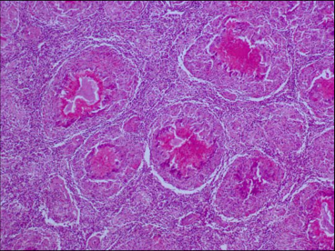 Figure 5
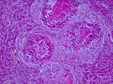 Figure 4
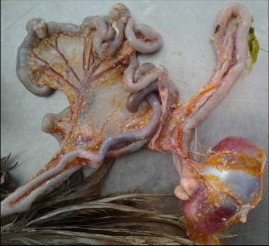 Figure 3
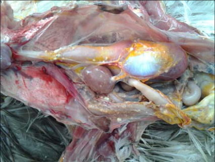 Figure 2
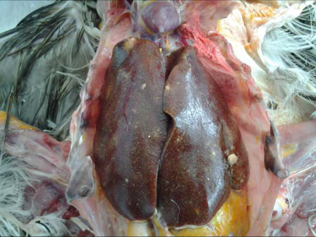 Figure 1
 Table 1
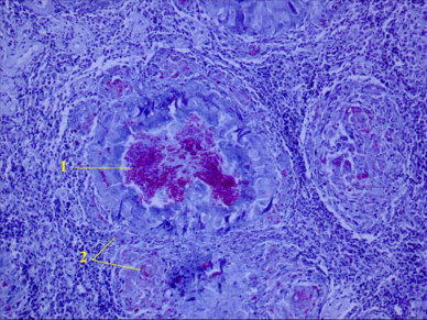 Figure 7
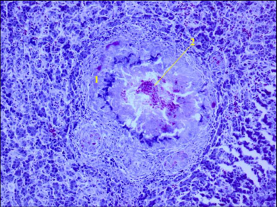 Figure 6
Introduction
Tuberculosis is a chronic infectious disease affecting several species of domestic and wild birds. Caused by the proliferation of different Mycobacterium species in the digestive tract, the liver and the spleen, this infection is mostly observed in zoological parks and in wild avian species (Del Pilar Silva et al., 2009 ; Stepień-Pyśniak et al., 2016 ; Zhu et al., 2016) [5,18,19] . Mycobacterium avium (serotypes 1, 2 and 3) and Mycobacterium genavense are the most incriminated pathogens in avian tuberculosis. Spread of the infection to human is possible, particularly in immuno-depressed patients. Hejlicek and Treml (1995) [11] classified sensitive avian species to experimental infection by Mycobacterium avium in four groups: very sensitive species (domestic fowl, sparrow, pheasant, partridge and seagull), little sensitive species (guinea fowl, turkey), resistant species (goose, duck) and very resistant species (pigeon, turtledove and crow). Infected birds are considered as the most important source of germ, which is issued from ulcerative lesions of intestines and excreted in feces to contaminate environment. Mycobacterium, such as M. avium, can resist during several years (more than 4 years) which can explain the reappearance of the disease in the same farm, even after elimination of all birds. Resistance of these germs is also considerable in variable temperatures. They can survive about 168 days to 244 days at 20°C and 37°C, respectively (Shalk et al., 1935; Schliesser and Weber, 1973) (in Fulton and Thoen, 2003) [6] . M. avium was isolated from incubated eggs of naturally infected breeder hens. However, correspondent chicks do not develop the disease. Dissemination of the bacteria can be assured by poultry infected carcasses. The aim of this article is to report and develop, for the first time, the anatomo-pathological aspects of a typical case of tuberculosis in backyard layer hen, diagnosed in the Avian Clinic of the National Veterinary School of Sidi Thabet (Tunisia).
Materials and Methods
Materials and Methods
Case history
Sporadic mortality was appeared in a free-range poultry flock, composed of 20 birds, with different provenance and age, reared outdoor. The 18 months old dead layer hen was very lean. It presented an allowance and a whitish diarrhea before death. Biosecurity measures in this little farming are poor, or even absent.
The procurement is based on the purchase of unknown provenance chickens.
Gross pathology and bacteriology analysis
The autopsy, was performed at the clinic of Avian Pathology of The National Veterinary School of Sidi Thabet (Tunisia). A Ziehl-Neelsen coloration was performed on different damaged organs.
Histopathological study
Histopathological analyzes were performed at the Histology department of the National Veterinary School of Sidi Thabet (Tunisia). Organ Harvesting (liver, spleen, intestines, pancreas, kidneys) are immediately emerged in a 10% formalin solution in order to fix them. After processing sections according to conventional methods of histological techniques, two types of stains are made, hematoxylin-eosin (HE) and the Ziehl-Neelsen.
Results
Results
Gross pathology and bacteriology
The necropsy exam revealed during the external examination, weight loss and traces of white diarrhea. At the opening of the thoraco-abdominal cavity, the liver appears enlarged, greenish-brown color and dotted with gray-yellowish nodules of variable size (2 to 5 mm), which are detected also within the organ. (Figure 1) These nodules are observed in the other viscera, mainly spleen, intestine, mesentery, gizzard, pancreas and kidneys (Figures 2 and3). The Ovary appears deformed and atrophied.
Histopathology
This study showed the presence of immunological granulomas (Koster follicles) in various tissues and viscera. Indeed, liver, spleen, intestines, kidney and pancreas are the site of multiple granulomas centered on an acidophilic disintegration necrosis range, surrounded by a macrophage and epitheloid reaction, with many small and irregular Langhans giant cells. The periphery was occupied by a fibroconnective hull and a few lymphocytes (Figures 4 and5). Clumps of acid-fast bacilli were noted in Ziehl-Neelsen stained sections. These bacilli were accumulated in the center of the granulomas and in the cytoplasm of macrophages (Figures 6 and7).
Discussion
Absent in commercial poultry in Tunisia, avian tuberculosis is rather observed in free-range flocks, where it be diagnosed in fowl, turkey, pheasant and, more rarely in goose and duck.
Several risk factors can favor the emergence of the disease in backyard poultry breeding. Indeed, unknown provenance of animals (local markets, other farms), mixture of avian and mammalian species in the same flock, possible contact between domestic poultry and wild birds and poor level of biosafety measures are considerable risk factors in the introduction and development of mycobacterium species in the free-range flocks (Hejlicek and Treml, 1993 [1,4,10,12] . Lesions caused by M. avium may be locolized or generalized in mammals, including cattle, rabbits and horses often present on farms in contact with infected birds (Lesslie and Birn, 1967; Gunnes et al., 1995) [8,14] . The germ can multiply in different tissues for a long time making the animal susceptible to any tuberculin. Clinical signs of avian tuberculosis are not pathognomonic. The infected animal passes through three phases: a latency phase, a development phase of lesions and finally phase of cachexia. The Ingestion of the tuberculosis bacillus causes an intestinal infection and promotes bacillemia. The liver is among the first organs affected, then the germ spreads to the other tissues. Avian tuberculosis is rarely observed in young birds, which are not necessarily more resistant to infection, but the disease is more common in adults. Indeed, the disease gradually settles over a long period. The lesions of avian tuberculosis are often less severe in young chickens than in older birds. Gross lesions described in our study were similar to those revealed by many authors in different bird species naturally infected by avian tuberculosis. In this way, lesions observed in the liver, the spleen and the intestines were in agreement with the results of Randall and Reece (1996) [17] , Gonzalez et al. (2002) [7] , Mayahi et al. (2013) [16] , Majeed et al. (2014) [15] and Zhu et al. (2016) [19] . However, contrarily to the descriptions of Beytut et al. (2001) [2] and Gonzalez et al. (2002) [7] , no lesions were identified in the lungs and in the heart and the infra-orbital sinus, respectively. In addition, we have not detect lesions in the conjunctiva and the joints as was described by Mayahi et al.
(2013) [16] on pigeons affected naturally by tuberculosis.
(Table 1) Inflammatory reaction noted in the intestine, related to the tuberculous granulomas, could explain the malabsorption and the disturbance of the digestive tract function, explaining growth retardation, weight loss and decrease in production. Histopathological lesions were those of a tuberculosis. They were characterized by the presence of multiple follicles of Koester with different size. Our observations were similar to those of many authors. Besides the examined viscera in this study, tuberculosis lesions can have other locations, including the bones, lungs, subcutaneous connective tissue, trachea and more rarely the nervous system in domestic and wild avian species (Hawkey et al., 1990 [2,9,17,19] .
| Bird | Liver | Spleen | Intes. | Pancr. | Kidney | Gizard | Lung | Heart | Loco. | Conj. | Sinus | Authors |
|---|---|---|---|---|---|---|---|---|---|---|---|---|
| BC | + | + | + | + | + | + | - | - | - | - | - | Our study |
| LH | + | + | + | - | - | - | - | + | - | - | + | Gonzalez et al. (2002) [7] |
| LH | + | + | + | - | - | - | + | - | - | + | - | Beytut et al. (2001)[2] |
| Pigeon | + | + | + | - | - | + | - | + | + | + | - | Mayahi et al. (2013)[16] |
| Pheasant | + | + | + | - | - | - | - | - | - | - | - | Kul et al. (2005) |
| BC: Backyard chicken; Conj. : Conjonctiva; LH: Layer hen: Intes. : Intestine; Loco. : Locomotor system; Pancr. : Pancrzas |
Table text extracted from the original PDF; superscript letters are shown inline. The original table image is below.
View original table image
The absence of macroscopic lesions on some viscera does not exclude the disease. In the same context, typical histological lesions of tuberculosis are mentioned on the lungs (Gonzalez et al., 2002) [7] and the intestines (Kul et al., 2005) [13] showing no lesion at the necropsy examination. The frequency and severity of lesions in the intestines, suggesting oral infection. Spread of Mycobacteria from the intestine promotes the liver and the spleen contamination (Bowes, 1993) [3] . Finally, the germ is excreted mainly through the droppings. These contribute to the contamination of the rearing environment and eggshell at the time of laying and thereafter.
Conclusion
Avian tuberculosis was diagnosed in an adult hen issued from a backyard flock. The cachexia, the chronic diarrhea and the lesions observed at autopsy allowed us to suspect this disease. The recourse to the histopathological examination revealed necrotic and granulomatous lesions and the presence of acidfast bacilli in various affected viscera. The existence of tuberculosis in backyard flocks is favored by the lack of biosecurity measures, the mixing of bird and animal species and the introduction in the farm of birds with unknown origin and sanitary status.
Conflict of interest
The authors declare that there is no conflict of interest.
Figures and tables from the paper
References
19 in the original paper- Abda S, Mamo G, Worku A, Ameni G. Preliminary study on avian tuberculosis and associated risks in domestic chickens at Shashemene district, Ethiopia. Journal of Biology and Medical Sciences. 2015; 3:13-23.
- Beytut E, Atabay HI, Akça A. Tuberculosis and sarcosporidiosis in the periorbital location in a hen. Kafkas Üniv. Vet Fak. Derg. 2001; 7:213-217.
- Bowes V. Avian tuberculosis in ostriches. Canadian Veterinary Journal. 1993; 34:758.
- Dhama K, Mahendran M, Tomar S. Pathogens transmitted by migratory birds: threat perceptions to poultry health and production. International Journal of Poultry Science. 2008; 7(6):516-525.
- Del Pilar Silva A, Leon CI, Guerrero MI, Neira R, Arias L, Rodriguez G. Avian tuberculosis of zoonotic importance at a zoo on the Bogotá Andean plateau (Sabana), Colombia. Canadian Veterinary Journal. 2009; 50:841-845.
- Fulton RM, Thoen CO. Tuberculosis, In: Saif YM, HJ Barnes, JR Glisson, FM Fadly, LR Mc Dougald and DE Swayne, Diseases of Poultry. 11 th Eds., Iowa State University Press, Ames, IA, USA, 2003, 836-844.
- Gonzalez M, Rodriguez-Bertos A, Gimeno I, Flores JM, Pizarro M. Outbreak of avian tuberculosis in 48-week-old commercial layer hen flock. Avian Diseases. 2002; 46(4):1055-1061.
- Gunnes G, Nord K, Vatn S, Saxegaard F. A case of generalized avian tuberculosis in a horse. Veterinary Record. 1995; 136:565-566.
- Hawkey C, Kock RA, Henderson GM, Cindery RN. Hematological changes in domestic fowl (Gallus gallus) and cranes (Gruiformes) with Mycobacterium avium infection. Avian Pathology. 1990; 19:223-234.
- Hejlicek K, Treml F. The occurrence of mycobacteriosis in free-living birds at different epizootiological situation of poultry tuberculosis (in Czech). Veterinary Medicine. 1993; 38:305-317.
- Hejlicek K, Treml F. Comparison of the pathogenesis and epizootiologic importance of avian mycobacteriosis in various types of domestic and free-living syntropic birds. Veterinary Medicine (Praha). 1995; 40(6):187-94.
- Kriz P, Kaevska M, Bartejsova I, Pavlik I. Mycobacterium avium subsp. avium found in raptors exposed to infected domestic fowl. Avian Diseases. 2013; 57(3):688-692.
- Kul O, Tunca R, Haziroglu R, Diker KS, Karahan S. An outbreak of avian tuberculosis in peafowl (Pavo cristatus) and pheasants (Phasianus colchicus) in a zoological aviary in Turkey. Veterinary Medicine of Czech. 2005; 50:446-450.
- Lesslie IW, Birn KJ. Tuberculosis in cattle caused by the avian type tubercle bacillus. Veterinary Record. 1967; 80:559-564.
- Majeed SK, Al-Sereah BA, Hamza FZ. Diagnosis of avian tuberculosis in a poultry farm in Wassit province (Case report). AL-Qadisiya Journal of Veterinary Medicine Science. 2014; 13:9-13.
- Mayahi M, Esmaeilzadeh S, Mosavari N, Asadollahi KP. Histopathological study of avian tuberculosis in naturally infected domestic pigeons with Mycobacterium avium subsp. avium. Iranian Journal of Veterinary Science and Technology. 2013; 5(1):45-56.
- Randall CJ, Reece RL. Color atlas of avian histopathology. Ed. Mosby-Wolfe, 1996, 232.
- Stepień-Pyśniak D, Puk K, Guz L, Wawrzyniak A, Marek A, Kosikowska U. Avian mycobacteriosis caused by Mycobacterium avium subspecies avium in four ornamental birds and in vitro drug sensitivity testing of isolates. Berl Munch Tierarztl Wochenschr. 2016; 129(12):65-71.
- Zhu DK, Song XH, Wang JB, Zhou WS, Ou XM, Chen HX, et al. Outbreak of avian tuberculosis in commercial domestic Pekin ducks (Anas platyrhynchos domestica). Avian Diseases. 2016; 60(3):677-680.