
Research article · 2016 · Full text
International Journal of Veterinary Sciences and Animal HusbandryAnalysis of rumen fluid in apparently healthy slaughtered cattle at Gondar Elfora abattoir
Volume 1(2) · Pages 37–42
Research areasEcology and biodiversity studies
Abstract
Author-writtenA cross sectional study was conducted from November 2013 to May 2014 in Gondar University, Amhara region, Northwest of Ethiopia to determine the physical, chemical and microbial characteristics of rumen fluid and to develop a base line data on healthy parameters of rumen fluid for therapeutic transfusion in clinical ruminal disorders from apparently healthy indigenous cattle of Ethiopia. Analysis was performed by physical, chemical and microscopical methods. Of the 384 rumen fluid samples of indigenous cattle examined color was found 54.7% yellowish brown, 25% brownish green, 11.2% greenish and 9.1% milky; consistency 88.8% Viscous, 4.7% watery and 6.5% frothy; odour 89.6% aromatic and 10.4% sour; sedimentation 95.6% were completed within 4-8 minute and 4.4% no appreciable sedimentation or floatation; pH 49% ranges from 5.5-6.5, 34.6% from 5.6 to7 and 6% greater than 7; complete cellulose digestion were observed 89.6% within 48-56hrs, 10.4% more than 56 hrs; methylene blue reduction achieved 65.4% within 3 minute, 30.2% within 6 minute and 4.4% more than 6 minute; glucose fermentation test revealed that 89.6% had gas formation and 10.4% with no gas formation; protozoan motility observed 13.8% as excellent motility, 75.4% with medium motility, 7.8% having sluggish motility and 6.0% with no motility; proportion of bacteria 92% with dominant gram negative bacteria and the remaining 8% of samples collected had dominant positive bacteria. This is the basic base line data for rumen content transfusion and for future study.
Summary
Source-checkedPlain-language reading of the full paper, prepared with AI and checked against the text. Each point links to the authors' own words.
Why it matters
- Rumen microflora is essential for converting plant fibers into milk and meat; disruptions to this process via rumen disorders can lower animal productivity. View evidence ↗
- Inactive rumens can potentially be treated through transfaunation, which involves providing rumen fluid from a healthy animal of the same species. View evidence ↗
- Analyzing rumen fluid is a key method for determining the current state of the rumen environment and its contents. View evidence ↗
The question
- The study aimed to investigate the physical, chemical, and microbial characteristics of rumen fluid in healthy cattle. View evidence ↗
- The researchers sought to establish baseline data for healthy rumen fluid to guide therapeutic transfusions for animals with ruminal disorders. View evidence ↗
What they did
- A cross-sectional study was performed from November 2013 to May 2014 at the Gondar ELFORA Abattoir in Ethiopia. View evidence ↗
- Samples were collected post-mortem from animals that showed no gross lesions during examination. View evidence ↗
- The rumen fluid was analyzed using a combination of physical, chemical, and microscopic techniques. View evidence ↗
What they found
- The majority of samples (88.8%) had a viscous consistency, while 6.5% were frothy and 4.7% were watery. View evidence ↗
- Rumen fluid odor was aromatic in 89.6% of the samples and sour in 10.4%. View evidence ↗
- Sedimentation was completed within 4-8 minutes for 95.6% of the samples, with 4.4% showing no appreciable sedimentation. View evidence ↗
- Rumen pH varied from 5.5 to over 7.0, with 49% between 5.5-6.5, 34.6% between 5.6-7, 10.4% below 5.5, and 6% above 7. View evidence ↗
- Complete cellulose digestion occurred within 48-56 hours in 89.6% of samples, while 10.4% took longer than 56 hours. View evidence ↗
- Methylene blue reduction occurred within 3 minutes for 65.4% of samples, within 6 minutes for 30.2%, and took over 6 minutes for 4.4%. View evidence ↗
- Glucose fermentation, indicated by gas formation, was positive in 89.6% of the samples. View evidence ↗
- Protozoan motility varied: 13.8% showed excellent motility, 22.1% medium, 7.8% sluggish, and 6.0% showed no motility. View evidence ↗
- Gram staining showed that 92% of the samples were dominated by gram-negative bacteria, while 8% were dominated by gram-positive bacteria. View evidence ↗
What it means
- The authors conclude that these findings provide baseline data to assess rumen fluid quality for therapeutic transfusions and the restoration of the rumen environment. View evidence ↗
- The authors recommend further research to establish baseline data for other breeds and agro-climatic regions due to potential variations in feed and microflora. View evidence ↗
Visual summary
3 visualsIllustrations explain the paper; charts are replotted from the paper's own table values.
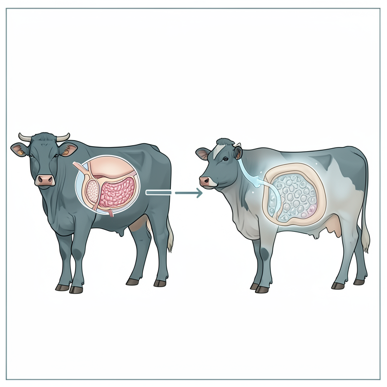
Study Goal Overview
AI-generated conceptual illustration (adult cattle and rumen fluid). It explains the idea and does not show study data.
View source evidence ↗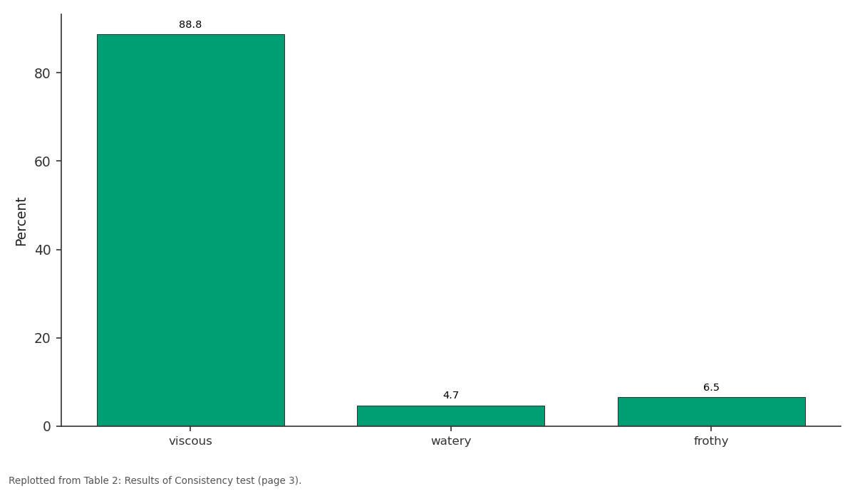
Percent: viscous vs watery vs frothy
Replotted from Table 2 (page 3) of the original paper. Every bar is the paper's own table value.
View source evidence ↗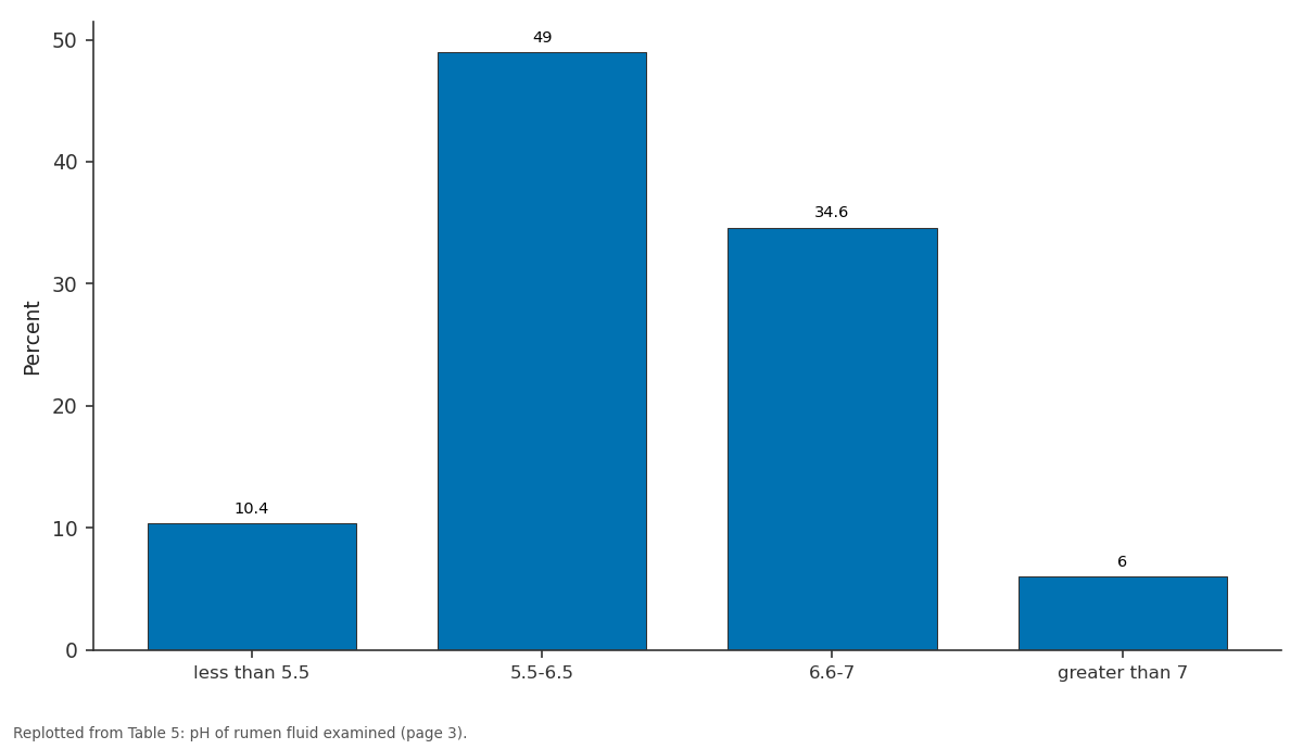
Percent: less than 5.5 vs 5.5-6.5 vs 6.6-7 vs greater than 7
Replotted from Table 5 (page 3) of the original paper. Every bar is the paper's own table value.
View source evidence ↗Figures & tables
From the original paper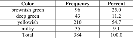 Table 1
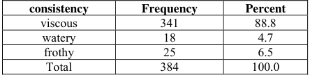 Table 2
 Table 3
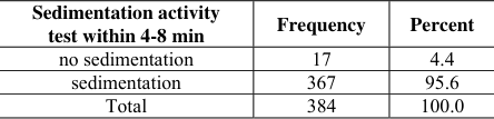 Table 4
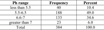 Table 5
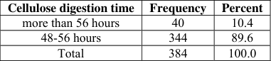 Table 6
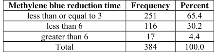 Table 7
 Table 8
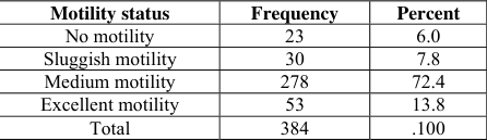 Table 9
 Table 10
Introduction
The majority of the world's rural poor, and a significant proportion of the urban poor, keep livestock and use them in a variety of ways that extend far beyond income generation. In many cases, livestock are a central component of smallholder risk management strategies (Bailey et al., 1999). Rumen micro flora plays important role in converting plant fibers into products like milk and meat and if this rumen digestion by microbes is affected due several rumen disorders, the production and productivity of animals are reduced. Rumen is a fermentative vat which allows the continuous development of microbial population, acting as a fermentation chamber due to having, anaerobiosis, buffer mean pH near neutral, presence of bacteria, protozoa and fungi, nutrient supplement and continuous removal of digesta and fermentation products, and constant osmotic pressure (Ruckebush, 1988) [30] . Rumen fluid is a mixture of all fluids present in rumen chamber as a consequence of digestive process and ingestion from the esophagus i.e. swallowing. The ingested fluids include saliva, water, and mucus that mix with all the digestive components, enzymes, carbohydrates, nitrogen and other suspended gases (Dehority, 2003) [10] . Rumen contents can be examined for the physical aspects like; color, odor, consistency and sedimentation activity test and the chemical characteristics such as; pH, glucose fermentation, cellulose digestion and methylene blue reduction test and also as for microbial tests like protozoan motility test and gram staining for rumen bacteria (Rosenberger, 1979). This examination of rumen fluid is often essential to assist in determination of the state of rumen environment and digesta after rumen fluid was obtained mainly from slaughtered animal and sometimes by rumen pump via stomach tube. The major relevant notice during collection is avoiding contamination of the sample with saliva (Radostitis et al., 2007) [28] .
The microbial protein synthesis in the rumen depends on the growth of microorganisms and on the efficiency of using energy and nitrogen substrates, which is the main constituent of animal's body and, therefore, vital for maintenance, growth and reproduction processes. The net result of these reactions going on in the rumen is responsible for the bioconversion of feed in to such form that is utilizable by the animal as a source of energy (Thauer, 1977) [33] . Color of rumen fluid of apparently healthy cattle varies according to feed type, from gray and olive to brownish green; pure green in grazing cattle, grey in those given fodder beet, yellowish-brown in those given maize silage or straw. Abnormal color like are milky grey can be observed in the time of acidosis (Rosenberger, 1979). Normal consistency of rumen fluid of cattle is slightly viscous. Extremely viscous samples may be composed chiefly of saliva, in which case another sample will have to be taken (Smith, 1996) [32] . Odor is normally aromatic and not repellent, depending on the odor of the particular feed eaten (hay, grass, roots, kale, silage, and so on). Abnormal odors include the repulsive musty, foul odor of protein decomposition, the penetrating acid odor of lactic acid from over eating readily-digestible carbohydrate, and the stale, indifferent odor of inactive rumen juice and the smell of abomasal contents indicative of pyloric obstruction (Rosenberger, 1979). The sedimentation activity time or sedimentation or floatation test provides a rapid evaluation of microbial activity. But the abnormal time; very rapid sedimentation with no floatation occurs in rumen acidosis, prolonged anorexia, and in time of inactive micro flora from indigestible roughages (Smith, 1996) [32] . The pH of rumen fluid ranges from 5.5-7.0 in apparently healthy cattle on a balanced ration. A pH paper with half unit sensitivity is sufficient to diagnose ruminal acidosis or alkalosis. Cattle on high carbohydrate diets have lower pH than those on roughage diets. Acid pH less than 5.5 in ruminants indicates ruminal acidosis while ruminal pH greater than 7 indicates ruminal alkalosis. Simple ruminal inactivity or anorexia results in ruminal alkalosis (Fubini, 2004) [15] . Cellulose digestion test is used to evaluate the cellulose digesting ability of rumen micro flora. The rumen harbors various types of cellulolytic bacteria which are active in degradation of the components of the feed. These bacteria identified in the rumen plays an important role in cellulose digestion and have been reported in high roughage diets (Church, 1993). The common features of bacteria found in the rumen of animals fed on high roughage are; majority of the bacteria are gram negative but the number of gram positive bacteria tends to increase on increasing high energy diets in the ration (Weimer et al, 1999). The ability of micro flora to ferment glucose is assessed by glucose fermentation test through measuring the volume of gas formed (Rosenberger, 1979). The rumen microbial ecosystem is a complex environment pre dominated by the bacterial population which is estimated to be between 10 9 -10 11 cells per gram of ruminal contents based on direct microscopic counts (Lee et al., 2000). Among these large bacterial populations majority of the bacterial species are obligate anaerobes or facultative anaerobes (Church, 1993). Gram stained smears from rumen fluid samples can be prepared for the identification of rumen bacteria. Mainly gram negative bacteria will dominate in physiologic rumen fluid but in ruminal acidosis gram positive streptococci and lactobacilli predominate (Williams and Coleman, 1997) [36] . An air dried smear of rumen fluid is stained by Gram's stain, or others are used for bacteria identification. Then the results of this staining will be interpreted based on the characteristics of a normal rumen flora the so called leading bacteria, the dominancy of one group of bacteria over the other can be determined (Smith, 1996) [32] . Rumen fluid of apparently healthy cattle has a motile fauna w hile the abnormal fluid have sluggish or no movement in case of acidosis (Smith, 1996) [32] . And hence this motility of ciliate protozoa is examined in a fresh film under magnifying microscope. And their motility is judged as highly mobile and very crowded (+++), motile and crowded (++), sluggish motility and low number (+), no or alive infusoria (0) (Rosenberger, 1979). Transfaunation of inactive rumen can be possible by providing ruminal fluid from a healthy ruminant of the same species.Cross species transfaunation may be of some benefit a s some species of ciliates are common to different ruminants. Ruminal contents may be obtained at a slaughtered house or from another animal that has been fitted with a ruminal canula. A potential to transmit certain disease exists, therefore, a tested donor or a herd mate of the patient is desirable (Smith, 1996) [32] . Considering the importance of rumen microbial digestion in ruminants in converting agricultural waste by products into valuable animal products, the present work is designed with the following objectives: To study the physical, chemical and microbial status of rumen fluid To develop a base line data on healthy parameters of rumen fluid for therapeutic transfusion in clinical ruminal disorder cases.
Materials and Methods
Materials and Methods
Study
Study
Study animals
The study included 384 indigenous breed cattle brought for sl aughter to Gondar ELFOR A battoir from November 2013 up to May 2014. These animals originated from different areas of Gondar and its surroundings. All the cattle slaughtered were males, adult and belonged to local breeds.
Study Design
A cross sectional study design was used in order to determine the physical, chemical and microbial characteristics of rumen fluid and to develop a base line data for normal rumen fluid for indigenous breed so that later it can be used for therapeutic purposes to treat those cases with ruminal disorders. Each week a two days visit was made and after ante mortem and postmortem examination rumen fluid was collected from the slaughtered cattle which was immediately transported to the laboratory for examination.
Sample size determination
The sample size required for this study was determined according to Thrusfield (2005). Since there was no previous work done in this study area, 50% prevalence was taken as expected prevalence for sample size determination of this study. The other determinants considered in sample size determination were 95% confidence interval and 5% desired absolute precision. Hence the sample size is estimated as follows: N=1.96 2 pexp (1-pexp) d 2 Where, N=required sample size Pexp=expected prevalence d 2 =desired absolute precision n=1.96 2 0.5(1-0.5) =384 (0.5) 2 From the confidence interval d=5%=0.5 Using the above formula, the minimum sample size calculated as 384.
Sampling methods
The animals were selected using systematic random sampling using regular interval to study rumen fluid analysis of cattle presented in abattoir for slaughter.
Study methodology
Ante mortem examination: In this abattoir ante mortem examination was performed before slaughter and during this time body condition scoring, tick infestation and any other abnormalities were observed and recorded. During ante mortem examination each animal was marked for the identification by writing a code number on its gluteal region by using unwashable ink. The animals which passed ante mortem examination were then selected for rumen fluid collection.
Post mortem rumen fluid collection : In the post mortem examination all the animals were examined for lesions. Those animals which did not reveal any gross lesion were selected for material collection. Immediately after slaughter in the evisceration stage, the fore stomachs were carefully removed from the abdominal cavity and opened and explored for abnormal contents as well as for the presence of any foreign materials by visualization and palpation. Then rumen fluid was taken by gentle squeezing of ingesta from the rumen manually. The rumen fluid so collected was immediately transported to pathology laboratory for further analysis.
Statistical Analysis
All data collected during the study period were entered and stored in Ms excel worksheet. Before subjected to statistical analysis, the data were thoroughly screened for errors and properly coded. For analysis SPSS Microsoft software version 16.0 was used. Descriptive statistical analysis such as table was used to summarize and present the data collected.
Results
Results
Physical characteristics 3.1.1 Color
Of 384 samples collected from apparently healthy cattle, the color of rumen fluid were yellowish brown in 210 (54.7%), brownish green in 96 (25%), greenish in 43 (11.2%) and milky in 35 (9.1%) animals, subjected for rumen fluid examination.
Odor
The odours of rumen fluid collected from 384 slaughtered cattle on examination were found to be aromatic in 344 (89.6%) and sour in 40 (10.4%) samples tested.
Chemical characteristics 3.2.1 pH
The pH of the rumen fluid was examined in 384 cases which varied from 5.5 to greater than 7 .0. The acidic pH of less than 5.5 was observed in 10.4% of cases; pH from 5.5-6.5 in 49.0%, and pH from 5.6 to7 in 34.6% of cases examined. the alkaline pH of greater than 7 was seen in 6% of animals.
Glucose Fermentation test
A total of 344 (89.6%) rumen fluid samples were positive for Glucose fermentation test which was characterized by gas formation and gas formation could not be seen in the remaining 40 (10.4%) samples collected from 384 healthy animals.
Examination of Protozoa and Bacteria 3.3.1 Protozoan motility
Protozoan motility was found to be varying, with excellent motility (+++) in 13.8%, medium motility (++) in 22.1%, sluggish motility (+) in 7.8% of samples examined while no motility (0) could be observed in the remaining 6.0% of samples.
Gram's stained rumen fluid smears
Gram staining test revealed 92% of samples revealed predominantly gram negative bacteria whereas the remaining 8% of samples examined, had mostly gram positive bacteria.
Discussion
Color: In most of the cases the colors of rumen fluid observed were yellow and brownish green which were in agreement with Rosenberger (1979) who has reported that color of rumen fluid of apparently healthy cattle varied according to feed type, from gray and olive to brownish green; pure green in grazing cattle, grey in those given fodder beet, yellowishbrown in those given maize silage or straw and the color may be abnormally milky in grain over feeding. In the present study, the examination of ruminal contents revealed that mostly these animals that were slaughtered must have been fed with grass, hay and concentrate. However, Jasmin et al.
(2011) reported that ruminal color was milky with rumen acidosis. The differences in the color of ruminal fluid observed may be due to the different proportion of these feeds, fed to the animals which were healthy in condition at the time of slaughter.
The rumen fluid from healthy slaughtered animals was observed to be mostly viscous in consistency. This finding concurred with Smith (1996) [32] who has stated that normal consistency of rumen fluid of cattle was slightly viscous in consistency and watery fluid was indicative of inactive bacteria and protozoa and in such abnormal cases sample may be frothy. The consistent viscous consistency observed in the present result is in the agreement with Smith (1996) [32] who reported that it could be due to the presence of active rumen micro flora. The viscous consistency was observed in most of the cases followed by frothy and watery rumen fluid respectively which indicate the normal rumen environment containing adequate population of ruminal flora, ingesta and varying quantities of digestive fluids.
In most of the cases of slaughtered cattle characteristics of rumen fluid observed had aromatic odors. Rosenberger (1979) has reported that the odor of ruminal fluid from healthy cattle would be normally aromatic and not repellent which supports the present result. This observation of aromatic odor in the ruminal fluids recorded in the present study may be presumed as normal odor for the indeginous breed of Ethiopia. The present study the rumen fluid from majority of cases revealed sedimentation within 4-8 minutes. Smith (1996) [32] has recorded no sedimentation in rumen fluid from cases of indigestion, anorexia and vagal indigestion In the present study also a few samples did not show any sedimentation which could be due to anorexia and indigestion prior to slaughter. However most of the samples showed sedimentation within 4-8 minutes. Hence it may be construed that the normal time required for sedimentation of rumen fluid is 4-8 minutes in local breeds.
In the present study, the pH of the rumen fluids examined was found to be mostly between 5.5-7.0. It has been reported that the pH of rumen fluid ranges from 5.5-7.0 in apparently healthy cattle on a balanced ration (Rosenberger, 1979). Slyter et al. (1970) have also stated that rumen microflora prefered a rumen pH between 6-6.7.2. A pH of 5.50 was considered as the cut-point between normal and abnormal by Nordlund and Garret and Garret et al, (1999) who reported it as the best cut-point to distinguish normal and fiber-deficient rations. Nordlund and Garret (1994) and Owens (1998) reported that the rumen fluid pH was dropped in animals when fed with large quantities of concentrate. In the present study, the rumen fluid pH was found to range from 5.5 to 7.0 which may be considered as normal base line value for the local breeds on available feeds.
In the present study the results indicated complete cellulose digestion within 48-56 hrs in 344 samples, while in 40 samples it was more than 56 hrs. The present study showed that most of the samples had active population of cellulose digesting rumen microflora which could digest the cellulose thread with the given time. However in 40 samples more time was taken for digestion of cellulose which may be due either inactivity or loss of partial population before examination of samples. Smith (1996) [32] opined that rumen fluids having fully active rumen micro flora would digest the cellulose within 48-56 hours. These bacteria identified in the rumen plays an important role in cellulose digestion and have been reported in high roughage diets (Church, 1993) In the present study a total of 344 rumen fluid samples were positive for Glucose fermentation test which was characterized by gas formation. However in 40 samples no gas formation could be observed. This gas formation could be attributed to the presence of fully active glucose fermenting rumen bacteria in the rumen fluid. Rosenberger (1979) has also stated that in physiologically healthy cattle the normal rate of gas formation was 1-2ml per hour which was in accordance with the results of the present study. Hence, the results of present study may be considered as normal.
The results of the present study revealed excellent and medium motility in most of the samples tested. Sluggish motility and no motility were also observed in a few samples. The excellent and medium motility observed in the study may be due to varying level and types of concentrate fed to animals. Franzolin and Dehority (1996) and Dehority and Orpin (1997) have also opinied that the concentration of protozoa in rumen contents generally decreased with the addition of concentrates to diets. Protozoan numbers typically increased when grain was included in forage-based diets (Hristov et al., 2001) and their number also may be sensitive to the type of grain fed or if mixed grains were included in the diet (Mendoza et al., 1999). The reason for low and no motility noticed in the present study may be due to reduction in number of protozoa following increased concentrate feeding. Inclusion of very high concentrate or grain in the diet might cause the diversity and number of protozoa to decline, a factor that may exacerbate a low ruminal pH and increase the risk of acidosis in cattle fed these types of diets. Based on the results of the present study, the base line value for motility of protozoa in the normal rumen fluid, may be considered as either excellent or medium motility.
The examination of gram stained rumen fluid smears revealed mostly gram negative bacteria in the rumen fluid of apparently healthy slaughtered indigenous cattle. Williams and Coleman, (1997) [36] stated that gram negative bacteria would normally dominate in physiologic rumen fluid. Hence, the predominance presence of Gram negative bacteria may be considered as normal bacterial flora of rumen fluid collected from healthy local cattle.
Conclusion and Recommendations
Based on the results of the present study, it may be concluded that the rumen fluids collected from normal, healthy, local breed cattle slaughtered at abattoir, on analysis revealed that the colors were yellowish and brownish green, viscous in consistency, aromatic in odor, sedimentation within 4-8 minutes, pH ranged from 5.5 to 7, complete cellulose digestion occurred within 48-56 hours, methylene blue reduction by rumen bacteria within 3 minutes, glucose fermentation by fermentative rumen bacteria characterized by the presence of gas formation, medium protozoan motility and predominant gram negative bacteria in most of rumen fluid samples. These finding may be considered as base line data for assessing the quality of rumen fluid before transfusion to treat ruminal disorders and to restore normal rumen environment after evacuation of ruminal contents following rumenotomy. Based on the above conclusions the following recommendations are forwarded: Further research should be undertaken to develop base line data for different breeds and in different agro climatic regions, as the type of feed, environmental micro flora may vary. The quality of rumen fluid should be examined before transfusion to other animals. The health status of donor animals should be checked to avoid transmission of diseases.
Figures and tables from the paper
| Color | Frequency | Percent |
|---|---|---|
| brownish green | 96 | 25.0 |
| deep green | 43 | 11.2 |
| yellowish | 210 | 54.7 |
| milky | 35 | 9.1 |
| Total | 384 | 100.0 |
Table text extracted from the original PDF; superscript letters are shown inline. The original table image is below.
View original table image
| consistency | Frequency | Percent |
|---|---|---|
| viscous | 341 | 88.8 |
| watery | 18 | 4.7 |
| frothy | 25 | 6.5 |
| Total | 384 | 100.0 |
Table text extracted from the original PDF; superscript letters are shown inline. The original table image is below.
View original table image
| Type of odor | Frequency | Percent |
|---|---|---|
| aromatic | 344 | 89.6 |
| sour | 40 | 10.4 |
| Total | 384 | 100.0 |
Table text extracted from the original PDF; superscript letters are shown inline. The original table image is below.
View original table image
| Sedimentation activity test within 4-8 min | Frequency | Percent |
|---|---|---|
| no sedimentation | 17 | 4.4 |
| sedimentation | 367 | 95.6 |
| Total | 384 | 100.0 |
Table text extracted from the original PDF; superscript letters are shown inline. The original table image is below.
View original table image
| Ph range | Frequency | Percent |
|---|---|---|
| less than 5.5 | 40 | 10.4 |
| 5.5-6.5 | 188 | 49.0 |
| 6.6-7 | 133 | 34.6 |
| greater than 7 | 23 | 6.0 |
| Total | 384 | 100.0 |
Table text extracted from the original PDF; superscript letters are shown inline. The original table image is below.
View original table image
| Cellulose digestion time | Frequency | Percent |
|---|---|---|
| more than 56 hours | 40 | 10.4 |
| 48-56 hours | 344 | 89.6 |
| Total | 384 | 100.0 |
Table text extracted from the original PDF; superscript letters are shown inline. The original table image is below.
View original table image
| Methylene blue reduction time | Frequency | Percent |
|---|---|---|
| less than or equal to 3 | 251 | 65.4 |
| less than 6 | 116 | 30.2 |
| greater than 6 | 17 | 4.4 |
| Total | 384 | 100.0 |
Table text extracted from the original PDF; superscript letters are shown inline. The original table image is below.
View original table image
| Glucose fermentation | Frequency | Percent |
|---|---|---|
| no gas formation | 40 | 10.4 |
| gas formation | 344 | 89.6 |
| Total | 384 | 100.0 |
Table text extracted from the original PDF; superscript letters are shown inline. The original table image is below.
View original table image
| Motility status | Frequency | Percent |
|---|---|---|
| No motility | 23 | 6.0 |
| Sluggish motility | 30 | 7.8 |
| Medium motility | 278 | 72.4 |
| Excellent motility | 53 | 13.8 |
| Total | 384 | .100 |
Table text extracted from the original PDF; superscript letters are shown inline. The original table image is below.
View original table image
| Type of bacteria | Frequency | percent |
|---|---|---|
| Dominant gram negative bacteria | 354 | 92 |
| Dominant gram positive bacteria | 30 | 8 |
| Total | 384 | 100 |
Table text extracted from the original PDF; superscript letters are shown inline. The original table image is below.
View original table image
References
37 in the original paper- Ayele S, Workalemahu A, Jabar MA, Belachew H. Lives tock Marketing in, Ethiopia. A Review of Structure, Performanceand Development Initiatives. Socio economic and Policy Research Working Paper. International Livestock Research Institute (ILRI), Nairobi, Kenya, 2003; 35.
- Beauchemin KA, McAllister TAY, Dong BI, Cheng Kj. Effects of mastication on digestion of whole cereal grains by cattle. Journal of Animal Science. 1994; 72:236.
- Beley D, Barrett CB, Little PD, Chabari F. Livestock markets and risk management among East African pastoralists. a review and research agenda. Pastoral risk management project technical report. 1999; 34:128-147.
- Boodur P, Sivaprakash BV, Kasaralivar VR, Dilip D. Indian poly vet. 2010; 11(2):184-188.
- Church DC. Digestive physiology and nutrition of ruminants. Second Edition. O.S.U. book stores, Inc., Corvallis, OR. USA, 1976, 1.
- Clarke RTJ. The gut and its mocro-organisms. Microbial ecology of the gut. Edited by R. T. J. Clarke and t. Bouchop. Academic Press, New York, USA, 1977, 3671.
- Cotta M. Utilization of nuclic acids by selenomonas ruminantium and other ruminal bacteria. Applied and Environmental Microbiology. 1990; 56:3867-3870.
- CSA. Federal democratic republic of Ethiopia central stastical inventory, stastical abstract, 2013.
- Dehority. In vitro growth and starch digestion by Entodinium exiguum as influenced by the presence or absence of live bacteria. J. Anim. Sci. 2001; 79:24652471.
- Dehority. The Rumen Microbiology. Nottingham university presss, Nottingham, UK, 2003, 20-300.
- Dehority BA, Microbes in the foregut of arctic ruminants. In: control of digestion and metabolism in ruminants. Prentice-hall, Englewood Cliffs, NJ, 1986, 307-325.
- Dehority BA. Ciliate protozoa in the rumen of Brazilian water buffalo, bubalus, bubalis Linnaeus. Journal of .Protozoan. 1989; 26:536-544.
- Dehority BA. Protozoa of the digestive tract of herbivorous mammals. Insect sci. Appl. 2001; 7:279-296.
- Dijkstra BJ, Tamminga S. Simulation of the effects of diet on the contribution of rumen protozoa to degradation of fiber in the rumen. Br. J. Nutr. 1995; 74:617-634.
- Fubini SL, Dichrome NG. Farm animal surgery, Saunders elsivier, USA, 2004, 115-116.
- Hobson PN. The rumen microbial ecosystem, Elsevier applied science, London, 1989; 3-7.
- Hristov ANM, Ivan LM, McAllister TA. Fermentation characteristics and ruminal ciliate protozoan populations in cattle fed medium-or high-concentrate barley-based diets. J. Anim. Sci. 2001; 79:515-524.
- Jasmin BH, Modesto RB, Schaer TP. Preoperative Ruminal pH Changes in Domestic Sheep (Ovis aries) housed in a biomedical research setting, Journal of Animal science. Assoc. Lab. Anim. Sci. 2011; 50(1):2732
- Jouany JP, Ushida K. The role of protozoa in feed digestion review. Asian Australasian J. Anim. Sci. 1999; 12:113-128.
- Leschine SB. Cellulose degradation in anaerobic environments. Annu. Rev. Microbiol. 1995; 49:399-426.
- McAllister TA, Bae HD, Jones GA, Chung KJ. Microbial attachment and feed digestion in the rumen. Journal of Animal .Science. 1994; 72:3004.
- Mendoza GD, Britton RA, Stock RA. Influence of ruminal protozoa on site and extent of starch digestion and ruminal fermentation. J. Anim. Sci. 1999; 71:1572.
- Mendoza GD, Britton RA, Stock RA. Effect of feeding mixtures of high moisture corn and dry-rolled grain sorghum on ruminal fermentation and starch digestion. Small Rumin. Res. 1999; 32:113-118.
- Morrison M, Miron J. Adhesion to cellulose by Ruminococcus albus: a combination of cellulosomes and Pilproteins. FEMS Microbiol. Lett. 2000; 185:109-115.
- Nagaraja TG, Towne G, Beharka AA. Moderation of ruminal fermentation by ciliated protozoa in cattle fed a high-grain diet. Appl. Environ. Microbiol. 1992; 58:2410-2414.
- Negus T. The Productivity and Profitability of beef cattle Technologies in Selected Villages of Ethiopia Msc Thesis presented to Addis Ababa University, Addis Ababa, Ethiopia, 2001.
- Panjarathinum R, Laxminarayana H. Studies on rumen micro flora in cows and buffaloes under different feeding regimes. 4. Distribution of bacteria and in vitro studies. Indian J. Animal. Science. 1997; 45:173-182.
- Radostitis OR, Gray CC, Blood DC, Hinchliff KW. Veterinary medicine. A text book of the disease of cattle, horses, sheep, pigs and goats, 10 th edition, Saunders Elsevier, Spain. 2007, 303-305.
- Rincon MT, Čepeljnik JC, Martin R, Lamed Y, Barak EA, Flint HHJ. Unconventional mode of attachment of the Ruminococcus flavefaciens cellulosomes to the cell surface. J. Bacteriol. 2005; 187:7569-7578.
- Ruckebush Y. Motility of gastro intestinal tract. In: The Ruminant animal, Digestive Physiology and Nutrition. Edited by D. C. Church. Prentice hall, Englewood cliffs, NJ. USA. 1988, 64-107.
- Shimizu M, Kinoshita M, Fujita J, Imai S. Rumen ciliate protozoan fauna and composition of the zebu cattle, Boss indicus and water buffalo, bubalus bubalis in Philippines. Bull. Nippon vet. Zootech. Coll. 1983; 32:83-88.
- Smith BP. Large animal internal medicine. Disease of horses, cattle, sheep and goats. 2 nd ed, Mosby, USA, 1996, 843-851.
- Thauer RK, Jumgermann K, Decker K. Energy conservation in chemotrophic anaerobic bacteria. Bacteriol. 1977; 91:100-180.
- Thrusfield M. Veterinary epidemiology. 2 nd ed. Black well science, Edinburgh, 1995, 182-189.
- Williams AG, Coleman GS. Hemicellulose degrading enzymes in rumen ciliate protozoa. Curr. Microbiol. 1985; 12:85-90.
- Williams AG, Coleman GS. The rumen protozoa. The Rumen Microbial Ecosystem (Ed. P. N. Hobson and C. S. Stewart). Blackie Academic and Professional Publishers, London, 1997, 73-139.
- Williams AG. The selectivity of carbohydrate assimilation in the anaerobic rumen ciliate Dasytricha ruminantium. J. Appl. Bacteriol. 1979; 47:511-520.
Cited by 1
From OpenAlex, OpenCitations and reference lists read by Scinovex
- Observações clínicas, hematologia e características do fluido ruminal de bovinos de corte recebendo dieta "Max Beef" grão inteiro ↗
Uila Almeida Aragão de Alcantara, Regina Nóbrega de Assis, Leonardo Magno de Souza, Rodolpho Almeida Rebouças, Carla Lopes de Mendonça, José Augusto Bastos Afonso et al. · Research Society and Development · 2020
Este estudo propôs caracterizar os efeitos da alimentação baseada na dieta do grão de milho inteiro na dinâmica de parâmetros clínicos e metabólitos bioquímicos de bovinos confinados. Foram utilizados 20 bovinos, sem raça definida, adultos, com peso médio de 300 kg, divididos em dois grupos experimentais. O grupo experimental (G1; n=15), composto por animais alimentado somente dieta “Max Beef” Grão…
❝ 3PDF