
Research article · 2017 · Full text
International Journal of Veterinary Sciences and Animal HusbandryA study on the effects of cottonseed cake on histopathological changes of liver in sheep
Volume 2(4) · Pages 10–13
Research areasResearch in Cotton CultivationRice Cultivation and Yield ImprovementAgricultural Systems and Practices
Abstract
Author-writtenHistopathological changes in liver of six male sheep were studied by feeding different levels (40%, 25% and 0%) of cottonseed cake in the total diets for two months. Liver of sheep from groups A and B revealed degeneration and necrosis of hepatocytes, degeneration of central vein and wider sinusoidal spaces. In the portal area, degeneration of branch of portal vein, hepatic arteriole and branch of bile duct, connective tissue infiltration and oedematous fluid in portal area were found. Congestion in the branch of portal vein and central vein was observed. Histopathological changes in liver of group A were more severe than that of group B. Feeding of 40% cottonseed cake in the diet caused more toxic effects to sheep than that of 25% cottonseed cake in the diet.
Summary
Source-checkedPlain-language reading of the full paper, prepared with AI and checked against the text. Each point links to the authors' own words.
Why it matters
- Cottonseed cake is widely used as a protein supplement in ruminant diets to improve productivity. View evidence ↗
- The use of cottonseed cake is limited due to the presence of gossypol, a toxic substance. View evidence ↗
- Long-term consumption of gossypol can negatively impact reproduction and growth in both male and female animals. View evidence ↗
The question
- The study aimed to investigate how feeding cottonseed cake affects the liver histopathology of local sheep in Myanmar. View evidence ↗
What they did
- The experiment used six male sheep, approximately 3 months old, weighing between 13 kg and 20 kg, housed in individual cages. View evidence ↗
- Sheep were divided into three groups: a control group (Group C) on a basal diet, Group A receiving 40% cottonseed cake (21.1 mg free gossypol/kg BW/day), and Group B receiving 25% cottonseed cake (13.2 mg free gossypol/kg BW/day). View evidence ↗
- The diets contained approximately 16% crude protein, consisting of 40% concentrate and 60% rice straw, provided at 3.5% of body weight over a two-month period. View evidence ↗
- Liver tissues were fixed in 10% formalin, stained with Harris Haematoxylin and Eosin (H&E), and analyzed using light microscopy. View evidence ↗
What they found
- Both groups fed cottonseed cake (A and B) showed degeneration of the central vein and hepatocytes. View evidence ↗
- Degeneration was observed in the portal area, specifically affecting the hepatic arteriole, the branch of the portal vein, and the branch of the bile duct, accompanied by oedematous fluid. View evidence ↗
- Necrosis and degeneration of liver cell cords, marked by the disappearance of nuclei, occurred in both Group A and Group B. View evidence ↗
- Infiltration of connective tissue was observed in the portal area of all sheep fed cottonseed cake. View evidence ↗
- Congestion occurred in the branch of the portal vein for Group B, while Group A exhibited congestion in the central vein and the disappearance of degenerated hepatocytes. View evidence ↗
What it means
- The authors concluded that gossypol in cottonseed cake leads to the degeneration and destruction of blood vessels in the liver. View evidence ↗
- The study concludes that a 40% inclusion of cottonseed cake in the diet is more toxic to the liver than a 25% inclusion. View evidence ↗
Visual summary
1 visualsIllustrations explain the paper; they do not show study data.
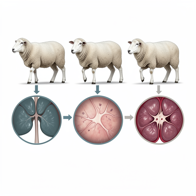
Dose-Response Relationship of Cottonseed Cake
AI-generated conceptual illustration (sheep liver tissue). It explains the idea and does not show study data.
View source evidence ↗Figures & tables
From the original paper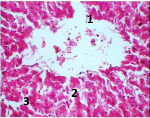 Figure 1
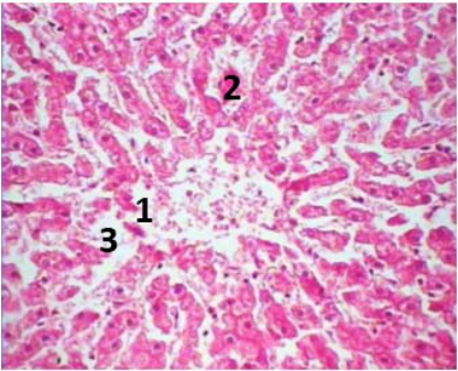 Figure 2
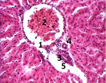 Figure 3
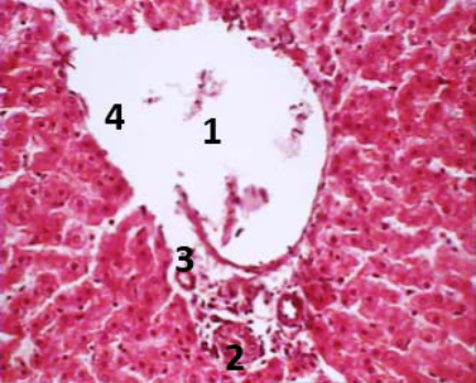 Figure 4
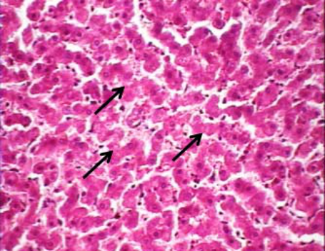 Figure 5
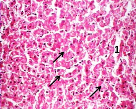 Figure 6
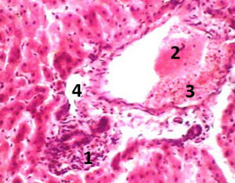 Figure 7
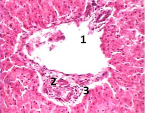 Figure 8
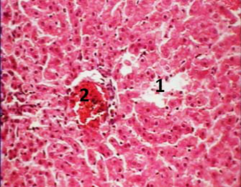 Figure 9
Introduction
Cottonseed cake, being rich in protein content, is used extensively for supplementing in the ruminant ration to increase productivity (Zahid et al., 2002) [18] . However, usage of cottonseed cake as animal feed for both ruminants and non-ruminants is restricted because of high levels of toxic substance, gossypol in cottonseed and cottonseed products (Matondi et al., 2007) [7] . Gossypol is a naturally occurring lipid soluble polyphenolic compound present in the seeds, stems and roots of cotton plant genus Gossypium (Price et al., 1993) [10] . Continuous feeding of diets containing gossypol can cause negative effects on growth performance and reproduction both in male and female (Santos et al., 2003) [13] . According to Randel et al. (1992) [11] , common clinical signs of gossypol toxicosis in monogastrics, preruminants and mature ruminants are similar and include labored breathing, dyspnea, decreased weight gain, anorexia, weakness and death after several days. In the present study, effects of feeding cottonseed cake on histopathological changes in liver of local sheep in Myanmar were studied.
Materials and Methods
Materials and Methods
Experimental animals and design
Six male sheep approximately 3 months of age and weighing from 13 kg to 20 kg were used in this experiment and kept in the individual cage. Primarily, all of the experimental animals were fed with rice straw and sesame cake as basal diet during the adaptation period for one month and also provided free access of drinking water. After adaptation period, sheep were randomly allocated into three groups (A, B and C) with two animals in each group. Group C was kept as control group and fed with basal diet, and groups A and B were fed 40% (21.1 mg free gossypol/kg body weight/day) and 25% (13.2 mg free gossypol/kg body weight/day) of cottonseed cake in the total diets, respectively. Diets were formulated isonitrogenously around 16% of crude protein. Concentrate contributed 40% and rice straw 60% of the feed given to sheep amounting to 3.5% of body weight. The experimental period was two months.
Microscopic examination
At the end of the experimental period, sheep from all groups; A, B and C were sacrificed by euthanization with excess dose of barbiturate via intravenous injection.
The liver was collected for tissue sections and then immediately fixed in 10% formalin. After that, tissue processing and staining were carried out. The samples were dehydrated in the series of ascending grades of alcohol followed by clearing in the changes of xylene, and then the tissues were infiltrated with different grades of melted paraffin in the automatic tissue processor. The tissues were then embedded in paraffin and finally the wax embedded specimens were sectioned at 3-4 µm thickness. The sections were floated on Luke-warm water in a floatation bath at 37 °C for stretching and then they were mounted on clean slides. Then glass slides were dried on a slide warmer at 37 °C. Tissue section slides were stained with Harris Haematoxylin and Eosin (H & E). Histopathological slides were examined under the light microscope and microphotographed with camera attached microscope.
Results
The observations in this study, the diets containing cottonseed cake at different inclusion levels induced varying degrees of histopathological changes in liver of sheep. Histopathological changes such as degenerated hepatocytes and degeneration of central vein were observed in both groups A and B. The sinusoidal spaces were wider than the normal and more distinct in group A (Figure 1 and2). Degenerations in the portal area such as degeneration of branch of portal vein and hepatic arteriole, degeneration of branch of bile duct and oedematous fluid in portal area were also found (Figure 3 and4). Moreover, liver cell cords were degenerated and necrosis characterized by disappearance of nucleus in both groups A and B (Figure 5 and6). Connective tissue infiltration was also seen in portal area of both groups fed with cottonseed cake (Figure 7 and8). In group B, congestion in branch of portal vein was observed (Figure 3 and7). In group A, congestion in central vein and disappearance of degenerated hepatocytes were revealed (Figure 9).
Discussion
In the present study, liver of sheep in both groups fed with cottonseed cake showed degeneration and necrosis of hepatocytes with widening of sinusoidal spaces (Figure 1 and 2). Moreover, degeneration and necrosis of liver cell cords characterized by disappearance of nucleus was also observed in both groups A and B (Figure 5 and6). This result was similar with the finding of El-Sharaky et al. (2010) [6] , who mentioned that the gossypol acetic acid treated male rats had serious degenerative changes in the liver. According to Ali and El-Sewedy (1984) [1] , gossypol was also hepatotoxic due to the depletion of hepatic glutathione. Glutathione was present in all types of living cells and was able to conjugate with a variety of chemically active compounds (Mitchell et al., 1976) [8] . Loss of cellular glutathione was indicative of cytotoxicity (Docks and Krishna, 1976) [5] . Sufficient depletion of cellular glutathione content allowed some reactive metabolites to react with cellular macromolecules leading to cellular injury and death (Docks and Krishna, 1976; Olson et al., 1980) [5,9] . There was infiltration of connective tissues around the blood vessels at the portal area of both groups A and B (Figure 7 and8). This was due to the destruction of the blood vessel walls and substitution of the damaged wall with the connective tissues. Sastry (1983) [14] reported that when irritant persist for long period, the body responds by producing excessive amount of connective tissue in the injury area. Moreover, sheep from both groups A and B exhibited degeneration of branch of portal vein and hepatic arteriole and also degeneration of central vein (Figure 1, 2, 3 and 4). Furthermore, proteinaceous fluid and RBCs in branch of portal vein (Figure 7) and oedematous fluid in the portal area (3, 4, 7 and 8) were also seen. These lesions were associated with the vascular wall injury and leakage of plasma protein into tissue space (Runnells et al., 1965) [12] . In addition, congestion in the branch of portal vein (Figure 3) in group B and congestion in central vein (Figure 9) in group A referred to as passive hyperaemia of the liver. Passive hyperaemia of the liver is caused by hindrance to the flow of blood in the hepatic vein, posterior vena cava and heart (Runnells et al., 1965) [12] . According to Wu et al. (1991) [16] , gossypol in cottonseed cake inhibited hydroxylase needed for synthesis of hydroxyproline. Szpak (2011) [15] reported that hydroxyproline is a major component of the protein collagen. The reduction in collagen content, or the more fragile character of the collagen in various organs, might induce such symptoms as vascular damage and capillary haemorrhage (Dewreede and Waymann, 1970) [4] . Furthermore, degeneration of branch of bile duct was found in Figure 3 and4. Yanwan et al. (1979) [17] reported that gossypol induced distension of endoplasmic reticulum and displacement of ribosomes from rough endoplasmic reticulum of hepatic cells. In addition, Band et al. (1989) [2] mentioned that gossypol inhibited protein synthesis in mammalian cells. That inhibition interfered the epithelial integrity (Chernoff, 2006) [3] .
Conclusion
From the present study, it could be concluded that the gossypol in cottonseed cake caused the degeneration and also destructions of blood vessel in liver of cottonseed cake treated groups. According to these lesions, inclusion of 40% cottonseed cake in the diet of sheep caused more toxic effect in liver than that of 25% in the diet.
Figures and tables from the paper
References
18 in the original paper- Ali SF, El-Sewedy SM. Effect of gossypol on liver metabolic enzymes in male rats. Toxicological Letters. 1984; 23:299-306.
- Band V, Hoffer AP, Band H, Rhinehardt AE, Knapp RC, Matlin SA et al. Antiproliferative effect of gossypol and its optical isomers on human reproductive cancer cell lines. Gynecologic Oncology. 1989; 32:273-277.
- Chernoff R. Systemic effects of nutrients on oral health, Geriatric Nutrition: The Health Professional's Handbook. 3 rd ed. USA, 2006, 193-195.
- Dewreede S, Waymann O. Effect of mimosine on the rat fetus. Teratology. 1970; 3:21-27.
- Docks EL, Krishna G. The role of glutathione in chloroform induced hepatotoxicity. Experimental Journal of Pathology. 1976; 24:13-22.
- El-Sharaky S, Newairy AA, Elguindy NM, Elwafa AA. Spermatotoxicity, biochemical changes and histological alteration induced by gossypol in testicular and hepatic tissues of male rats. Food and Chemical Toxicology. 2010; 48:3354-3361.
- Matondi GHM, Masama E, Mpofu IDT, Muronzi FF. Effect of feeding graded levels of cottonseed meal on goat erythrocyte membrane osmotic fragility. Livestock Research and Rural Development. 2007; 19:1-10.
- Mitchell JR, Hinson JA, Nelson SD. Glutathione and drug induced tissue lesions, Glutathione: Metabolism and function. Raven Press, New York, 1976, 357-367.
- Olson RD, McDonald JD, Vanboxtel CJ, Boerth RC, Harbison RD, Stonim AE et al. Regulatory role of glutathione and soluble sulfhydryl groups in the toxicity of Adriamycin. Journal Pharmacological Experimental Therapeutics. 1980; 215:450-545.
- Price WD, Lovell RA, McChesney DG. Naturally occurring toxins in feedstuffs: center for veterinary medicine perspective. Journal of Animal Science. 1993; 71:2556-2562.
- Randel RD, Chase Jr CC, Wyse SJ. Effects of gossypol and cottonseed products on reproduction of mammals. Journal of Animal Science. 1992; 70:1628-1638.
- Runnells RA, Monlux WS, Monlux AW. Principles of Veterinary Pathology. 7 th ed. The Iowa State University Press, 1965, 628-635.
- Santos JE, Villasenor M, Robinson PH, De Peters EJ, Holmberg CA. Type of cottonseed and level of gossypol in diets of lactating dairy cows: plasma gossypol, health and reproductive performance. Journal of Dairy Science. 2003; 86:892-905.
- Sastry GA. Veterinary Pathology. 6 th ed. CBS Publisher and Distributor Delhi, 1983, 240-284.
- Szpak P. Fish bone chemistry and ultrastructure: implications for taphonomy and stable isotope analysis. Journal of Archaeological Science. 2011; 38(12):33583372.
- Wu YW, Chik CL, Albertson BD, Linehan WM, Knazek RA. Inhibitory effects of gossypol on adrenal function. Acta Endocrinology. 1991; 124:672-678.
- Yanwan W, Shudong Z, Sher-pu S. Ultrastructural observation of the toxic effect of gossypol on rats. Acta Anatomica Sinica. 1979; 1:03-05.
- Zahid IA, Lodhi LA, Rehman N, Akhtar MS. Effects of gossypol on micrometry of teddy male goats. Pakistan Veterinary Journal. 2002; 22:101-104.