
Research article · 2016 · Full text
International Journal of Veterinary Sciences and Animal HusbandryStudy on lungworm infection in small ruminants: Prevalence and risk factors in and around Gondar Town, Northwest, Ethiopia
Volume 1(2) · Pages 47–52
Research areasTuberculosis Research and EpidemiologyZoonotic diseases and public healthHelminth infection and control
Abstract
Author-writtenA cross-sectional study was conducted from November 2011 to April 2012 to estimate the prevalence of lung worm infection in small ruminants, to identify lung worm species and the potential risk factors in and around Gondar town, North West Ethiopia. Fecal and post mortem examination were conducted in 601 (112 goats and 489sheep) and 73 (15 goats and 58 sheep), respectively. The overall prevalence of 43.76% and 46.57% was found by fecal and post mortem examination respectively. There was a perfect agreement between fecal and post mortem examination (Kappa=0.82).When the prevalence was calculated by considering different risk factors, the lung worm prevalence was significant (p<0.05) between sex with the high prevalence of female (46.77%) than males (34.87%) and different peasant association, but it was not significant (p>0.05) between species of animals, among different age groups, body condition score, month, management system and housing condition of the animal. The prevalence of lung worm was high in Dictyocaulus filaria among the three lung worm species and Protostrongylus rufescens has the lowest one, there was a significant difference (p<0.05) among them. In conclusion, this study indicated that lung worm has a high prevalence in the study area which implicate the need of control and prevention intervention.
Summary
Source-checkedPlain-language reading of the full paper, prepared with AI and checked against the text. Each point links to the authors' own words.
Why it matters
- Lungworms inhabit the lower respiratory system of ruminants and cause significant economic losses. View evidence ↗
- In sheep and goats, parasitic pneumonia is most frequently caused by three species: Dictyocaulus filaria, Muellerius capillaris, and Protostrongylus rufescens. View evidence ↗
The question
- The study aimed to estimate the prevalence of lungworm infection, identify the species present, and determine potential risk factors in small ruminants around Gondar town, Ethiopia. View evidence ↗
What they did
- The researchers examined 601 animals (112 goats and 489 sheep) via fecal analysis and 73 animals (15 goats and 58 sheep) via post-mortem examination. View evidence ↗
- Fecal samples were screened for first-stage (L1) lungworm larvae using a modified Baermann technique. View evidence ↗
What they found
- The overall prevalence of lungworm infection was 43.76% based on fecal exams and 46.57% based on post-mortem exams. View evidence ↗
- Female animals had a significantly higher prevalence of infection (46.8%) compared to males (34.9%). View evidence ↗
- Prevalence varied significantly by peasant association (PA), with the highest rate in Debarka (56.75%) and the lowest in Tseda (30.30%). View evidence ↗
- In post-mortem examinations, young animals under one year old showed a significantly higher prevalence (75%) than adults (38.6%). View evidence ↗
- In sheep, D. filaria was the most common species (52.1%), whereas in goats, M. capillaries was dominant (56%). View evidence ↗
- The modified Baermann technique demonstrated a sensitivity of 79% and a specificity of 100% compared to post-mortem examination. View evidence ↗
What it means
- The authors conclude that the high prevalence and variety of lungworm species pose a threat to small ruminant production in the study area. View evidence ↗
- The authors recommend two strategic anthelmintic treatments: one at the start of the dry season and one at the end of the long rainy season. View evidence ↗
- The authors note that the association between sex and infection needs further verification because the sample size for males was very small. View evidence ↗
Visual summary
3 visualsIllustrations explain the paper; charts are replotted from the paper's own table values.
Study Workflow
AI-generated conceptual illustration (sheep and goats). It explains the idea and does not show study data.
View source evidence ↗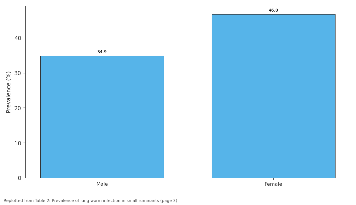
Prevalence (%): Male vs Female
Replotted from Table 2 (page 3) of the original paper. Every bar is the paper's own table value.
View source evidence ↗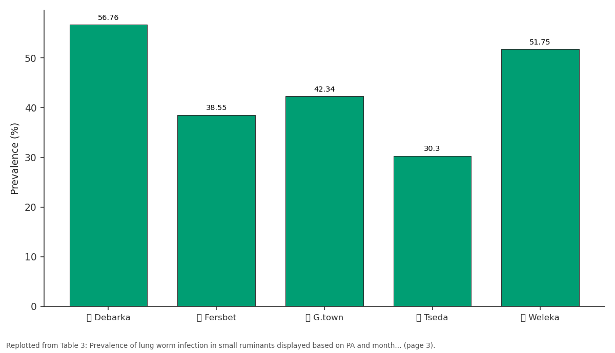
Prevalence (%): Debarka vs Fersbet vs G.town vs Tseda vs Weleka
Replotted from Table 3 (page 3) of the original paper. Every bar is the paper's own table value.
View source evidence ↗Figures & tables
From the original paper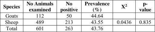 Table 1
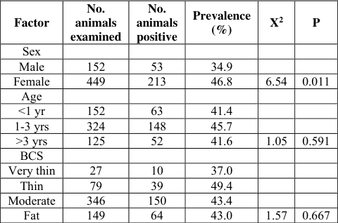 Table 2
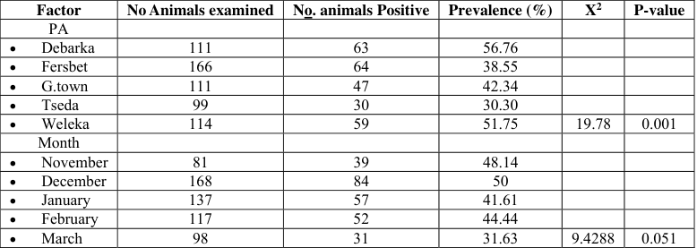 Table 3
 Table 4
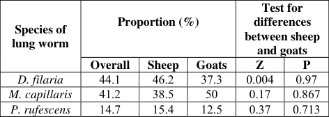 Table 8
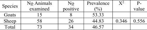 Table 5
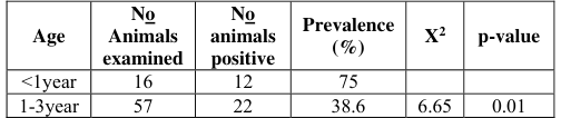 Table 6
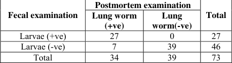 Table 9
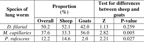 Table 7
Introduction
Ethiopia has numerous livestock resource with a total contribution of 15% Gross domestic product and 33% of the agricultural output. Current estimate of shows that there are 41.5 million heads of cattle, 28.2 million of sheep and goats,5.8 million of equine species, a million camels and over 42 million of poultry (DACA, 2006) [9] . sheep and goats provide as much as 30% the meat and milk consumed in sub-Saharan Africa and are found on small holding through the continent (ILCA, 1990) [18] . Health livestock represents one of mans most valuable renewable resource. They provide high quality edible protein, fiber of all types, leaser and arrange of useful by products, as well as providing an important ways of generating and storing wealth. Supplying motive power and fuel in developing countries. However, the demands on such live stock resource are extreme. Not only do live stock production system Have to cope with the demands of the ever increasing human population, super imposed on this is an even greater and legitimate requirement for them to contribute to improving the level of human nutrition by making live stock products more freely and cheaply available. This needs accomplished in situations were the land available to raise live stock being progressively reduced by the greater imperatives and priorities associated with housing and cropping (Waller, 1997) [35] . Helminthes parasites of ruminants are ubiquitous in many tropical and sub-tropical environments of the world providing nearly perfect condition for their survival and development. Among them lung worm which inhabit the lower respiratory system impose significant economic impact (Hansen and perry, 1996) [14] . Dictyocaulidae and /or certain Metastrongylidae are kwon to exist in East Africa (Ethiopia, Kenya and Tanzania) and South Africa (Shah-fischer and Say, 1989) [31] . Parasitic or Verminous pneumonia of sheep and goats most commonly are caused by infection with Dictyocaulus filaria, Muellerius capillaris, or Protostrongylus rufescens. In contrast to the acute viral and bacterial pneumonias, which result in bronchopneumonia affecting the anterior ventral portion of the lung (Kahn et al., 2005) [20] .
International Journal of Veterinary Sciences and Animal Husbandry
Dictyocaulus filaria is the most important lung worm of sheep and is commonly associated with chronic syndrome of coughing and unthriftiness with usually affects lambs and kids (Urquhart et al., 1996) [38] . Muellerius capillaris is a common lung worm of sheep and goats but infestation rarely leads clinical diseases (Jones et al., 1996) [19] . According to Pugh, 2002 [26] ; D. filaria is the most pathogen. M. capillaris is the most common and the least pathogenic, and P. rufescens is intermediate in pathogenicity. Infestation with Dictyocaulus filaria, Muellerius capillaris and Protostrongylus rufescens are all encountered in lambs 4-6 months of age are most severely affected but sheep of all ages are susceptible to lung worm infestation (Blood et al., 1983) [6] . Dictyocaulus filaria have direct life cycle where as Protostrongylus and Muellerius have in direct life cycles and rely on variety of snails and slugs to serve as intermediate hosts (Kahn et al., 2005) [20] . The life cycle of helminthes parasite of lungs are completed through the passage of their eggs or larvae up on the air way to the pharyngeal region, from which they are swallowed and passed to the out side environment in the faeces. Because of their dependence on fecal transmission, parasitism of lungs and air ways is often diagnosed by microscopic examination of faeces. The baermann technique can used to recover the larvae of lung worm from faeces (Hendrix, 1998) [15] . Pasture provide the link between the free living and parasitic phase of nematode parasite of grazing livestock. At different level of growth, pastures species may facilitate, or impede survival of free living population, the establishment of parasite burden and lessen or intensify the effect parasites in livestock (Waller, 1997) [35] . There for the objective of the study are (1).To estimate the prevalence of lung worms (2).To identify the potential risk factors(3).To identify the species of lung worms (D. filaria, M. capillaris and P. rufescens).
Materials and Method
Materials and Method
Study area
The study was conducted in and around Gondar town, which is the center of north Gondar zone in Amhara regional state. The town is located at the North-west part of Ethiopia at a distance of about of 748 km from the capital of Ethiopia. The average altitude of the area is 1967m above sea level and the latitude and longitude of the area is 12.4 0 N and 27.3 o E, respectively. The topography of Gondar is generally marked by the presence of numerous mountains, plateaus, hilly and sloppy area and rivers and the main topographic category of the area is Weina degas (mid land). The average rain fall of the area is estimated about 1000 mm and the short rains occur during March, April, and May, while the long rainy season extends from June to September. The average maximum and minimum daily temperature is 22-30.7 0 C and 12.3-17 0 C, respectively (NGZ ARDO, 2009) [24] .
Study animal
The study animals were randomly selected small ruminants (sheep and goats) in the study area, which are kept under the traditional husbandry system. Both sexes and all age groups were included during sampling. Generally the proportion of males to females was small in any flock and the number of goats is small in the area. These animals originated from peasant association in and around Gondar town.
Study Design and Examination Method
A cross sectional study was carried to determine the prevalence of lung worm infection in small ruminants, to identify lung worm species and the potential risk factors in and around Gondar town from October, 2011 to April, 2012. The study was conducted based on Fecal and post mortem examination. Species of animals, age groups, body condition score, month, management system and housing condition of the animal were considered as potential risk factors
Sample size and sampling procedure
The sampling size for the study was determined by using simple random sampling technique (Thrusfield, 2005) [35] . During calculation of the sample size, an expected prevalence of 37% was taken based information from a previous study at Dessie and Kombolcha (Regassa et al., 2009) [29] . The following formula was used to calculate the sample size:
The sample size calculated was 358 but in order to increase the accuracy of the study the number of the animals was increased to 601 (112 goats and 489 sheep).
Study methodology
Coprological examination: Fecal samples were taken directly from the rectum of randomly selected animals using disposable glove. Then the samples were placed in labeled plastic sampling bottles separately and taken to the laboratory in cool box. In the laboratory, the fecal sample was screened for the presence of lung worm larvae (L1) by using the modified Baermann technique (Kassi, 1999; Zajac and Comboy, 2006) [21,39] . The procedure was as follows: 10gram of feces was placed in a piece of double-layer cheese cloth, which was gathered around the sample so that it was fully enclosed. Use a rubber band to fasten the cloth, passé through the rubber band by two applicator sticks, which rest the edge of glass suspended the sample. Dip the feces with nylon in to conical glass filled with Luke warm water. Allow the feces to stand at 3-4 hours and discard the feces with nylon collect the material at the bottom of hollow stem Petri dish, examine with 10x objective lens and then transfer to microscopic slide to identify the species by using pipette. The species of lung worm was identified based on the morphological features given for each species (Soulsby, 1982; Taylor et al., 2007) [33,34] . When collecting faecal samples, information regarding the species of the animals, sex, age, body condition, feeding system, housing condition, and date of the sampling and origin of the animal were properly recorded on the provided format.
Postmortem examination: In addition to coprological examination, postmortem examination was carried out in a total of 73 animals (15 goats and 58 sheep) slaughtered at Restaurants to investigate the presence of adult lung worms.
In the mean time the age and species of the slaughtered animals were recorded. Procedures: lungs from slaughtered animals were palpated for protostrongylidae nodules. If the nodules were present the area was trimmed of and worms extracted from the tissue by gently compressing small non calcified nodules or a part of large nodules between two glass slides and carefully teasing the worm away from the tissue using thumb forceps. Air passage was opened starting from the trachea down to the small bronchi with fine blunt pointed scissors to detect the presence of adult Dictyocaulidae (Kassai, 1999) [21] .
The sensitivity and specificity of the modified Baermann technique
In this study a trial was made to evaluate the ability of the modified Baerman technique to detect lungworm infected and non infected animals correctly in relation to the gold standard postmortem examination method. For this the same 73 sheep and goats were subjected to both coprological and postmortem examinations. The results of the two tests were placed in a two by two contingency table and then comparisons were made. The sensitivity of the modified Baerman technique was calculated as the proportion of the number of animals tested positive by both tests divided by the total number of animals in which lungworms were detected in postmortem examination (a/a+c). Specificity was calculated as the proportion of the number of animals tested negative by both examinations divided by the total number of animals in which lungworms were not detected in postmortem examination (d/b+d) (Thrusfield, 2005) [35] .
Data analysis
The data collected from the study animals during both coprological and post mortem examinations were recorded on specially designed formats. The data were then coded and entered to computer Microsoft excel spreadsheet. All the statistical analyses were performed using STATA version 9 software. The prevalence was computed as the number of animals affected by the lungworms divided by the total number of animals tested multiplied by 100%. The association of the prevalence of lung worm infection with potential risk factors such as species, age, sex, BCS, month of sampling, feeding system, housing condition and PA was analyzed by using chi-square (X 2 ) test.
Results
Results
Coprological examination
Out of the total 601 small ruminants (489 sheep and 112 goats) examined by modified Baermann technique, 263 (43.79%) animals were found to be infected with lung worms. The prevalence of lungworm infection was not significantly (P>0.05) different between goats (44.64%) and sheep (43.55%) (Table 1). The results of the analysis of the prevalence of lung worm infection with the sex, age and BCS of sheep and goats are presented in Table 2. Accordingly, the infection was found to be significantly (P<0.05) associated with the sex of the animals. The prevalence was higher in females (46.8%) than males (34.9%). Age and BCS were not significantly associated with the prevalence of lung worm infection (P>0.05 in both cases). This study showed a significant (P = 0.001) variation in the prevalence of lungworm infection between the different PAs from which animals were sampled. The highest prevalence was observed in Debarka (56.75%) followed by Weleka (51.75%) and the lowest was observed at Tseda PA (30.30%). However, the occurrence of lung worm infection was not significantly (P>0.05) affected by the month of sampling (Table 3). A test conducted to see the association between the prevalence of lung worm infection and the feeding and housing condition of the animals showed no significant association with both factors (P>0.05 for each factor) (Table 4).
| Factor | No. animals examined | No. positive | Prevalence (%) | X2 | P |
|---|---|---|---|---|---|
| Feeding system | |||||
| Free ranging | 566 | 243 | 42.93 | ||
| Free ranging with supplementary feed | 35 | 20 | 57.14 | 2.70 | 0.100 |
| Housing condition | |||||
| In door | 103 | 54 | 52.43 | ||
| Out door | 498 | 209 | 41.97 | 3.79 | 0.051 |
Table text extracted from the original PDF; superscript letters are shown inline. The original table image is below.
View original table image
| Factor | No Animals examined | No. animals Positive | Prevalence (%) | X2 | P-value |
|---|---|---|---|---|---|
| PA | |||||
| Debarka | 111 | 63 | 56.76 | ||
| Fersbet | 166 | 64 | 38.55 | ||
| G.town | 111 | 47 | 42.34 | ||
| Tseda | 99 | 30 | 30.30 | ||
| Weleka | 114 | 59 | 51.75 | 19.78 | 0.001 |
| Month | |||||
| November | 81 | 39 | 48.14 | ||
| December | 168 | 84 | 50 | ||
| January | 137 | 57 | 41.61 | ||
| February | 117 | 52 | 44.44 | ||
| March | 98 | 31 | 31.63 | 9.4288 | 0.051 |
Table text extracted from the original PDF; superscript letters are shown inline. The original table image is below.
View original table image
| Factor | No. animals examined | No. animals positive | Prevalence (%) | X2 | P |
|---|---|---|---|---|---|
| Sex | |||||
| Male | 152 | 53 | 34.9 | ||
| Female | 449 | 213 | 46.8 | 6.54 | 0.011 |
| Age | |||||
| <1 yr | 152 | 63 | 41.4 | ||
| 1-3 yrs | 324 | 148 | 45.7 | ||
| >3 yrs | 125 | 52 | 41.6 | 1.05 | 0.591 |
| BCS | |||||
| Very thin | 27 | 10 | 37.0 | ||
| Thin | 79 | 39 | 49.4 | ||
| Moderate | 346 | 150 | 43.4 | ||
| Fat | 149 | 64 | 43.0 | 1.57 | 0.667 |
Table text extracted from the original PDF; superscript letters are shown inline. The original table image is below.
View original table image
| Species | No Animals examined | No positive | Prevalence (%) | X2 | p- value |
|---|---|---|---|---|---|
| Goats | 112 | 50 | 44.64 | ||
| Sheep | 489 | 213 | 43.55 | 0.0436 | 0.835 |
| Total | 601 | 263 | 43.76 |
Table text extracted from the original PDF; superscript letters are shown inline. The original table image is below.
View original table image
Postmortem examination
Postmortem examination of lungworms was performed on 73 small ruminants slaughtered at restaurants of which 34(46.57%) animals were found to be infected with different species of lung worms. As in coprological examination, there was no significant (P>0.05) difference in prevalence between sheep and goats, although it tends to be higher in goats (Table 5). Unlike the coprological examination, there was a significant (P<0.05) association between prevalence of lung worm and age of the animals. The prevalence was considerably higher in young animals <1yr (75%) than adult animals (38.6%) (Table 6).
| Age | No Animals examined | No animals positive | Prevalence (%) | X2 | p-value |
|---|---|---|---|---|---|
| <1year | 16 | 12 | 75 | ||
| 1-3year | 57 | 22 | 38.6 | 6.65 | 0.01 |
Table text extracted from the original PDF; superscript letters are shown inline. The original table image is below.
View original table image
| Species | No Animals examined | No positive | Prevalence (%) | X2 | P- value |
|---|---|---|---|---|---|
| Goats | 15 | 8 | 53.33 | ||
| Sheep | 58 | 26 | 44.83 | 0.346 | 0.556 |
| Total | 73 | 34 | 46.57 |
Table text extracted from the original PDF; superscript letters are shown inline. The original table image is below.
View original table image
Species identification
In coproplogical examination, three species of lung worms' namely D. filaria, M. capillaries and P. rufescens were identified in both sheep and goats. In sheep, D. filaria was the most prevalent (52.1%) while P.rufescens was the least (14.6%). However in goats, M. capillaries was the dominant species (56%) and P. rufescens was the least (2%). There was a significant variation in the proportions of M. capillaries and P. rufescens between sheep and goats but no significant difference was observed for D. filaria (Table 7). In postmortem examination the same three species of lungwrms were identified as in coprological examination. Similar to coprological examination, D. filaria and M. capillaris were the dominant species observed in sheep and goats, respectively while P. rufescens was the least prevalent in both species of animals (Table7).
| Species of lung worm | Proportion (%) | — | — | Test for differences between sheep and goats | — |
|---|---|---|---|---|---|
| Overall | Sheep | Goats | Z | P-value | |
| D. filarial | 50.2 | 52.1 | 42.0 | 1.13 | 0.259 |
| M. capillaries | 37.6 | 33.3 | 56.0 | 2.82 | 0.005 |
| P. rufescens | 12.2 | 14.6 | 2.0 | 2.21 | 0.027 |
Table text extracted from the original PDF; superscript letters are shown inline. The original table image is below.
View original table image
Evaluation of the sensitivity and specificity of the modified Baermann technique
In this study a trial was made to evaluate the sensitivity and specificity of the modified Baerman technique to detect the larvae (L1) of lungworms in the faeces in relation to the gold standard postmortem examination. According based on the given formula, the sensitivity and specificity of the modifief Baermann technique computed as 79% and 100% respectively (Table 8)
| Species of lung worm | Proportion (%) | — | — | Test for differences between sheep and goats | — |
|---|---|---|---|---|---|
| Overall | Sheep | Goats | Z | P | |
| D. filaria | 44.1 | 46.2 | 37.3 | 0.004 | 0.97 |
| M. capillaris | 41.2 | 38.5 | 50 | 0.17 | 0.867 |
| P. rufescens | 14.7 | 15.4 | 12.5 | 0.37 | 0.713 |
Table text extracted from the original PDF; superscript letters are shown inline. The original table image is below.
View original table image
Discussion
The present study revealed an over all prevalence of 43.76% lungworm infection in small ruminants in and around Gondar town by coprological examination. This finding is relatively lower than the prevalence reported by some previous studies: 53.6% in six districts of Wello (Alemu et al., 2006) [2] , 49% in and around Debre Birhan (Habtamu, 2010) [13] and 58% in Assela (Wondowoson, 1992) [36] . However it is higher than the 36.7% report from Dessie and Kombolcha districts (Regassa et al., 2009) [29] and 33.83% from Gondar town (Addis et al., 2011). The reason for such variation may by the different agro ecological conditions of the country, animal husbandry system and the competence of the investigator to detect the larvae of the parasite in the faeces. The observed prevalence is slightly lower than the prevalence obtained from postmortem examination (46.57%); however the sample size used for the latter was very small. In both coprological and postmortem examinations, there was no significant variation (P>0.05) in the prevalence of lungworm infection between goats and sheep, although it tended to be higher in goats. When the feeding behavior of the two species is considered, naturally goats are browsers and thus less exposed to helminthes than sheep, which are used to grazing close to the ground and consequently more exposed to the infective larvae on pasture. However due to deterioration of browsing plants associated with expansion of agricultural activities and deforestation, nowadays it is common to see goats grazing on communal pastures along with sheep and therefore both species are equally exposed to lungworm infection. The present finding is contrary to previous studies which reported a significantly higher prevalence in goats than sheep (Alemu et al., 2006; Addis et al., 2011) [2] or in sheep than goats (Regassa et al., 2010). In the present study three species of lungworms were identified in both sheep and goats in both coprological and postmortem examinations; however with different proportions. D. filaria and M. capillaries were the most prevalent species identified in sheep and goats, respectively while P. rufescens was the least dominant in both species of animals. This finding is inconsistent with previous studied (Alemu et al., 2006; Regassa et al., 2010) [2] where M. capillaries was reported to be the most dominant species in both sheep and goats. However it is quite in agreement with the result of Addis et al. (2010) [1] . The marked difference in the proportion between D. filaria and the other two species (M. capillaris and P. rufescens) is associated with the differences in life cycle of these lung worms. D. filaria, has a direct life cycle and also takes less time to reach the infective stage and after ingestion, the larvae can appear in the feces in a few weeks (Soulsby, 1982) [33] . Compared with D. filaria, the transmission of P. rufescens and M. capillaris is epidemiologically complex vent involving the animal host, parasite and intermediate host. Further more, the development of 1 st stage to infective stage larvae in the snail takes 12 to14 days and the prepatent period in the final host reaches 30 to 40 days. The probabilities of infection, transmission and reinfection would therefore be much lower compared with D. filaria (Urquhart et al., 1996) [38] . Similar to a previous study (Alemu et al., 2006) [2] , the present study indicated that females animals were significantly (p<0.05) more susceptible to lung worm infection than males. However, the study carried out by Regassa et al., (2010) showed no significant variation. There are two possible explanations for the observation of higher prevalence in females than males. The first is that the resistance of infection in females might have been abrogated at time of parturition and early lactation. This periparturient relaxation of resistance in females results in inability to expel adult worms (Craig, 1998). The other possible reason could be the dominance of the numbers of females over males. The total number of female and male animals sampled in the current study was 449 (74.7%) and 152 (25.3%), respectively. The number of females is almost three times that of males. This big variation in sample size between females and males might have caused such difference in prevalence. Therefore, further study is required to verify this fact. Age of the animals was not significantly (P>0.05) associated with the prevalence of lung worm infection. Almost an equal level of infection was observed between young (41.4%) and older (41.6%) animals and the prevalence in the adult age group (45.7%) was slightly higher than that of the other age groups. This finding is inconsistent with that of previous studies (Alemu et al., 2006; Regassa et al., 2010) [2] where the infection tended to increase with increasing of age. Contrary to all these reports, Addis et al. (2010) [1] have reported a decrease in prevalence with increasing of age. In contrast to some studies which reported significant association of lungworm infection with poor BCS (Sisay, 2008) [32] , the prevalence of lung worm infection in the present study was not significantly (P>0.05) associated with the BCS of the animals. Out of the total 601 animals examined only 106 (17.6%) scored thin to very thin BCS while the majority (82.4%) of animals sampled scored moderate to fat BCS. The small sample size of thin and very thin animals compared to the larger size of moderate and fat animals in the present study could be a possible reason for the absence of association. It was reported that animals with low body condition scores appear to be less compatible in getting ride-off lung worm infection than animals that have high body condition score (Kimberling, 1988) [22] . In this study the prevalence of lung worm infection was significantly (P = 0.001) associated with the peasant association from which samples were taken. Such variation may be the result of altitudinal differences between the PAs, accessibility to anthelmintic medication and the level of awareness of the owner. The monthly dynamics of lung worm infection during the study period showed that the prevalence was relatively higher in November and December, then declined from January through March; however, the monthly variation was not significant (P>0.050). This finding is inconsistent with that of others (Alemu et al., 2006; Regassa et al., 2010) [2] who reported a significant monthly variation with a sharp decline from November to March associated with the progress of the dry period. The test made to evaluate the diagnostic capability of the modified Baermann technique has revealed that the sensitivity and specificity of the method to be 79% and 100%, respectively. This shows that when the Barman technique is applied to test sheep and goats for lungworm infection it misses only 21 infected animals out of 100 but it never misses a non infected animal.
Conclusion and Recommendation
The current study showed 43.76% and 46.57% of prevalence of lung worm infection in small ruminants in coprological and postmortem examination, respectively. All the three important lung worm species: D. filaria, M. capillaris and P. rufescens were identified in both sheep and goats. The observation of such a level infection in a season known to be dry in Ethiopia suggests the existence of suitable ecological conditions in the study area for the survival and perpetuation of the parasite throughout the year. Furthermore, the observed high prevalence coupled with the presence of all the important lungworm species reminds that the parasite could be a threat for small ruminant production in the area. This study has signified that sex (being female) of the animals and peasant associations are the two important risk factors associated with the occurrence of lungworm infection in small ruminants in the study area. However, the association of infection with sex requires further verification as the number of male animals involved in the study is very small compared to females. Furthermore, the absence of association between lungworm infection and species of animal, age, body condition and month of the year should be confirmed through a further research.
Based on the above facts the following recommendations are forwarded Two strategic treatments using broad spectrum or specific anthelmintics, one at the beginning of dry season and the other at the end of long rainy season, should be applied to reduce the high level of infection in the area, Grazing of small ruminants in wet areas should be avoided, that is a favorable habitant for the development of the lung worm's larvae, The snail and slug intermediates host creep up on plants in early morning and evening there fore, animals should not be allowed to graze in such times. It is quite difficult to establish the seasonality of the infection on the basis of the facts and figures presented by this study. There fore, an additional investigation is required on the prevalence and intensity of this parasite during the rest of the years.
Figures and tables from the paper
| Fecal examination | Postmortem examination | — | Total |
|---|---|---|---|
| Lung worm (+ve) | Lung worm(-ve) | ||
| Larvae (+ve) | 27 | 0 | 27 |
| Larvae (-ve) | 7 | 39 | 46 |
| Total | 34 | 39 | 73 |
Table text extracted from the original PDF; superscript letters are shown inline. The original table image is below.
View original table image
References
39 in the original paper- Addis M, Fromsa A. Study on the Prevalence of Lung Worm Infection in Small Ruminants in Gondar Town, Ethiopia. Vet. Res. 2010; 4(3):85-89.
- Alemu A, Gelaye E, Ayelet G, Zeleke A. Study on Small Ruminants Lung Worm Infectionin North East Ethiopia. Veterinary Parasitology. 2006; 142:330-33.
- Ayalew W, Frecher LJ, Malo R, Beauregard C. Study on the Incidence of Preliminary Survive of Sheep Helminthiasis in Kembebit, 1993.
- Atiken ID. Diseases of Sheep.4 th ed.Scot land, UK: Black Well, Moredium, 2007, 336-338.
- Ball weber L. The Practical Veterinarian, Veterinary Parasitology. Mississippi State: BH, 2002, 219-225.
- Blood DC, Radostitis OM, Henderson JA. Veterinary Medicine. A text book of the disease of cattle, sheep, pigs, goats and horse. 6 th ed. British: ELBS TINDALL and BAILLIERE, 1983, 921-922.
- Coste R, Smith AJ. The Tropical Agriculturalist; Animal Health Specific Disease. 1 st ed. London and Oxford: CTA, Macmillan Eduction LTD. 1994; 2:147-150.
- Craig TM. Epidemiology of Internal Parasite, Effect of Climate and Host on Reproductive Cycle on Parasite Survival. In: Small Animal for Mixed Ruminant Practitioner, Western Veterinary Conference, Las vegas, Nevada, 1998.
- DACA. Drug Administration and Control Authority of Ethiopia. Standard Treatment Guidelines for Veterinary Practices. Ethiopia: Chamber Printing House, 2006, 2.
- Devendra C, Mcleroy GB. Goat and Sheep Production in the Tropics 2 nd . England: ESBS. 1997, 218-219.
- Foreyt WJ. Veterinary Parasitology: Reference Manual.5 th ed.IowaState University Press: Black well, 2001, 88.
- Fraser CM. The Merck Veterinary Manual. A Hand Book of Diagnosis and Therapy for the Veterinarian. 4 th ed. USA: MERCK and INC.RAHWAY, N.J, 1973, 699-701.
- Habtamu T. Prevalence of Ovine Lung Worm in and around Debre Birhan. DVM thesis. Gondar University FVM. Gondar, Ethiopia, 2010.
- Hansen J, Perry B. The Epidemiology, Diagnosis and Control of Helminth Parasites of Ruminants, Nairobi, Kenya: ILRAD, 1996, 38.
- Hendrix CM. Diagnosti Veterinary Parasitology. 2 nd ed. MOSBY, INC. Auburn, Alabama, 1998.
- Hindson JC, Winter AC. Manual of Sheep Disease. 2 nd ed. UK: Black Well Science, 2002, 196-198.
- Howard JC. Current Veterinary Therapy: Food Animal Practice. 2 nd ed. Mexico: WB, SAUNDERS, 1986, 673674.
- ILCA. International live stock center for Africa. Annual Report Program High Lands ILCA Addis Ababa, Ethiopia, 1990.
- Jones TC, Hunt RD, King NW. Veterinary pathology. 6 th ed.USA: LIPPINCOTT WILLIAMS and WILKINS, 1996, 631-634.
- Kahn CM, BM, MA. Respiratory Disease of Sheep and Goats. In: Merck Veterinary Manual. 9 th ed. USA: MERCK and CO. INC, 2005, 1181-1230.
- Kassai T. Veterinary Helminthology. 1 st ed. British: BH, 1999, 191-198.
- Kimberling CV. Disease of Sheep. 3 rd ed. Lea and Febieger, Philadelphia, 1988, 29-31.
- McCgavin MD, Zachary JF. Parasitic Pneumonia of Sheep and Goats. In: Pathologic Basis Veterinary Disease 4 th ed. China: MOSBY ELSEVIER, 2002, 534-536.
- NGZARDO. North Gondar Zone Agricultural and Rural Development Office. Annual Report of North Gondar Zone Cooperative Office, 2009.
- Peacock C. Improving Goat Production in the Tropics. A Manual of Development Workers. 1 th ed. UK: Oxfam in Association with FARM Africa, 1997, 35-36.
- Pugh DG. Sheep and Goat Medicine.1 th ed. USA: SAUNDERS an Imprint of Elsevier, 2002, 123-124.
- Radostitis OM, Bood DC, Gay CC. Veterinary Medicine: A text book of the disease of cattle, sheep, goats pigs and horse. 8 th ed. London: Bailliere Tindall. 1994; (2):12511252.
- Radostitis OM, Gay CC, Hinchcliff KM, Constable PB. Veterinary Medicine. A text book of the disease of cattle, sheep, goats pigs and horse.10 th ed. Spain: SAUNDERS ELSEVIER, 2007, 1568-1569.
- Regassa A, Toyeb M, Abebe R, Megresa B, Mekibib B, Mekuria S. Lung Worm Infection in Small Ruminants: Prevalence and Associated Risk Factors in Dessie and Kombolcha Districts, North Eastern Ethiopia. Veterinary Parasitology. 2009; 169:144-148.
- Saxena CB, Ral P, Shrivastava VP. Veterinary Post Mortem Examination Laboratory Manual.1 st .ed.Delhi, VLKAS publishing House PVT.LTD, 1998, 112.
- Shah-fischer M, Say R. Manual of Tropical Veterinary Parasitology.2 nd ed. UK: ELBS, 1989, 54-85.
- Sisay GB. Study on the Prevalence of Lung Worms of Small Ruminants in and Around Assela. DVM Thesis. Hawassa University, FVM. Hawassa, Ethiopia. Smith, M.C. and Sherman. D.M. (1994): Goat Medicine. Walter Kluewer Company, Philadelphia, 2008.
- Soulsby EJL. Helminthes, Arthropods, and Protozoa of Domesticated Animals. 7 th ed. London: Bailliere Tindall, 1982, 262-274.
- Taylor MA, Coop RL, Wall RL. Parasite of Respiratory System. In: Veterinary Parasitolog. 3 rd ed. UK: Black Well, 2007, 195-198.
- Thrusfield M. Veterinary Epidemiology. 2 nd ed. UK: Black Well Science. 180-181. Waller PJ (1997).Sustainable Helminth Control of Ruminants in Developing Countries. ELSEVIER. Veterinary Parasitology. 2005; 7(l):195-207.
- Wondwoson T. Prevalence of lung worm in Ovine in and around Assela. DVM. Thesis. FVM. Addis Ababa University. Debre Zeit, Ethiopia, 1992.
- Urquhart GM, Armour J, Duncan AM, Dunn AM, Jennings FW. Veterinary Parasitology. 1 th ed. UK: ELBS, 1987, 57-58.
- Urquhart GM, Armour J, Duncan AM, Dunn AM, Jennings FW. Veterinary Parasitology. 2 nd ed. Scot land: Black Well Science, 1996, 41-60.
- Zajac AM, Conboy GA. Veterinary Clinical Parasitology. 7 th ed. USA: Black Well, 2006, (2):12-13.
Cited by 4
From OpenAlex, OpenCitations and reference lists read by Scinovex
- Lungworm infection in small ruminants in Ethiopia: Systematic review and meta-analysis ↗
Kassahun Asmare, Berhanu Sibhat, Aynalem Haile, Desie Sheferaw, Kassaye Aragaw, Mesele Abera et al. · Veterinary Parasitology Regional Studies and Reports · 2018
❝ 12 - Prevalence and pathological features of ovine lungworm infection in slaughtered animals in Nangarhar Province of Afghanistan ↗
Assadullah Samadi, Najibullah Faizi, Ahmad Jan Abi, Abdul Razaq Irshad, Nabil Hailat · Comparative Clinical Pathology · 2019
❝ 10 - Prevalence of Protostrongylus sp. in ruminants in a semi-arid region of Northeastern Brazil ↗
Lucia Oliveira de Macedo, Karlla Keyla Ferreira dos Santos, Renata Silva Brito, Gílcia Aparecida de Carvalho, Rafael Antonio Nascimento Ramos · Arquivo Brasileiro de Medicina Veterinária e Zootecnia · 2022
Lungworms are an important group of nematodes that infect domestic and wild animals throughout the world (Panuska, 2006;Bezerra-Santos et al., 2020).In general, these parasites colonize the lower respiratory tract, resulting in a wide plethora of injuries and, occasionally, causing fatal pneumonia (Panayotova-Pencheva and Alexandrov, 2010).In livestock animals, aside from their pathogenic implications, infections by lungworms also cause significant economic losses,…
❝ 0PDF - Epidemiología molecular de las enfermedades parasitarias del tracto respiratorio en pequeños rumiantes del departamento de Boyacá ↗
Díaz Anaya, Adriana María · 2019
❝ 0