
Research article · 2016 · Full text
International Journal of Veterinary Sciences and Animal HusbandrySlaughter house surveillance for tuberculosis among cattle in Ri-Bhoi district of Meghalaya
Volume 1(1) · Pages 14–15
Research areasAnimal Disease Management and EpidemiologyLivestock Management and Performance ImprovementViral Infections and Vectors
Abstract
Author-writtenThe study was carried out to investigate bovine tuberculosis by necropsy inspection in different abattoirs of Meghalaya. Gross visual lesions were found in 26 carcasses. Suspected 39 tissue samples were collected from 26 carcasses. Pre-culture stain revealed 30 (76.92%) and culture growths were from 31 (79.49%) tissue samples. Highest lesions were recorded in lymph nodes (48.71%) followed by lungs (28.20%) and liver (23.07%). Pre-scapular (47.37%) and retropharyngeal (31.58%) contribute more lesions than other lymph nodes. Visual inspection may serve as good screening method for tuberculosis infected carcasses although pre culture staining and culture of bacteria from suspected lesion samples is necessary to have a concluding remark about the infection. The study also confirms the endemic status of bovine tuberculosis in these areas.
Summary
Source-checkedPlain-language reading of the full paper, prepared with AI and checked against the text. Each point links to the authors' own words.
Why it matters
- Bovine tuberculosis (BTB) is a significant infectious disease affecting domesticated and some captive wild animals in many developing nations. View evidence ↗
- M. bovis is estimated to cause approximately 5% of all tuberculosis infections in humans. View evidence ↗
- Eating beef from infected cattle poses a public health risk because cooking does not always eliminate M. bovis infection. View evidence ↗
The question
- The study aimed to investigate the prevalence and nature of bovine tuberculosis infection in abattoirs within the Ri-Bhoi district of Meghalaya. View evidence ↗
What they did
- The researchers sampled 120 animals from slaughterhouses, selecting those based on their debilitating health conditions. View evidence ↗
- All carcasses underwent gross necropsy inspection to identify visible lesions suspected of being tuberculosis. View evidence ↗
- Pre-culture staining using the Ziehl-Neelson (ZN) method was used on all tissue samples to detect acid-fast bacteria (AFB). View evidence ↗
- Mycobacterial culture was performed by inoculating samples onto Lowenstein Jensen (LJ) slants, which were incubated at 37°C and monitored weekly for eight weeks. View evidence ↗
What they found
- Based on slaughterhouse surveillance, the prevalence rate of bovine tuberculosis in the study area was 15%. View evidence ↗
- Among the organs examined, lymph nodes had the highest frequency of lesions (48.71%), followed by lungs (28.20%) and liver (23.07%). View evidence ↗
- Among the lymph nodes involved, the prescapular lymph nodes showed the highest rate of infection at 47.37%. View evidence ↗
- Of the 39 suspected lesion samples, 30 (76.92%) were positive by pre-culture stain and 31 (79.49%) were positive by culture. View evidence ↗
- Five animals had visible lesions that did not show mycobacterial growth or staining, suggesting these lesions may have been caused by parasitic infestation. View evidence ↗
What it means
- The authors conclude that while visual inspection is a useful screening tool, pre-culture staining and bacterial culture are necessary for a definitive diagnosis of infection. View evidence ↗
- The study confirms that bovine tuberculosis is endemic in the Ri-Bhoi district of Meghalaya. View evidence ↗
- The authors note that the manual maceration and homogenization of tissue samples prior to decontamination might have affected the culture conditions. View evidence ↗
Figures & tables
From the original paper Table 1
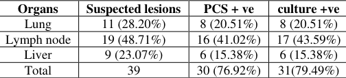 Table 2
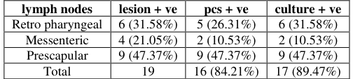 Table 3
Introduction
In many developing countries bovine tuberculosis (BTB) is a major infectious disease among domesticated animals and certain captive wild animals. It is estimated that M. bovis is responsible for about 5% of all TB infection in human (Michel et al. 2010) [5] . India possesses more than 16% of world cattle population. Cattles are also considered as natural host of M. bovis. Milk from healthy lactating cows had been reported to shed M. bovis bacilli (Danbirni et al. 2010) [3] . Ingestion of beef from infected cattle can be a major threat to public health as cooking may not always be an effective against M. bovis infection (van der Merwe et al. 2009) [7] . Abattoirs, butcher shops provide an ideal environment as a monitoring point for the screening of carcasses for BTB. Aerosol exposure to M. bovis is considered to be the most frequent route of infection of cattle, but infection may be occurred by contaminated material. The present study was undertaken for the purpose to investigate the infection of Bovine TB in abattoirs in Ri-Bhoi district of Meghalaya. In Meghalaya cattle are reared mostly for milk and livelihood. Predominantly local indigenous cattle constitutes more than 70% and others are jersey crossbred in Ri-Bhoi district.
Materials and Methods
Materials and Methods
Gross necropsy
All the carcasses were inspected for any gross visible lesion suspected of tuberculosis. Organs and tissue samples were collected from all the carcasses for further analysis. In this study, an animal was considered positive on necropsy if 1 or more lymph nodes or other tissues contained focal or multifocal abscesses or granulomas.
Pre-culture staining (PCS)
Ziehl-Neelson (ZN) staining for the detection of acid-fast bacteria (AFB) was performed on all tissue samples. A sample was considered positive for tuberculosis if there was evidence of granulomatous inflammation associated with focal necrosis or mineralization and/or if there was identification of AFB on the ZN stain.
Mycobacterial culture and species identification
Fresh and stored tissue samples were macerated and decontaminated using NALC and inoculated on to Lowenstein Jensen (LJ) media. Briefly, approximately 1g of tissue exhibiting gross visible lesions was sliced and homogenized and then subjected for decontamination. The supernatant was discarded and the pellet formed re-suspended in 300µl of phosphate buffered saline (140mM NaCl, 26mM KCl, 10.0 mM Na2HPO4 and 1.7 mM KH2PO4). Then the re-suspended pellets were inoculated in duplicates onto LJ slants (one incorporating glycerol and the other pyruvate). LJ slants were incubated at 37 o C and observed weekly for eight weeks. Using a sterile 0.1 µl plastic loop, the re-suspended pellets were spread and fixed at 80 o C (for 10 min) onto a labelled slide. The slides were subjected for staining with modified ZN stain.
Results and Discussion
A total of 120 animals were pooled from slaughter house based on their debilitating health condition. Suspected visual lesions were observed in 20 carcasses, of which 16 were positive for mycobacteria in pre culture ZN stain and 18 positive in subsequent culture (Table 1). However, because a few of the animal each had more than one organ presenting lesions, samples of suspicious organs were obtained from 26 animals. In terms of organ involvement, the majority lesions were found in lymph nodes (48.71%) followed by lungs (28.20%) and liver (23.07%) (Table 2). Out of 19 lymph nodes, prescapular lymph nodes contribute more (Table 3). The high proportion of these lymph nodes with lesions suggestive of tuberculosis indicates that most routes of infection could be through aerosol infection or injuries or ulcer in the neck, shoulder, thorax or chest cavity (Atiadeve et al. 2014) [2] . Prevalence rate was found to be 15% in this area based on slaughter house surveillance. The apparent animal prevalence with lesions suggestive of tuberculosis is comparable with Stefan et al. (2009) [6] . A distribution of lesions by organs shows that lymph nodes were the most infected followed by lung tissue and liver. Even though Bovine TB lesions are often found in the pulmonary region, other organs can equally be affected (Guitierrez et al. 1993) [4] . In 5 animals, although lesions are found but did not show any stain and culture growth, which may be due to parasitic infestation. The presence of visible lesions in any organs may not always be linked to mycobacterial infections as lesions may also cause by parasitic infestation or intracellular agents and this may lead to erroneous judgement by the meat inspector (Asseged et al. 2004) [1] . In the context of slaughter surveillance or carcass inspection, isolation of mycobacteria from bovine tissues can be difficult because there should first exist gross visible lesions. It also be noted that not all infected bovines may exhibit gross lesions. Preparation of sample by manual maceration and homogenisation of tissue before decontamination may also affect the culture condition. Due to economic loss most farmers sell infected/sick animals or send to slaughter house. These areas are considered as endemic for BTB which also reflected in this study. Microscopic examination of specimens taken from the abattoir were consistently scored highest for the presence of AFB implying a greater mycobacterial load, probably emanating from very sick animals (Atiadeve et al. 2014) [2] . This might be the reason of high incidence of tuberculosis in these areas. Transmission to neighbouring herd at market area and contamination through slaughter utensils are also recorded.
| lymph nodes | lesion + ve | pcs + ve | culture + ve |
|---|---|---|---|
| Retro pharyngeal | 6 (31.58%) | 5 (26.31%) | 6 (31.58%) |
| Messenteric | 4 (21.05%) | 2 (10.53%) | 2 (10.53%) |
| Prescapular | 9 (47.37%) | 9 (47.37%) | 9 (47.37%) |
| Total | 19 | 16 (84.21%) | 17 (89.47%) |
Table text extracted from the original PDF; superscript letters are shown inline. The original table image is below.
View original table image
| Organs | Suspected lesions | PCS + ve | culture +ve |
|---|---|---|---|
| Lung | 11 (28.20%) | 8 (20.51%) | 8 (20.51%) |
| Lymph node | 19 (48.71%) | 16 (41.02%) | 17 (43.59%) |
| Liver | 9 (23.07%) | 6 (15.38%) | 6 (15.38%) |
| Total | 39 | 30 (76.92%) | 31(79.49%) |
Table text extracted from the original PDF; superscript letters are shown inline. The original table image is below.
View original table image
| — | Total | Positive carcasses |
|---|---|---|
| Visual lesions (suspected) | 120 | 26 (21.67%) |
| pre culture stain | 16 (13.33%) | |
| Culture | 18 (15.0 %) |
Table text extracted from the original PDF; superscript letters are shown inline. The original table image is below.
View original table image
Proficient visual inspection may serve as good screening method for tuberculosis infected carcasses although pre culture stain and/or culture of bacteria may give brief idea about the infection.
Acknowledgement
Authors are thankful to ICAR, New Delhi for funding the "Outreach Project on Zoonotic diseases" and Director of Research (Veterinary) for necessary facilities to carry out the research. Due acknowledgement is also extended to the abattoir workers for providing samples.
References
7 in the original paper- Asseged B, Woldesenbet Z, Yimer E, Lemma E. Evaluation of Abattoir Inspection for the diagnosis of Mycobacterium bovis infection in cattle at Addis Ababa Abattoir. Trop. Anim. Health Prod. 2004; 36:537-546.
- Atiadeve SK, Gyamfi OK, Mensah EM, Galyuon IKA, Owusu D, Bonsu FA et al. Slaughter surveillance for tuberculosis among cattle in three metropolitan abattoirs in Ghana. J. Vet. Med. Anim. Health, 2014; 6:198-207.
- Danbirni S, Sackey ABK, Ayo JO, Bawa EK, Kudi AC, Okayeto SO et al. Exposure and Shedding in Milk of Mycobacterium bovis in dairy herds using One-Step antigen rapid bovine tuberculosis antibodies test and Ziehl-neelsen stain. Vet. Res. 2010; 3:38-42.
- Guitierrez MC, Garcia MJF. Comparison of ZiehlNeelsen staining and immune-histochemistry for the detection of Mycobacterium bovis in Bovine and Caprine tuberculosis lesions. J. Comp Pathol. 1993; 109:361-370.
- Michel AL, Muller B, Van Helden PD. Mycobacterium bovis at the animal-human interface: a problem, or not? Vet. Microbiol. 2010; 140:371-381.
- Stefan B, Fridessa R, Habtamu M, Gadisa E, Mengistu A, Yamuah L et al. The Burden of Mycobacteria Disease in Ethiopian Cattle: Implications for Public Health. PloS ONE, 2009; 4:344-348.
- Van der Merwe M, Bekker JL, van der Merwe P, Michel AL. Cooking and Drying as Effective Mechanisms in Limiting the Zoonotic effect of Mycobacterium bovis in Beef JS. Afr. Vet. Assoc. 2009; 80:142-145. (En.).
Cited by 7
From OpenAlex, OpenCitations and reference lists read by Scinovex
- Bovine tuberculosis in India: The need for One Health approach and the way forward ↗
Harini Ramanujam, Kannan Palaniyandi · One Health · 2023
Bovine tuberculosis (bTB) is a chronic granulomatous infectious illness in cattle. The etiological agent of bTB is Mycobacterium bovis. However, other members belonging to the Mycobacterium tuberculosis complex, like M. tuberculosis, M. africanum, M. caprae, M. orygis, and M. microti are known to cause bTB in cattle. There are 303.76 million bovines in India, and it is the largest producer…
❝ 43PDF - A review on bovine tuberculosis in India ↗
Ahmed Kabir Refaya, Gunapati Bhargavi, Noelin Chinnu Mathew, Ananthi Rajendran, Radhika Krishnamoorthy, Soumya Swaminathan et al. · Tuberculosis · 2020
❝ 43 - Role of abattoir monitoring in determining the prevalence of bovine tuberculosis: A systematic review and meta‐analysis ↗
Harini Ramanujam, Kannan Thiruvengadam, Ranjani Singaraj, Kannan Palaniyandi · Transboundary and Emerging Diseases · 2021
Bovine tuberculosis (bTB) is one of the major zoonotic concerns of the world, as milk and meat from cattle are major products for human consumption. Bovine tuberculosis not only affects the health of cattle and poses an imminent zoonotic threat, but also causes significant economic loss in both developed and developing countries. This systematic review reports the prevalence of Mycobacterium…
❝ 14PDF - Challenges in diagnosing bovine tuberculosis through surveillance and characterization of Mycobacterium species in slaughtered cattle in Kolkata ↗
Molla Zakirul Haque, CHANCHAL GUHA, Ayan Mukherjee, Sukhen Samanta, Partha Sarathi Jana, Ujjwal Biswas et al. · BMC Veterinary Research · 2024
BACKGROUND: Tuberculosis in cattle is caused by Mycobacterium tuberculosis complex (MTBC) species. Apart from MTBC, different Nontuberculous Mycobacteria (NTM) species have also been isolated from cattle. The presence of NTM infection in bovines makes the diagnosis of bovine tuberculosis (bTB) a cumbersome task. Therefore, a cross sectional study was conducted to isolate and characterize different Mycobacterium spp. from a slaughterhouse…
❝ 6PDF - Recovery of Mycobacterium tuberculosis Complex Isolates Including Pre–Extensively Drug-Resistant Strains From Cattle at a Slaughterhouse in Chennai, India ↗
Harini Ramanujam, Ahmed Kabir Refaya, Kannan Thiruvengadam, N. Pazhanivel, Devika Kandasamy, Ashok Kumar Shanmugavel et al. · Open Forum Infectious Diseases · 2024
Abstract Background India has the highest global burden of human tuberculosis (TB) and the largest cattle herd with endemic bovine TB (bTB). However, the extent of cross-species transmission and the zoonotic spillover risk, including drug-resistant Mycobacterium tuberculosis complex (MTBC) strains circulating in cattle, remain uncharacterized. Methods To address this major knowledge gap, we investigated tissue samples from 500 apparently healthy…
❝ 5PDF - Surveillance and Species Identification of Mycobacteria in Cattle from Abattoirs of Assam and Meghalaya ↗
Acheenta Gohain Barua, Koushik Kakoty, Pranjal Moni Nath, Himangshu Raj, Jyoti Pawan Chutia, Pranab Koch · International Journal of Current Microbiology and Applied Sciences · 2019
The study was carried out to investigate bovine tuberculosis and diagnostic potential of IFN-γ assay and necropsy inspection in different abattoirs of Assam and Meghalaya, including its species identification. A total number of 234 animals were screened by IFN-γ and necropsy inspection. IFN-γ was performed according to kit procedures and carcasses were inspected for any gross visible lesion. Species identification…
❝ 0PDF - Haematological and biochemical parameters altered in tuberculin reactor cattle ↗
AG Barua, Chayanika Goswami, Hanumanthrao G. Raj, Rumi Saikia Bora, P Kakoty · International Journal of Chemical Studies · 2018
Tuberculosis is a chronic granulomatous infectious zoonotic disease prevalent in developing countries resulting high mortality and economic loss. The present study was taken up to understand the correlation between different haematological features in the typical cases of tuberculosis in dairy herds with that of comparative tuberculin positive, inconclusive and negative reactors. The study was carried out in unorganized dairy farms